Distal cervical caries in the mandibular second molar - Surgical ...
Distal cervical caries in the mandibular second molar - Surgical ...
Distal cervical caries in the mandibular second molar - Surgical ...

Create successful ePaper yourself
Turn your PDF publications into a flip-book with our unique Google optimized e-Paper software.
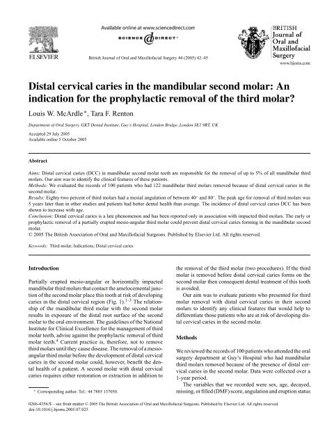
British Journal of Oral and Maxillofacial Surgery 44 (2005) 42–45<br />
<strong>Distal</strong> <strong>cervical</strong> <strong>caries</strong> <strong>in</strong> <strong>the</strong> <strong>mandibular</strong> <strong>second</strong> <strong>molar</strong>: An<br />
<strong>in</strong>dication for <strong>the</strong> prophylactic removal of <strong>the</strong> third <strong>molar</strong><br />
Louis W. McArdle ∗ , Tara F. Renton<br />
Department of Oral Surgery, GKT Dental Institute, Guy’s Hospital, London Bridge, London SE1 9RT, UK<br />
Accepted 29 July 2005<br />
Available onl<strong>in</strong>e 5 October 2005<br />
Abstract<br />
Aims: <strong>Distal</strong> <strong>cervical</strong> <strong>caries</strong> (DCC) <strong>in</strong> <strong>mandibular</strong> <strong>second</strong> <strong>molar</strong> teeth are responsible for <strong>the</strong> removal of up to 5% of all <strong>mandibular</strong> third<br />
<strong>molar</strong>s. Our aim was to identify <strong>the</strong> cl<strong>in</strong>ical features of <strong>the</strong>se patients.<br />
Methods: We evaluated <strong>the</strong> records of 100 patients who had 122 <strong>mandibular</strong> third <strong>molar</strong>s removed because of distal <strong>cervical</strong> <strong>caries</strong> <strong>in</strong> <strong>the</strong><br />
<strong>second</strong> <strong>molar</strong>.<br />
Results: Eighty-two percent of third <strong>molar</strong>s had a mesial angulation of between 40 ◦ and 80 ◦ . The peak age for removal of third <strong>molar</strong>s was<br />
5 years later than <strong>in</strong> o<strong>the</strong>r studies and patients had better dental health than average. The <strong>in</strong>cidence of distal <strong>cervical</strong> <strong>caries</strong> DCC has been<br />
shown to <strong>in</strong>crease with age.<br />
Conclusion: <strong>Distal</strong> <strong>cervical</strong> <strong>caries</strong> is a late phenomenon and has been reported only <strong>in</strong> association with impacted third <strong>molar</strong>s. The early or<br />
prophylactic removal of a partially erupted mesio-angular third <strong>molar</strong> could prevent distal <strong>cervical</strong> <strong>caries</strong> form<strong>in</strong>g <strong>in</strong> <strong>the</strong> <strong>mandibular</strong> <strong>second</strong><br />
<strong>molar</strong>.<br />
© 2005 The British Association of Oral and Maxillofacial Surgeons. Published by Elsevier Ltd. All rights reserved.<br />
Keywords: Third <strong>molar</strong>; Indications; <strong>Distal</strong> <strong>cervical</strong> <strong>caries</strong><br />
Introduction<br />
Partially erupted mesio-angular or horizontally impacted<br />
<strong>mandibular</strong> third <strong>molar</strong>s that contact <strong>the</strong> amelocemental junction<br />
of <strong>the</strong> <strong>second</strong> <strong>molar</strong> place this tooth at risk of develop<strong>in</strong>g<br />
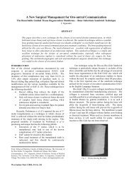
<strong>caries</strong> <strong>in</strong> <strong>the</strong> distal <strong>cervical</strong> region (Fig. 1). 1–3 The relationship<br />
of <strong>the</strong> <strong>mandibular</strong> third <strong>molar</strong> with <strong>the</strong> <strong>second</strong> <strong>molar</strong><br />
results <strong>in</strong> exposure of <strong>the</strong> distal root surface of <strong>the</strong> <strong>second</strong><br />
<strong>molar</strong> to <strong>the</strong> oral environment. The guidel<strong>in</strong>es of <strong>the</strong> National<br />
Institute for Cl<strong>in</strong>ical Excellence for <strong>the</strong> management of third<br />
<strong>molar</strong> teeth, advise aga<strong>in</strong>st <strong>the</strong> prophylactic removal of third<br />
<strong>molar</strong> teeth. 4 Current practice is, <strong>the</strong>refore, not to remove<br />
third <strong>molar</strong>s until <strong>the</strong>y cause disease. The removal of a mesioangular<br />
third <strong>molar</strong> before <strong>the</strong> development of distal <strong>cervical</strong><br />
<strong>caries</strong> <strong>in</strong> <strong>the</strong> <strong>second</strong> <strong>molar</strong> could, however, benefit <strong>the</strong> dental<br />
health of a patient. A <strong>second</strong> <strong>molar</strong> with distal <strong>cervical</strong><br />
<strong>caries</strong> requires ei<strong>the</strong>r restoration or extraction <strong>in</strong> addition to<br />
∗ Correspond<strong>in</strong>g author. Tel.: 44 7885 137050.<br />
<strong>the</strong> removal of <strong>the</strong> third <strong>molar</strong> (two procedures). If <strong>the</strong> third<br />
<strong>molar</strong> is removed before distal <strong>cervical</strong> <strong>caries</strong> forms on <strong>the</strong><br />
<strong>second</strong> <strong>molar</strong> <strong>the</strong>n consequent dental treatment of this tooth<br />
is avoided.<br />
Our aim was to evaluate patients who presented for third<br />
<strong>molar</strong> removal with distal <strong>cervical</strong> <strong>caries</strong> <strong>in</strong> <strong>the</strong>ir <strong>second</strong><br />
<strong>molar</strong>s to identify any cl<strong>in</strong>ical features that would help to<br />
differentiate those patients who are at risk of develop<strong>in</strong>g distal<br />
<strong>cervical</strong> <strong>caries</strong> <strong>in</strong> <strong>the</strong> <strong>second</strong> <strong>molar</strong>.<br />
Methods<br />
We reviewed <strong>the</strong> records of 100 patients who attended <strong>the</strong> oral<br />
surgery department at Guy’s Hospital who had <strong>mandibular</strong><br />
third <strong>molar</strong>s removed because of <strong>the</strong> presence of distal <strong>cervical</strong><br />
<strong>caries</strong> <strong>in</strong> <strong>the</strong> <strong>second</strong> <strong>molar</strong>. Data were collected over a<br />
1-year period.<br />
The variables that we recorded were sex, age, decayed,<br />
miss<strong>in</strong>g, or filled (DMF) score, angulation and eruption status<br />
0266-4356/$ – see front matter © 2005 The British Association of Oral and Maxillofacial Surgeons. Published by Elsevier Ltd. All rights reserved.<br />
doi:10.1016/j.bjoms.2005.07.025
L.W. McArdle, T.F. Renton / British Journal of Oral and Maxillofacial Surgery 44 (2005) 42–45 43<br />
Fig. 3. Mean DMF score <strong>in</strong> our study compared with mean DMF score<br />
reported <strong>in</strong> Adult Dental Health Survey (1998).<br />
Fig. 1. Radiograph of distal <strong>cervical</strong> <strong>caries</strong> <strong>in</strong> <strong>the</strong> <strong>mandibular</strong> <strong>second</strong> <strong>molar</strong><br />
with associated impacted mesio-angular third <strong>molar</strong>.<br />
of <strong>the</strong> third <strong>molar</strong>, proximity of <strong>the</strong> third <strong>molar</strong> to <strong>the</strong> amelocemental<br />
junction of <strong>the</strong> <strong>second</strong> <strong>molar</strong>, dental chart<strong>in</strong>g of<br />
associated buccal teeth, and presence of an erupted ipsilateral<br />
maxillary third <strong>molar</strong>.<br />
The mesial angulation of <strong>the</strong> third <strong>molar</strong> tooth was calculated<br />
by measur<strong>in</strong>g <strong>the</strong> angle of <strong>in</strong>tersection between <strong>the</strong><br />
<strong>mandibular</strong> occlusal plane and <strong>the</strong> occlusal plane of <strong>the</strong> third<br />
<strong>molar</strong>. Trac<strong>in</strong>g paper was attached to <strong>the</strong> dental panoramic<br />
radiograph and <strong>the</strong> <strong>mandibular</strong> occlusal plane was drawn.<br />
This plane was def<strong>in</strong>ed as a l<strong>in</strong>e through <strong>the</strong> tips of <strong>the</strong> cusps<br />
of <strong>the</strong> <strong>mandibular</strong> pre<strong>molar</strong> and <strong>molar</strong> teeth. The occlusal<br />
plane of <strong>the</strong> third <strong>molar</strong> was <strong>the</strong>n drawn through <strong>the</strong> tips of<br />
<strong>the</strong> cusp of <strong>the</strong> third <strong>molar</strong>. The angle of <strong>in</strong>tersection between<br />
<strong>the</strong>se two planes equates to <strong>the</strong> mesial tilt of <strong>the</strong> tooth from<br />
<strong>the</strong> occlusal perpendicular and this angle was def<strong>in</strong>ed as <strong>the</strong><br />
tooth’s mesial angulation.<br />
Results<br />
There were 59 men and 41 women; 78 patients had distal <strong>cervical</strong><br />
<strong>caries</strong> <strong>in</strong> a s<strong>in</strong>gle <strong>mandibular</strong> <strong>second</strong> <strong>molar</strong> and 22 had<br />
distal <strong>cervical</strong> <strong>caries</strong> <strong>in</strong> <strong>the</strong> <strong>mandibular</strong> <strong>second</strong> <strong>molar</strong>s on both<br />
sides. In total 122 <strong>mandibular</strong> third <strong>molar</strong>s were extracted due<br />
to distal <strong>cervical</strong> <strong>caries</strong> <strong>in</strong> <strong>the</strong> <strong>second</strong> <strong>molar</strong>—62 left-sided<br />
and 60 right-sided.<br />
The median age of <strong>the</strong> group was 30 years (range 18–64<br />
years, Fig. 2). Dental disease was measured by calculat<strong>in</strong>g<br />
<strong>the</strong> DMF score (decayed, miss<strong>in</strong>g, or filled); 39 patients had<br />
a DMF score of 5 or less; 36 between 6 and 10, and 24 of 11<br />
or more.<br />
All 122 third <strong>molar</strong>s were partially erupted and radiographic<br />
exam<strong>in</strong>ation showed that 119 teeth were <strong>in</strong> contact<br />
with <strong>the</strong> <strong>second</strong> <strong>molar</strong> tooth at, or close to, <strong>the</strong> amelocemental<br />
junction. There was no radiographic evidence of contact<br />
<strong>in</strong> three teeth.<br />
Mesial angulations of <strong>the</strong> third <strong>molar</strong> fell <strong>in</strong>to three groups;<br />
100 (82%) had an angulation of between 40 ◦ and 80 ◦ ;12<br />
(10%) less than 40 ◦ , and 10 (8%) greater than 80 ◦ .<br />
An upper maxillary third <strong>molar</strong> that could contribute to<br />
food pack<strong>in</strong>g was associated with 80 <strong>mandibular</strong> third <strong>molar</strong>s<br />
(65%), and was absent <strong>in</strong> 42.<br />
In 110 (90%) of <strong>the</strong> 122 teeth <strong>the</strong> buccal segments were<br />
complete with both first and <strong>second</strong> pre<strong>molar</strong>s, and first and<br />
<strong>second</strong> <strong>molar</strong>s be<strong>in</strong>g present. In n<strong>in</strong>e a pre<strong>molar</strong> had been<br />
lost and <strong>in</strong> three <strong>the</strong> first <strong>molar</strong> had been lost.<br />
Discussion<br />
In this study <strong>the</strong> DMF score was used as a measure of dental<br />
health. In calculat<strong>in</strong>g <strong>the</strong> score <strong>the</strong> <strong>second</strong> <strong>molar</strong> tooth was<br />
excluded from <strong>the</strong> calculation if distal <strong>cervical</strong> <strong>caries</strong> was its<br />
only lesion. We assumed that distal <strong>cervical</strong> <strong>caries</strong> <strong>in</strong> that<br />
Fig. 2. Age distribution of patients.
44 L.W. McArdle, T.F. Renton / British Journal of Oral and Maxillofacial Surgery 44 (2005) 42–45<br />
Fig. 4. Patients <strong>in</strong> our study were older than those reported by Brickley and Shepherd.<br />
tooth is specific and would not develop <strong>in</strong> <strong>the</strong> absence of <strong>the</strong><br />
impacted third <strong>molar</strong>.<br />
The mean DMF score for patients with distal <strong>cervical</strong><br />
<strong>caries</strong> was half <strong>the</strong> mean score for similar age groups <strong>in</strong> <strong>the</strong><br />
general population (Fig. 3). 6 This confutes <strong>the</strong> notion that<br />
susceptibility to distal <strong>cervical</strong> <strong>caries</strong> <strong>in</strong> <strong>second</strong> <strong>molar</strong> teeth<br />
is l<strong>in</strong>ked to a high susceptibility to dental <strong>caries</strong> <strong>in</strong> general. 1–3<br />
The <strong>in</strong>cidence of distal <strong>cervical</strong> <strong>caries</strong> <strong>in</strong> <strong>the</strong> <strong>second</strong> <strong>molar</strong><br />
is relatively low (2% 5 ) and does not attract much attention<br />
<strong>in</strong> dental journals. Pericoronitis is, however, <strong>the</strong> most common<br />
<strong>in</strong>dication for <strong>the</strong> removal of <strong>mandibular</strong> third <strong>molar</strong>s<br />
and occurs <strong>in</strong> relatively young adults. 3,5,6,9 We suggest that<br />
people with low DMF scores have a high standard of oral<br />
hygiene, so m<strong>in</strong>imis<strong>in</strong>g <strong>the</strong> risk of pericoronitis associated<br />
with <strong>the</strong> third <strong>molar</strong>. This would lead to <strong>the</strong> retention of <strong>the</strong><br />
tooth and <strong>the</strong> development of distal <strong>cervical</strong> <strong>caries</strong> <strong>in</strong> <strong>the</strong><br />
<strong>second</strong> <strong>molar</strong>. With good oral hygiene <strong>the</strong> pericoronal tissues<br />
would be easily ma<strong>in</strong>ta<strong>in</strong>ed, however, <strong>the</strong> contact area<br />
between <strong>the</strong> <strong>second</strong> and third <strong>molar</strong>s would be relatively <strong>in</strong>accessible<br />
with consequent long-term plaque accumulation.<br />
The development of dental <strong>caries</strong> is a relatively slow process<br />
and would occur over a protracted period of time at <strong>the</strong> contact<br />
po<strong>in</strong>t on <strong>the</strong> <strong>second</strong> <strong>molar</strong>. Consequently, <strong>the</strong>se patients<br />
are older than <strong>the</strong> usual patients who have third <strong>molar</strong>s<br />
removed.<br />
Normally, <strong>the</strong> age at which third <strong>molar</strong>s are removed is<br />
between 25 and 28 years. 5,7–10 The median age of patients<br />
<strong>in</strong> this study was 30 years, suggest<strong>in</strong>g that DCC seems to<br />
post-date o<strong>the</strong>r diseases affect<strong>in</strong>g third <strong>molar</strong>s by almost 5<br />
years. 5,8,9 This is illustrated by compar<strong>in</strong>g <strong>the</strong> age distributions<br />
of our study with that of Brickley and Shepherd<br />
(Fig. 4). 5 From <strong>the</strong> graph, a lateral shift to <strong>the</strong> right is apparent<br />
that demonstrates a peak <strong>in</strong>cidence for third <strong>molar</strong> removal<br />
attributable to DCC that is 5 years later than for all third<br />
<strong>molar</strong> removal 5 . The data suggest that <strong>the</strong> development of<br />
distal <strong>cervical</strong> <strong>caries</strong> <strong>in</strong> <strong>the</strong> <strong>second</strong> <strong>molar</strong> is a protracted process<br />
that develops over time and <strong>in</strong>creases with cont<strong>in</strong>ued<br />
exposure to <strong>the</strong> oral cavity. Fur<strong>the</strong>r confirmation of this correlation<br />
with age was published by Bruce et al. who showed<br />
that <strong>the</strong> <strong>in</strong>cidence of distal <strong>cervical</strong> <strong>caries</strong> as an <strong>in</strong>dication<br />
for removal of third <strong>molar</strong>s <strong>in</strong>creased significantly with age<br />
(Fig. 5). 11<br />
The true <strong>in</strong>cidence of distal <strong>cervical</strong> <strong>caries</strong> <strong>in</strong> <strong>mandibular</strong><br />
<strong>second</strong> <strong>molar</strong>s may be as high as 5%. This is because most<br />
studies report large numbers of third <strong>molar</strong>s that are removed<br />
Fig. 5. Percentage of patients <strong>in</strong> each age group who had distal <strong>cervical</strong><br />
<strong>caries</strong> of <strong>second</strong> <strong>molar</strong>s as <strong>the</strong> <strong>in</strong>dication for removal of third <strong>molar</strong>s (Bruce<br />
et al. 11 ).<br />
for prophylactic reasons. 5,7–11 Consequently, this distorts <strong>the</strong><br />
true <strong>in</strong>cidence of disease processes that account for removal<br />
of third <strong>molar</strong>s.<br />
Ano<strong>the</strong>r factor that is associated with <strong>the</strong> risk of develop<strong>in</strong>g<br />
distal <strong>cervical</strong> <strong>caries</strong> is <strong>the</strong> angulation of <strong>the</strong> third <strong>molar</strong><br />
tooth and <strong>the</strong> po<strong>in</strong>t of contact that <strong>the</strong> third <strong>molar</strong> makes with<br />
<strong>the</strong> <strong>second</strong> <strong>molar</strong>. We found that a mesial angulation between<br />
40 ◦ and 80 ◦ was common. Of 122 third <strong>molar</strong>s extracted, 100<br />
(82%) had an angulation with<strong>in</strong> this range. <strong>Distal</strong> <strong>cervical</strong><br />
<strong>caries</strong> <strong>in</strong> <strong>second</strong> <strong>molar</strong> teeth did, however, sometimes occur<br />
at angulations outside <strong>the</strong> 40–80 ◦ range.<br />
<strong>Distal</strong> <strong>cervical</strong> <strong>caries</strong> is responsible for a small percentage<br />
of third <strong>molar</strong>s removed. To suggest that <strong>the</strong> risk of develop<strong>in</strong>g<br />
distal <strong>cervical</strong> <strong>caries</strong> is low is wrong. If pericoronitis<br />
were not an important reason for removal of third <strong>molar</strong>s,<br />
<strong>the</strong>n a large number of <strong>the</strong>se teeth would be reta<strong>in</strong>ed later<br />
<strong>in</strong>to life, and if <strong>the</strong>y were reta<strong>in</strong>ed <strong>the</strong>n <strong>the</strong> <strong>in</strong>cidence of distal<br />
<strong>cervical</strong> <strong>caries</strong> of <strong>the</strong> <strong>second</strong> <strong>molar</strong> as an <strong>in</strong>dication would<br />
rise accord<strong>in</strong>gly—as alluded to by Bruce et al. 11 The removal<br />
of <strong>mandibular</strong> third <strong>molar</strong>s <strong>in</strong> young adults for pericoronitis<br />
removes <strong>the</strong> ma<strong>in</strong> causal factor (<strong>the</strong> third <strong>molar</strong>) of distal <strong>cervical</strong><br />
<strong>caries</strong> of <strong>the</strong> <strong>second</strong> <strong>molar</strong>. With <strong>the</strong> third <strong>molar</strong> absent<br />
<strong>the</strong> exposure of <strong>the</strong> distal root surface of <strong>the</strong> <strong>second</strong> <strong>molar</strong> is<br />
corrected and so prevents <strong>the</strong> development of distal <strong>cervical</strong><br />
<strong>caries</strong> <strong>in</strong> <strong>second</strong> <strong>molar</strong> tooth.<br />
Conclusion<br />
<strong>Distal</strong> <strong>cervical</strong> <strong>caries</strong> occurs <strong>in</strong> <strong>the</strong> <strong>second</strong> <strong>molar</strong> <strong>in</strong> <strong>the</strong> presence<br />
of a mesio-angular impacted third <strong>molar</strong>. This type<br />
of <strong>caries</strong> has not been reported <strong>in</strong> relation to any o<strong>the</strong>r<br />
cl<strong>in</strong>ical scenario. It appears to develop as a relatively late
L.W. McArdle, T.F. Renton / British Journal of Oral and Maxillofacial Surgery 44 (2005) 42–45 45<br />
phenomenon, with patients tend<strong>in</strong>g to be 4–5 years older<br />
than average and hav<strong>in</strong>g better dental health than average.<br />
A mesial angulation of <strong>the</strong> third <strong>molar</strong> <strong>in</strong> <strong>the</strong> range of 40–80 ◦<br />
with a contact po<strong>in</strong>t <strong>in</strong> <strong>the</strong> region of <strong>the</strong> amelocemental<br />
junction of <strong>the</strong> <strong>second</strong> <strong>molar</strong> tooth are features that also characterise<br />
this group of patients.<br />
This study suggests that <strong>the</strong> non-<strong>in</strong>tervention approach<br />
<strong>in</strong> terms of manag<strong>in</strong>g disease-free partially erupted mesioangular<br />
<strong>mandibular</strong> third <strong>molar</strong>s may ultimately be detrimental<br />
to <strong>the</strong> dental health of <strong>the</strong> patient. Rout<strong>in</strong>e screen<strong>in</strong>g for<br />
DCC will have no benefit as once <strong>the</strong> disease process is identified<br />
<strong>the</strong> consequences are apparent. The development of<br />
DCC <strong>in</strong> <strong>the</strong> <strong>second</strong> <strong>molar</strong> will necessitate a restorative and<br />
possible endodontic procedure to conserve <strong>the</strong> <strong>second</strong> <strong>molar</strong><br />
tooth <strong>in</strong> addition to <strong>the</strong> removal of <strong>the</strong> third <strong>molar</strong> and, <strong>in</strong><br />
some cases, <strong>the</strong> extraction of <strong>the</strong> <strong>second</strong> <strong>molar</strong> tooth will be<br />
<strong>in</strong>dicated also.<br />
It could be suggested that <strong>the</strong> early, prophylactic, removal<br />
of a partially erupted mesio-angular <strong>mandibular</strong> third <strong>molar</strong><br />
would prevent DCC affect<strong>in</strong>g <strong>the</strong> <strong>second</strong> <strong>molar</strong>. The authors<br />
suggest, <strong>the</strong>refore, that fur<strong>the</strong>r studies are undertaken to<br />
establish whe<strong>the</strong>r <strong>the</strong> non-<strong>in</strong>tervention approach is potentially<br />
flawed and whe<strong>the</strong>r consideration should be given to<br />
<strong>the</strong> prophylactic removal of partially erupted mesio-angular<br />
third <strong>molar</strong> teeth.<br />
References<br />
1. van der L<strong>in</strong>den W, Cleaton-Jones P, Lownie M. Diseases and lesions<br />
associated with third <strong>molar</strong>s. Review of 1001 cases. Oral Surg Oral<br />
Med Oral Pathol Oral Radiol Endod 1995;79:142–5.<br />
2. Current cl<strong>in</strong>ical practice and parameters of care: <strong>the</strong> management of<br />
patients with third <strong>molar</strong> teeth. Faculty of Dental Surgery of <strong>the</strong> Royal<br />
College of Surgeons of England; September 1997.<br />
3. Knutsson K, Brehmer B, Lysell L, Rohl<strong>in</strong> M. Pathoses associated with<br />
<strong>mandibular</strong> third <strong>molar</strong>s subjected to removal. Oral Surg Oral Med<br />
Oral Pathol Oral Radiol Endod 1996;82:10–7.<br />
4. National Institute for Cl<strong>in</strong>ical Excellence. Guidance on <strong>the</strong> removal of<br />
wisdom teeth; March 2000.<br />
5. Brickley MR, Shepherd JP. An <strong>in</strong>vestigation of <strong>the</strong> rationality of lower<br />
third <strong>molar</strong> removal, based on USA National Institutes of Health criteria.<br />
Br Dent J 1996;180:249–54.<br />
6. Office for National Statistics. Adult Dental Health Survey, oral health<br />
<strong>in</strong> <strong>the</strong> United K<strong>in</strong>gdom. London: HMSO; 1998.<br />
7. Chiapasco M, De Cicco L, Marrone G. Side effects and complications<br />
with third <strong>molar</strong> surgery. Oral Surg Oral Med Oral Pathol<br />
1993;76:412–20.<br />
8. de Boer PJ, Raghoebar M, Stegenga B, Scheon PJ, Boer<strong>in</strong>g G. Complications<br />
after third <strong>molar</strong> extraction. Qu<strong>in</strong>tessence Int 1995;26:779–84.<br />
9. Nordenram Å, Hult<strong>in</strong> M, Kjellman O, Ramström G. Indications for surgical<br />
removal of <strong>the</strong> <strong>mandibular</strong> third <strong>molar</strong>. Swed Dent J 1987;11:23–9.<br />
10. Lysell L, Rohl<strong>in</strong> M. A study of <strong>in</strong>dications used for removal of <strong>the</strong><br />
<strong>mandibular</strong> third <strong>molar</strong>. Int J Oral Maxillofac Surg 1988;17:161–4.<br />
11. Bruce RA, Frederickson GC, Small GS. Age of patients and morbidity<br />
associated with <strong>mandibular</strong> third <strong>molar</strong> surgery. J Am Dent Assoc<br />
1980;101:240–5.