IDSA July Aug 2006 - Modern Dentistry Media
IDSA July Aug 2006 - Modern Dentistry Media
IDSA July Aug 2006 - Modern Dentistry Media

Create successful ePaper yourself
Turn your PDF publications into a flip-book with our unique Google optimized e-Paper software.
CLINICAL<br />
CORONECTOMY: AN ALTERNATIVE THERAPY FOR<br />
THE SYMPTOMATIC, IMPACTED THIRD MOLAR<br />
REPORT OF 9 CASES<br />
NIKA VAFAEI 1 , CARLO FERRETTI 2<br />
Key words: impacted third molar, coronectomy<br />
The mandibular third molar remains the tooth most prone<br />
to impaction and interestingly, the incidence of<br />
impaction appears to be increasing. 1 Mechanisms for<br />
impaction of third molars remain unproven supposition, 2 but<br />
the result is the failure of eruption into a normal functional<br />
position. 2 Problems which may arise due to tooth impaction<br />
(be it partial or full) include pericoronitis, cheek biting, pressure<br />
on adjacent teeth causing pain, food impaction in the area,<br />
buccal or lingual eruption, pericoronal infection, caries,<br />
periodontal problems with associated teeth, and association<br />
with pathological lesions such cysts and tumours. 2 Thus, the<br />
obligatory surgical removal of wisdom teeth remains a<br />
common procedure in dental practice. Prophylactic removal to<br />
avoid the aforementioned problems is often performed but it<br />
is a persistent source of discussion. Adding to the controversy<br />
is the proposed association between third molar impaction,<br />
anterior incisor crowding and atypical facial pain. 2, 3<br />
Irrespective of the motive, the surgical removal of wisdom<br />
teeth may be associated with several post-operative<br />
complications. The most commonly observed complications<br />
include pain, edema, acute alveolar osteitis, infection,<br />
mandibular fracture, damage to adjacent teeth, and<br />
haemorrhage. 1, 4-7 Possibly the most concerning complication is<br />
1, 2,4,6,8<br />
temporary or permanent sensory nerve damage.<br />
The development of a post-operative complication is<br />
influenced by operator, patient, and tooth associated factors.<br />
There is a strong correlation between the degree of impaction,<br />
the type of impaction (i.e. vertical, mesioangular, distoangular<br />
1. Niko Vafaei, BDS,<br />
Public Service, Community Service, Kwa-Zulu Natal, South Africa<br />
2. Carlo Ferretti, BDS, MDent, FCD(SA), MFOS<br />
Private Practice – Bedford Gardens Clinic, Bedfordview, Johannesburg,<br />
South Africa<br />
Senior Specialist – Division of Maxillofacial and Oral Surgery, Chris Hani<br />
Baragawanath Hospital and University of the Witwatersrand,<br />
Johannesburg, South Africa<br />
Corresponding author:<br />
Dr Carlo Ferretti<br />
P.O Box 75471, Gardenview, 2047, South Africa • Tel: 0027 11 615 9595<br />
Facsimile: 0027 11 616 8649 • E-mail: ferretti@mweb.co.za<br />
and horizontal), and the anatomical relation of the roots to the<br />
inferior alveolar nerve canal and postoperative complications.<br />
A preexisting infection or a pathological lesion in or around the<br />
tooth also increases the risk. 7, 9 An important iatrogenic factor<br />
is surgeon experience. Several studies have confirmed the<br />
inverse relationship between surgeon experience and<br />
complications. 9-13 Surgical technique as well as the use of<br />
certain instrumentation are potential risk factors, in particular<br />
with regard to nerve damage. 9-13<br />
Lingual nerve (LN) and inferior alveolar nerve damage (IAN)<br />
is largely attributed to the anatomical proximity of the<br />
impacted third molar to these nerves. Both the IAN and the LN<br />
arise from the posterior branch of the mandibular nerve which<br />
in turn is a branch of cranial nerve V, the trigeminal nerve. The<br />
IAN consists predominantly of sensory fibres with only a few<br />
motor fibres (distributed via the mylohyoid nerve to the<br />
mylohyoid muscle and the anterior belly of the digastric<br />
muscle). The lower molar and premolar teeth and adjacent<br />
parts of the gingiva are supplied by the IAN, and its terminal<br />
branches supply sensation to the ipsilateral lower lip via the<br />
mental nerve. The course of the IAN within the mandibular<br />
canal proceeds anteriorly from the medial aspect of the midramus<br />
(at the lingula) along with the inferior alveolar artery<br />
(together they are referred to as the inferior alveolar<br />
neurovascular bundle) in the intraosseous inferior alveolar<br />
canal. It is here that its course approximates the third molar to<br />
varying degrees. The nerve proceeds anteriorly in its bony canal<br />
within the body of the mandible just apical to the lower molars<br />
and premolars to emerge from the mental foramen as the<br />
terminal mental nerve branch which innervates the skin of the<br />
ipsilateral chin and the lower lip. The smaller incisive branch is<br />
the intraosseous anterior continuation of the nerve and<br />
supplies the canine and incisor teeth. 14 The lingual nerve runs<br />
its course anterior to the inferior alveolar nerve, proceeding<br />
anteriorly in the soft tissues lingual to the third molar and<br />
supplies the mucous membrane of the anterior two-thirds of<br />
the tongue. 14<br />
Important predictors of neural injury include the use of<br />
lingual retractors, particular surgical techniques such as vertical<br />
6 INTERNATIONAL DENTISTRY SA VOL. 10, NO. 2
CLINICAL<br />
1A<br />
1B<br />
1C<br />
1D<br />
1E<br />
1F<br />
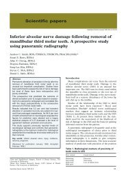
Figure 1 – 32 year old male patient with partially erupted, vertically impacted 38 (A) which on panoramic radiographs demonstrates an intimate relationship<br />
of the root apices to the inferior alveolar canal (B). The crown is exposed by means of a mucoperiosteal flap (C) and the crown resected (D). The retained<br />
roots (black arrows indicating the tooth margin) are reduced to a sub-crestal level (white arrows indicate the osseous socket margin). This leaves<br />
undisturbed, retained root fragments below the socket margin allowing for bone formation above the roots.<br />
tooth sectioning and ostectomy, anatomical variations of<br />
nerve, and lingual root angulation. The mechanism of nerve<br />
injury (compression, stretching or complete transsection) is a<br />
strong determinant of sensory alteration and recovery 15 , as is<br />
increased patient age. The latter is due to the increased<br />
difficulty of surgery and the decreased potential for repair and<br />
regeneration in older patients. 15,16 The reported incidence of LN<br />
and IAN damage following third molar extraction ranges<br />
INTERNATIONAL DENTISTRY SA VOL. 10, NO. 2 7
CLINICAL<br />
between 0 and 23 %. 11 More recent data has however<br />
reported the incidence to be in a lower range of 0-3.6%. 10-13,<br />
16, 17<br />
Whether temporary or permanent, iatrogenic nerve<br />
damage following extraction of the lower third molar is a<br />
common cause of litigation and patient dissatisfaction in<br />
dental practice. 10 This complication can be mitigated to some<br />
degree by preoperative risk assessment. The three most<br />
common radiographic features suggesting close proximity of<br />
an impacted third molar to the IAN include diversion of the<br />
canal, interruption of the canal walls, and darkening of the<br />
12, 18, 19<br />
root. The advent of cone beam computed tomography<br />
has significantly increased the accuracy of preoperative<br />
assessment but its expense precludes it from widespread<br />
availability and routine use.<br />
The decision to remove impacted third molars is the<br />
culmination of a complex algorithm which must evaluate the<br />
reasons for removal and weigh these against the potential risk<br />
factors. The fundamental tenet of surgical exodontia is that<br />
the prophylactic removal of any tooth which has a high risk for<br />
complications (whether due to local conditions or systemic<br />
factors) cannot be condoned. Once a decision has been made<br />
that the benefits of surgical removal of a tooth outweigh any<br />
potential complications it is incumbent upon the practitioner<br />
to select a surgical technique with the lowest potential<br />
complication rate. Ecuyer and Debien 20 first described a<br />
technique, which they termed coronectomy, involving the<br />
resection of the crown of a tooth with deliberate retention of<br />
the roots. 18,19 This technique may be considered when an<br />
impacted lower third molar must be removed and key<br />
radiographic findings show close relation of the tooth roots<br />
with the inferior alveolar nerve canal, in order to mitigate the<br />
risk of IAN damage during tooth extraction. 18,19 The procedure<br />
requires transection of the tooth just below its crown and<br />
reduction of the remaining root fragments below the lingual<br />
and buccal plates (Fig. 1), allowing for bone formation superior<br />
to these roots. 21 Following retention of vital roots all pulps<br />
survive 18 , however root mobility must be avoided during the<br />
coronectomy procedure as these will become a source of<br />
infection. 18,21 Exclusion criteria for coronectomy include a<br />
tooth with active infection extending to its roots, periapical<br />
pathology associated with the tooth, and mobile teeth.<br />
Postoperatively, although retained root movement is<br />
unpredictable, they appear to migrate away from the canal<br />
(Fig. 2) allowing removal at a later stage (if required) with<br />
substantially lower risk of nerve damage. 21 Complications<br />
associated with coronectomy include osteitis, unintentional<br />
root mobility with subsequent infection, and temporary<br />
sensory disturbance of the lip related to the technique and<br />
damage caused by inappropriate burr usage. 18<br />
Recently this technique has been subject to closer scrutiny<br />
and several studies have been published on the topic. In a<br />
study by Pogrel et al 21 , forty-one patients had 50 lower third<br />
molars treated by coronectomy. There were no cases of inferior<br />
alveolar nerve damage; there was however one case of<br />
transient lingual nerve involvement, probably due to lingual<br />
retractor use. One patient required subsequent removal of the<br />
roots of both lower third molars because of failure to heal, and<br />
one patient required subsequent removal of a root because of<br />
subsequent migration to the surface. Root migration was<br />
noted in approximately 30% of patients over a 6 month<br />
period. 21 O’Riordan 18 conducted a retrospective study of 52<br />
patients who were operated over a 10 year period. 3 of 52<br />
patients had to have the roots removed subsequent to the<br />
coronectomy procedure due to pain or infection. Neural<br />
complications included 3 cases of temporary sensory<br />
disturbance of the lower lip which the author attributes to<br />
pressure transmitted to the nerve when splitting the crown<br />
from the root, or a slight elevation of the root when splitting.<br />
One case of prolonged anesthesia of the lip was noted, due to<br />
bur damage. 18 Finally, a prospective randomized study by<br />
Renton et al 19 of 128 patients requiring operations on<br />
mandibular third molars which had radiographic evidence of<br />
proximity to the inferior alveolar canal nerve. Patients were<br />
randomly assigned to either the extraction [n = 102] or the<br />
coronectomy [n = 94] group. Some roots were dislodged<br />
during intended coronectomy and were therefore removed,<br />
resulting in two subgroups (successful coronectomy n = 58,<br />
and failed coronectomy n = 36). Nineteen nerves were<br />
damaged (19%) after extraction, none after successful<br />
coronectomy, and three (8%) after failed coronectomy<br />
(p = 0.01). The incidence of dry socket infection was similar in<br />
the three groups (10/102, 10%, 7/58, 12%, and 4/36, 11%,<br />
respectively). The incidence of acute localized osteitis was<br />
found in 10–12% in all groups. Follow up of the coronectomy<br />
procedure after 13 months showed five root segments had<br />
started to migrate. 19<br />
Case reports<br />
To the above mentioned cases we add our own experience<br />
with a further 9 patients. All patients were offered<br />
coronectomy if clinical examination revealed an impacted third<br />
molar which has been repeatedly symptomatic and<br />
radiographic examination suggested a high risk of inferior<br />
alveolar injury. All patients (4 male and 5 female with ages<br />
ranging from 19 to 36 years of age) were given a detailed<br />
account of all the treatment options and the principles of<br />
8 INTERNATIONAL DENTISTRY SA VOL. 10, NO. 2
CLINICAL<br />
2A<br />
a<br />
2B<br />
b<br />
2C<br />
c<br />
Figure 2 – Panoramic radiograph of 22 year old female patient presenting with persistent pain associated with horizontally<br />
impacted teeth (A, a). Due to high risk of IAN injury (note darkening of canal as it crosses the roots) bilateral coronectomy was<br />
performed (B, b). 1 year follow-up (C, c) shows significant root fragment migration away from the IAN, ossification anterior to<br />
the roots and absence of periapical pathology<br />
coronectomy, and were operated by the same surgeon. 3 of<br />
these patients had both lower wisdom teeth treated by<br />
coronectomy; the remaining 6 patients had a single wisdom<br />
tooth treated by coronectomy. This resulted in 12 teeth<br />
planned for coronectomy, of which one tooth required<br />
complete removal due to inadvertent dislodgement of the<br />
roots during surgery. Post operative follow-up period ranged<br />
from 3 months to 1 year. None of the patients developed post<br />
surgical lingual and labial anaesthesia and thus far none have<br />
developed infection requiring subsequent root removal. Long<br />
term radiographic follow-up demonstrated considerable root<br />
migration away from the inferior alveolar canal (Fig. 2)<br />
Discussion<br />
Justifiably, the new technique of coronectomy is advocated<br />
with caution and some surgeons have expressed resistance to<br />
the adoption of this treatment alternative as it is contrary to<br />
the dogma of exodontia. Given its recent emergence, a<br />
10 INTERNATIONAL DENTISTRY SA VOL. 10, NO. 2
CLINICAL<br />
significant limitation is the lack of long term follow up, in<br />
particular with regard to the potential risk of an intentionally<br />
retained root. It is proposed that the roots may become a<br />
source of infection, leading to an apical periodontitis following<br />
pulp necrosis, which could spread to the inferior alveolar canal<br />
given the root proximity. 22 Questionable outcomes also include<br />
the variable rate of root migration, periodontal status in the<br />
region, and the need for repeated radiographic and clinical<br />
evaluation, and a possible second operation to remove<br />
symptomatic roots. 23 Though not justifying the routine use of<br />
this procedure, Garcia-Garcia mentions that following<br />
breakage of the apex during conventional wisdom tooth<br />
extraction in close proximity to the IAN, the roots should<br />
probably not be removed. 24 It must be said that these<br />
contrarian views are anecdotal and no well structured study<br />
we are aware of has supported these views. On the contrary,<br />
the aforementioned studies and our experience corroborate<br />
that coronectomy is a treatment alternative with a very low<br />
complication rate. Should root removal subsequently become<br />
necessary, the root migration that follows coronectomy may<br />
decrease the risk of neural injury as the retained roots are no<br />
longer intimately associated with inferior alveolar<br />
neurovascular bundle.<br />
Given the unpredictability of lower third molar removal, it<br />
is not always possible to avoid potential injury. However,<br />
awareness of the various associated risk factors makes it<br />
possible to minimize consequences, if not prevent them all<br />
together.<br />
IAN involvement during lower third molar extraction is a<br />
cause for concern as it is both a clinical and medico legal<br />
issue. 21 The coronectomy technique diminishes the possibility<br />
of nerve injury thus avoiding patient dissatisfaction, and also<br />
offers a less traumatic approach than conventional third molar<br />
removal. Whilst widespread acceptance of coronectomy rightly<br />
awaits the results of longer term follow-up studies, the<br />
preliminary results are encouraging, and the practitioner who<br />
routinely removes impacted wisdom teeth should consider this<br />
surgical option in selected patients.<br />
References<br />
1. Chaparro-Avendaño AV. Morbidity of third molar extraction in<br />
patients between 12 and 18 years of age. 2005; 10(5):422-3.<br />
2. Hamasha AA. Reasons for Third Molar Teeth Extraction in<br />
Jordanian Adults. J Contemp Dent Pract <strong>2006</strong>; (7)5:088-095.<br />
3. Al-Balkhi KM . The Effect of Different Lower Third Molar<br />
Conditions on the Re-Crowding of Lower Anterior Teeth in the<br />
Absence of Tight Interproximal Contacts One-Year Post Orthodontic<br />
Treatment: A Pilot Study. J Contemp Dent Pract 2004; 3:066-073.<br />
4. Huang IY, Wu CW. The displaced lower third molar: a literature<br />
review and suggestions for management. J Oral Maxillofac Surg 2007;<br />
65 (6): 1186-90.<br />
5. Lacasa JM, Jimenez JA. Prophylaxis versus pre-emptive treatment<br />
for infective and inflammatory complications of surgical third molar<br />
removal: a randomized, double blind, placebo controlled, clinical trial<br />
with sustained release amoxicillin/clauvanic acid. Int J Oral Maxillofac<br />
Surg 2007; 36(4): 321-7.<br />
6. Wagner KW, Schoen R. Complicated late mandibular fracture<br />
following third molar removal. Quintessence Int 2007; 38(1): 63-5.<br />
7. Woldenburg Y, Gatot I. Iatrogenic mandibular fracture associated<br />
with third molar removal: Can it be prevented Medical Oral Patol Oral<br />
Cir Bucal. 2007; 12(1): e70-2.<br />
8. Song F, O’Meara S. The effectiveness and cost-effectiveness of<br />
prophylactic removal of wisdom teeth. Health Technology Assessment<br />
2000; 4(15): 1-55.<br />
9. Blondeau F, Daniel N. Extraction of impacted mandibular third<br />
molars: postoperative complications and their risk factors. Journal of<br />
Canada Dental Association 2007; 73(4):325.<br />
10. Valmaseda-Castellon E, Berini-Aytes L. Lingual nerve damage<br />
after third lower molar surgical extraction. Oral Surg Oral Med Oral<br />
Path Oral Radiol Endod 2000; 90(5): 567-73.<br />
11. Bataineh A. Sensory nerve impairment following mandibular<br />
third molar surgery. J Oral Maxillofac Surg 2001; 59:1012-1017.<br />
12. Ban Guan Tay A, Ser Go W. Effect of exposed inferior alveolar<br />
neurovascular bundle during surgical removal of impacted lower third<br />
molars. J Oral Maxillofac Surg 2004; 62: 592-600.<br />
13. Rehman K, Webster K. Links between anaesthetic modality and<br />
nerve damage during lower third molar surgery. British Dental Journal<br />
2002; 192(1): 43-45.<br />
14. Moore, K. L. Clinically Oriented Anatomy. 3rd Ed. Philadelphia<br />
Lippincott Williams & Wilkins, 1992. Pgs 663-668, 863-868.<br />
15. Jerjes W, Moles DR. Permanent sensory nerve impairment<br />
following third molar surgery: a prospective study. Oral Surg Oral Med<br />
Oral Pathol Oral Radiol Endod <strong>2006</strong>; 102: e1-e7.<br />
16. Queral-Godoy E, Berini-Aytes L. Incidence and evolution of<br />
inferior alveolar nerve lesions following lower third molar extraction.<br />
Oral Surg Oral Med Oral Path Oral Radiol Endod 2005; 99(3): 259-64.<br />
17. Valmaseda-Castellon E, Berini-Aytes L. Lingual nerve damage<br />
after third lower molar surgical extraction. Oral Surg Oral Med Oral<br />
Path Oral Radiol Endod 2001; 92: 377-83.<br />
18. O’Riordan BC. Coronectomy (intentional partial odontectomy of<br />
lower third molars). Oral Surg Oral Med Oral Path Oral Radiol Endod<br />
2004; 98(3): 274-80.<br />
19. Renton T, Hankins M. A randomized controlled clinical trial to<br />
compare the incidence of injury to the inferior alveolar nerve as a result<br />
of coronectomy and removal of mandibular third molars. Br J Oral<br />
Maxillofac Surg 2005; 43: 7-12.<br />
20. J. Ecuyer and J. Debien. Deductions operatoires. Actualités<br />
Odonto-Stomatologiques 1984 ; 148 : 695–701.<br />
21. Pogrel MA. Coronectomy: A technique to protect the inferior<br />
alveolar nerve. J Oral Maxillofac Surg 2004; 62: 1447-1452.<br />
22. Garcia-Garcia, A. Coronectomy: a questionable procedure. J<br />
Oral Maxillofac Surg 63:723-725, 2005.<br />
23. Assael, LA. “Coronectomy: A time to ponder or a time to act”<br />
J Oral Maxillofac Surg 2004; 62(12): 1445-1446.<br />
24. Garci-Garcia, A. “Is coronectomy really preferable to<br />
extraction” Br J Oral Maxillofac Surg. <strong>2006</strong>; 44(1): 75.<br />
12 INTERNATIONAL DENTISTRY SA VOL. 10, NO. 2