A New Surgical Management for Oro-antral ... - Exodontia.Info
A New Surgical Management for Oro-antral ... - Exodontia.Info
A New Surgical Management for Oro-antral ... - Exodontia.Info

You also want an ePaper? Increase the reach of your titles
YUMPU automatically turns print PDFs into web optimized ePapers that Google loves.
A <strong>New</strong> <strong>Surgical</strong> <strong>Management</strong> <strong>for</strong> <strong>Oro</strong>-<strong>antral</strong> Communication<br />
The Resorbable Guided Tissue Regeneration Membrane – Bone Substitute Sandwich Technique<br />
C Ogunsalu<br />
ABSTRACT<br />
This paper describes a new technique <strong>for</strong> the closure of oro-<strong>antral</strong> fistula/communication, in which<br />
both hard tissue (bone) and soft tissue closure is achieved. The sandwich technique utilizes a suitable<br />
bone grafting material sandwiched between two sheaths of Biogide ® (a resorbable membrane) <strong>for</strong> the<br />
hard tissue closure of oro-<strong>antral</strong> communication post traumatic exodontia. The bone grafting material<br />
utilized <strong>for</strong> this case was Bio-oss. The result obtained was excellent with regeneration of sufficient<br />
bony tissue to allow placement of an endosseous implant. This sandwich technique is a simple and<br />
excellent technique <strong>for</strong> the closure of oro-<strong>antral</strong> communication, especially when subsequent<br />
placement of endosseous implant is considered without the need of donor site surgery <strong>for</strong> bone<br />
grafting. The otorhinolaryngologists and oral and maxillofacial surgeons should find this technique<br />
very useful in the closure of oro-<strong>antral</strong> fistulae.<br />
INTRODUCTION<br />
One of the clinical complications encountered by oral<br />
surgeons is oro-<strong>antral</strong> communication (OAC) with<br />
progressive <strong>for</strong>mation of oro-<strong>antral</strong> fistula (OAF). The<br />
incidence of this complication may vary from 0.31% to<br />
3.8% after simple extraction of maxillary teeth (1, 2).<br />
Buccal sliding flap, palatal flap, soft palate flap and related<br />
modifications are the various modalities available <strong>for</strong> the<br />
management of OAC or OAF (3-10). These techniques have<br />
the following shortfalls:<br />
• Buccal sliding flap reduces the depth of the<br />
vestibular sulcus, hence need <strong>for</strong> a vestibuloplasty<br />
• Only soft tissue closure is achieved, hence the need<br />
of complex hard tissue (bone) grafting when<br />
endosseous implant is considered<br />
• Severe pain and scarring in palatal flaps; the palatal<br />
denuded area takes too long to heal.<br />
Recently, Yoshimasa et al (11) reported the use of 3 rd<br />
molar transplantation as a technique in which closure of<br />
OAC is achieved without the need <strong>for</strong> further prosthodontic<br />
treatment of the single tooth missing in the region. This<br />
proposed modality of treatment by Yoshimasa et al is<br />
promising and unique but has the following pitfalls:<br />
• Extraction of the 3 rd molar, which is to be<br />
transplanted, may lead to any of the known<br />
complications of 3 rd molar extraction.<br />
• OAF cannot be closed in this manner.<br />
• Root canal treatment of the transplanted tooth is<br />
indicated, and this should be continuously<br />
monitored <strong>for</strong> failure.<br />
Our technique using the Bio-oss-Bio-Gide Sandwich<br />
technique is particularly unique because it excludes all the<br />
above shortfalls and further has the advantage of concurrent<br />
bone tissue regeneration in the OAC/OAF site, which will<br />
enable the placement of an endosseous implant in future<br />
without the need <strong>for</strong> complex maxillary sinus lift procedure.<br />
This is the first reported case of the sandwich technique<br />
utilized <strong>for</strong> the closure of oro-<strong>antral</strong> communication (OAC)<br />
in the English Language literature.<br />
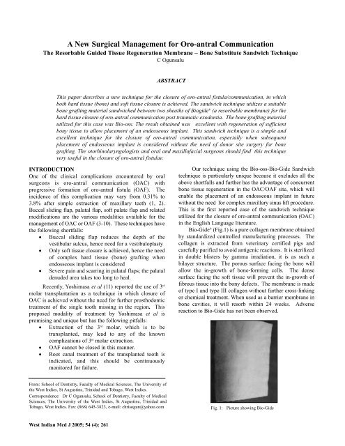
Bio-Gide ® (Fig.1) is a pure collagen membrane obtained<br />
by standardized controlled manufacturing processes. The<br />
collagen is extracted from veterinary certified pigs and<br />
carefully purified to avoid antigenic reactions. It is sterilized<br />
in double blisters by gamma irradiation, it is as such a<br />
bilayer structure. The porous surface facing the bone will<br />
allow the in-growth of bone-<strong>for</strong>ming cells. The dense<br />
surface facing the soft tissue will prevent the in-growth of<br />
fibrous tissue into the bony defects. The membrane is made<br />
of type I and type III collagen without further cross-linking<br />
or chemical treatment. When used as a barrier membrane in<br />
bone cavities, it will resorb within 24 weeks. Adverse<br />
reaction to Bio-Gide has not been observed.<br />
From: School of Dentistry, Faculty of Medical Sciences, The University of<br />
the West Indies, St Augustine, Trinidad and Tobago, West Indies.<br />
Correspondence: Dr C Ogunsalu, School of Dentistry, Faculty of Medical<br />
Sciences, The University of the West Indies, St Augustine, Trinidad and<br />
Tobago, West Indies. Fax: (868) 645-3823, e-mail: chrisogun@yahoo.com<br />
Fig. 1: Picture showing Bio-Gide<br />
West Indian Med J 2005; 54 (4): 261
262<br />
Bone Substitute<br />
Bio-Oss ® is a safe and effective bone graft material.<br />
Under the election microscope, Bio-Oss looks very similar<br />
to human bone. Because of this similarity to human bone,<br />
Bio-Oss is highly successful in helping new bone to <strong>for</strong>m. It<br />
is prepared from specially processed bovine sources. Since<br />
it is highly purified bone, no allergic reaction or infection<br />
has been observed following its use.<br />
Case Report<br />
A 40-year-old Jamaican male was referred to the Cornwall<br />
Dental Centre by his Dentist, with a history of recently<br />
created OAC, following the extraction of the right maxillary<br />
second molar. Clinical examination and panoramic<br />
tomogram confirmed the presence of the oro-<strong>antral</strong><br />
communication (Fig. 2).<br />
Based on the author’s previous use of Biogran-Bio-<br />
Gide Sandwich technique <strong>for</strong> the reconstruction of the<br />
maxillary sinus floor following tumour surgery (12, 13), and<br />
the excellent outcome in terms of the quality and quantity of<br />
bone regenerated (13), the sandwich technique <strong>for</strong> bone<br />
regeneration was used <strong>for</strong> the first-time in the closure of this<br />
OAC. This procedure was done on the same day of<br />
presentation and the immediate post-operative radiograph<br />
revealed adequate bone height and stabilization of the bone<br />
in the sub<strong>antral</strong> region in the location of the OAC (Fig. 3).<br />
Technique<br />
Some cancellous granules of Bio-Oss ® were sandwiched<br />
between sheaths of appropriately trimmed Bio-Gide ® which<br />
were previously sutured together in three sides using 3/0<br />
vicrly (resorbable) suture. The fourth side was then<br />
adequately closed using the same suture after the Bio-oss ®<br />
had been inserted, thus creating a closed sandwich. The<br />
sandwich is prepared in such a way that it has a smooth side<br />
which is marked with “up” and a rough side (Fig. 4). A full<br />
thickness mucoperiosteal flap was raised in relation to the<br />
buccal aspect of the edentulous area of #17 extending from<br />
#16 to the mid-point of tooth #18.<br />
The prepared sandwich was tucked into the OAC in<br />
such a way that it <strong>for</strong>med a convexity towards the sinus and<br />
a concavity towards the alveolar bone. The rough surface of<br />
the sandwich is faced to the alveolar bone and additional<br />
bio-oss is filled into this concavity. Marginal alveolectomy<br />
Fig. 3: DPT immediately post repair of oro-<strong>antral</strong> communication with the<br />
sandwich technique – note good quality and quantity of repair<br />
Fig. 4: Picture showing the Bio-Oss-Bio-Gide open sandwich<br />
is per<strong>for</strong>med and flaps repositioned and sutured in place<br />
whilst achieving primary closure. The suturing was made<br />
water-tight. Postoperative orthopanthomogram was taken<br />
(Fig. 3) to radiologically quantify the amount of bone<br />
grafting/augmentation and closure of oro-<strong>antral</strong> fistula.<br />
Outcome<br />
A follow-up radiograph at eight months showed the creation<br />
of a new maxillary sinus at the bony floor and sub<strong>antral</strong><br />
bone of good quality and height that can permit the<br />
placement of an endosseous implant (Fig. 5).<br />
Fig. 2: DPT showing an oro-<strong>antral</strong> communication in the right maxilla<br />
Fig. 5:<br />
DPT showing the repaired area eight months post surgery – note<br />
the maintained good quality and quantity of bone
Ogunsalu<br />
263<br />
DISCUSSION<br />
The sandwich technique in the closure of oro-<strong>antral</strong><br />
communication/fistula is new and promising. Ogunsalu et al<br />
first used this technique in 2000 in the reconstruction of the<br />
maxillary sinus floor and alveolus post excision of a bone<br />
destroying lesion without the need of bone graft donor site.<br />
In their classic papers (12, 13), the authors suggested other<br />
possible application of this technique to include<br />
reconstruction of orbital floor, closure of oro-<strong>antral</strong> fistula,<br />
frontal sinus ablation and reconstruction of table,<br />
reconstruction of bony cleft defects and mastoid ablation.<br />
As no donor site surgery is necessary, this is an<br />
advantageous technique in terms of time saving, cost and,<br />
more importantly, less discom<strong>for</strong>t to the patient during and<br />
after surgery. Furthermore, both bony (hard tissue) and soft<br />
tissue closure is achieved <strong>for</strong> oro-<strong>antral</strong> communication in<br />
contrast to only soft tissue closure obtained by buccally<br />
sliding flap and palatal flaps. The reconstructed bony tissue<br />
regenerated from this technique will also be able to receive<br />
an endosseous implant.<br />
In their recent publication, Khan et al (14) concluded<br />
that SPECT offers a simple, reproducible, objective and<br />
physiologic approach to studying the osseointegration<br />
process that occurs after placement of endosseous implant.<br />
This method can also be utilized <strong>for</strong> the measurement of<br />
osteoblastic activity index following the sandwich technique.<br />
This will enable us to predict/determine how soon<br />
endosseous implants can be placed after such technique has<br />
been utilized in regenerating bone.<br />
The main objectives of the current clinical approaches<br />
to tissue replacement and reconstruction are to alleviate<br />
discom<strong>for</strong>t and to restore mechanical stability and function.<br />
The current modalities <strong>for</strong> treating lost tissues include the<br />
utilization of autogenous grafts, allografts, synthetic<br />
materials and xynografts. Although all these modalities of<br />
treatment have been successful, each of them has its own<br />
limitation or shortfall. One of the main problems with<br />
autogeneous graft is the fact that humans do not have a<br />
significant store of excess tissue <strong>for</strong> transplantation. Also,<br />
relating to replacement of lost bone, donor site morbidity,<br />
anatomic and structural problems and increased level of bone<br />
resorption during healing are among the problems with<br />
autogeneous bone grafting.<br />
The sandwich technique thus offers a promising<br />
approach to replacement of lost bone without the above<br />
mentioned limitations. This technique can also be used<br />
successfully in the closure of typical post extraction oro<strong>antral</strong><br />
fistula as long as the ingrowing oral tissue into the<br />
fistula is removed appropriately.<br />
CONCLUSION<br />
The sandwich technique in the management of oro-<strong>antral</strong><br />
communication or fistula, in this single case, has yielded<br />
excellent results in terms of hard tissue and soft tissue<br />
closure. Furthermore, it has regenerated more than enough<br />
bone to enable placement of an endosseous implant in<br />
position #17.<br />
REFERENCES<br />
1. Punwutikorn J, Waikakul A, Pairuchvej V. Clinically significant<br />
<strong>Oro</strong><strong>antral</strong> Communications – a study of incidence and site. Int J Oral<br />
Maxillofac Surg 1994; 23: 19-21.<br />
2. Hirata Y, Kino K, Nagoya S, Miyamoto R, Yoshimasu H, Amagasa T.<br />
A clinical investigation of oro-maxillary sinus per<strong>for</strong>ation due to tooth<br />
extraction. Kokubyo Gakkai Zasshi 2001; 68: 249-53.<br />
3. Killey HC, Kay LW. An analysis of 250 cases of oro-<strong>antral</strong> fistula<br />
treated by the buccal flap operation. Oral Surg Oral Med Oral Pathol<br />
1967; 24: 726-39.<br />
4. Ziemba RB. Combined buccal and reverse palatal flap <strong>for</strong> closure of<br />
oral-<strong>antral</strong> fistula. J Oral Surg 1972; 30: 727-9.<br />
5. Von Wowern N. Treatment of oro-<strong>antral</strong> fistulae. Arch Otolaryngol<br />
1972; 96: 99-104.<br />
6. Choukas NC. Modified palatal flap technique <strong>for</strong> closure of oro<strong>antral</strong><br />
fistulas. J Oral Surg 1974; 32: 112-3.<br />
7. Haanaes HR, Pedersen KN. Treatment of oro<strong>antral</strong> communication. Int<br />
J Oral Surg 1974; 3: 124-32.<br />
8. Ehrl PA. <strong>Oro</strong><strong>antral</strong> communication. Epicritical study of 175 patients,<br />
with special concern to secondary operative closure. Int J Oral Surg<br />
1980; 9: 351-8.<br />
9. Yih WY, Merrill RG, Howerton DW. Secondary closure of oro<strong>antral</strong><br />
and oronasal fistulas: a modification of existing techniques. J Oral Surg<br />
1980; 9: 351-8.<br />
10. Kraut RA, Smith RV. Team approach <strong>for</strong> closure of oro<strong>antral</strong> and<br />
oronasal fistule. Atlas Oral Maxillofac Surg Clin North Am 2000; 8:<br />
55-75.<br />
11. Kitagawa Y, Sano K, Nakamura M, Ogasawara T. Use of third molar<br />
transplantation <strong>for</strong> closure of the oro<strong>antral</strong> communication after tooth<br />
extraction: A report of 2 cases. Oral Surg Oral Med Oral Path Oral<br />
Radiol Endod 2003; 95: 409-15.<br />
12. Ogunsalu C, Jadusign W, Lewis A. Sinus floor and alveolar floor<br />
reconstruction using synthetic bioactive glass (Biogran) Sandwich – an<br />
innovation in bone regeneration and pre-implant reconstructive<br />
surgery. BJ Oral Maxillofacial Surg 2000; 38: 645-55.<br />
13. Ogunsalu C, Shirly S, Lewis A, Jadusign W. Sinus floor and alveolar<br />
floor reconstruction using synthetic bioactive glass (Biogran)<br />
Sandwich – an innovation in bone regeneration and pre-implant<br />
reconstructive surgery. Intern Mag Oral Implantol: In Press.<br />
14. Khan O, Archibald A, Thompson E. The role of quantitative single<br />
photo emission computerized tomography (SPECT) in the osseous<br />
integration process of dental implants. Oral Surg Oral Med Oral Pathol<br />
2000; 90: 228-32.