BME 327 - Biomedical Engineering
BME 327 - Biomedical Engineering
BME 327 - Biomedical Engineering

You also want an ePaper? Increase the reach of your titles
YUMPU automatically turns print PDFs into web optimized ePapers that Google loves.
<strong>BME</strong> <strong>327</strong>: Magnetic Resonance Imaging<br />
2013 Winter Quarter<br />
Lecture Time and Room: TuTh 9:30 am-10:50 pm, Tech M120<br />
Instructors:<br />
Michael Markl, PhD, Associate Professor of Radiology and <strong>BME</strong><br />
Tel: 312-695-1799<br />
E-mail: mmarkl@northwestern.edu<br />
Office hr: Tu, Th 9:30-10:50 am<br />
Room: Tech-M120<br />
Guest-Instructor: Prof. Fluckiger, NWU Radiology<br />
Prerequisite:<br />
General Physics and<br />
<strong>BME</strong> 305 (<strong>Biomedical</strong> Signals Analysis)<br />
or permission of instructor<br />
Required Textbooks:<br />
Principles of Magnetic Resonance Imaging<br />
by D. G. Nishimura<br />
(Available for purchase in Norris Center Bookstore)<br />
References:<br />
Magnetic Resonance Imaging: Physical Principles and Sequence Design<br />
by E. Mark Haacke, et al.<br />
1999 John Wiley & Sons, Inc.<br />
(<strong>Engineering</strong> Library)<br />
Principles of Magnetic Resonance Imaging: A Signal Processing Perspective<br />
by Zhi-Pei Liang et al.<br />
2000 The Institute of Electrical and Electronic Engineers, Inc.<br />
(<strong>Engineering</strong> Library)<br />
Magnetic Resonance Imaging: Theory and Practice<br />
by M. T. Vlaardingerbroek, et al.<br />
1999 Springer-Verlag, Berlin.<br />
(Medical School Library)
Grading System<br />
Item % of final grade Comments<br />
Homework 30% Assigned once per week, scored 0-10<br />
Midterm 25% open book, scored 0-100<br />
Final 40% open book, scored 0-100<br />
Presentation 5% open book, scored 0-5<br />
Grading scale:<br />
>=93 A<br />
90-93 A-<br />
87-90 B+<br />
83-87 B<br />
80-83 B-<br />
77-80 C+<br />
73-77 C<br />
70-73 C-<br />
Course Management System (CMS) will be used for passing out assignments, solutions,<br />
class notes, handouts, and announcements.<br />
Course Description:<br />
Magnetic resonance imaging (MRI) is an emerging technique that has found widespread<br />
applications in medical imaging research and clinical applications in recent years. It has the<br />
potential to provide a variety of information about the human body, including anatomy,<br />
function, blood flow, and metabolism. This course will provide fundamentals and basic<br />
concepts of magnetic resonance imaging and their applications to disease diagnosis.<br />
This course will first introduce the basic physics of MRI, including magnetic<br />
moments and resonance, nuclear spin interactions with applied magnetic fields, and<br />
magnetic relaxation. The second portion of the course will discuss basic concepts of image<br />
formation, including radiofrequency pulse excitation, magnetic field gradients, imaging<br />
equation, Fourier Transform, k-space, and two-dimensional spatial encoding. The final<br />
portion of the course will introduce practical imaging methods and applications, such as<br />
image artifacts, fast imaging methods, signal-to-noise, contrast-to-noise, resolution, MR<br />
imaging of heart and blood vessels, and MR imaging of the neural system.<br />
At the end of the class, students are expected to have (1) a basic but systematic<br />
understanding of the MRI fundamentals; (2) a basic understanding of major technical<br />
issues in MRI; (3) general knowledge of clinical applications of MRI. This will prepare<br />
students to perform advanced research on MR imaging in the future.
Course Objectives:<br />
The students should:<br />
1. understand the basic physics of magnetic resonance imaging, including:<br />
a. nuclear spin interactions with applied magnetic fields<br />
b. magnetic resonance<br />
c. magnetic relaxations: Bloch equation<br />
2. understand the fundamentals of image formation, including:<br />
a. radiofrequency pulse excitation<br />
b. phase of magnetization as a function of magnetic field gradients<br />
c. frequency and phase encoding<br />
d. Fourier transform relationship between image and k-space: Imaging equation<br />
e. basic signal sampling requirements for magnetic resonance imaging and image<br />
artifacts if the Nyquist sampling rate is not met (aliasing)<br />
f. resolution<br />
g. image contrast (T1, T2, and spin-density weighted imaging)<br />
3. understand main factors affecting images, including:<br />
a. magnetic resonance signal and contrast as functions of tissue parameters<br />
b. effects of field inhomogeneities on images<br />
c. sources of image noise<br />
d. effects of contrast agents on image signal intensity<br />
e. definition and formation of echoes in spin-echo and gradient-echo sequences<br />
4. learn to determine k-space trajectories from magnetic field gradient waveforms and<br />
vise versa,<br />
5. learn to analyze changes in signal-to-noise and contrast-to-noise ratios as functions of<br />
imaging parameters,<br />
6. become aware of basic clinical applications of magnetic resonance imaging in<br />
diagnosing diseases of heart, blood vessels, body, and neural systems.
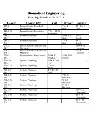
Syllabus for <strong>BME</strong> <strong>327</strong>: Magnetic Resonance Imaging<br />
1 01/08 Introduction, overview, basic concepts of MRI<br />
2 01/10 Basic mathematics related to MR Physics<br />
3 01/15<br />
4 01/17<br />
5 01/22<br />
6 01/24<br />
Spin physics: Nuclear Spin, interactions with applied<br />
magnetic fields, rf-excitation, FID<br />
T1, T2, T2* Relaxation, Bloch equations, Chemical shift<br />
& MR spectroscopy<br />
Imaging principles: magnetic field gradients, spatial<br />
localization, frequency encoding, imaging equation<br />
Imaging principles: Fourier transform, slice selection,<br />
phase encoding, echoes, k-space<br />
7 01/29 Fundamental MRI techniques: Spin echo<br />
8 01/31 Fundamental MRI techniques: Gradient echo<br />
9 02/05<br />
10 02/07<br />
Lab: Introduction to MRI hardware, tour of imaging<br />
facility (Prof. Fluckiger at CTI)<br />
Imaging principles: k-space, finite sampling, aliasing,<br />
resolution, Mid-term review<br />
11 02/12 Midterm Exam<br />
12 02/14<br />
13 02/18<br />
14 02/21<br />
15 02/26<br />
16 02/28<br />
Imaging principles: rf-excitation revisited, slice<br />
selection, inversion recovery<br />
Imaging considerations: contrast manipulation, noise,<br />
steady state. saturation pulses<br />
Imaging considerations: field inhomogeneity, T2*,<br />
contrast agent<br />
Imaging considerations: MR-signal phase, phasecontrast<br />
MRI<br />
Imaging considerations: Fast imaging, parallel imaging,<br />
3D imaging, final review<br />
Nishimura: Chaps.<br />
1 & 3<br />
Nishimura: Chaps.<br />
2<br />
Nishimura: Chap.<br />
4, 6.1<br />
Nishimura: Chap.<br />
4<br />
Nishimura: Chap.<br />
5<br />
Nishimura: 5.2,<br />
5.3, 2.2, 6.2<br />
Nishimura: 7.1,<br />
Handout<br />
Nishimura: 7.3,<br />
Handout<br />
Nishimura: 2.4,<br />
5.6.2<br />
Nishimura: 5.7<br />
Nishimura: Chap.<br />
6 Handout<br />
Nishimura: 7.2 -<br />
7.5<br />
Nishimura: 7.1.<br />
Handout<br />
Handout<br />
17 03/05 Advanced applications I: cardiovascular None<br />
18 03/07 Advanced applications II: TBD None<br />
19 03/12 Advanced applications III: TBD None<br />
20 03/14 Student presentations Papers