Synthesis and characterization of DOPA-PEG conjugates - Phillip B ...
Synthesis and characterization of DOPA-PEG conjugates - Phillip B ...
Synthesis and characterization of DOPA-PEG conjugates - Phillip B ...

Create successful ePaper yourself
Turn your PDF publications into a flip-book with our unique Google optimized e-Paper software.
<strong>Synthesis</strong> <strong>and</strong> Characterization <strong>of</strong> <strong>DOPA</strong>-<strong>PEG</strong> Conjugates<br />
Xiaoping Zeng, Eric Westhaus, Nicole Eberle, Bruce Lee, <strong>and</strong><br />
<strong>Phillip</strong> B. Messersmith*<br />
Northwestern University<br />
Medical School <strong>and</strong> Department <strong>of</strong> Biomedical Engineering<br />
303 E. Chicago Ave.<br />
Chicago, IL 60611<br />
Introduction<br />
Mussel adhesive proteins (MAPs) are remarkable underwater adhesive<br />
materials which form tenacious bonds between certain marine organisms <strong>and</strong><br />
the substrates upon which they reside. 1 These proteins are secreted by the<br />
organisms as fluids which undergo a crosslinking or hardening reaction which<br />
leads to the formation <strong>of</strong> a solid adhesive plaque. One <strong>of</strong> the unique features<br />
<strong>of</strong> MAPs is the presence <strong>of</strong> L-3,4-dihydroxyphenylalanine (<strong>DOPA</strong>), an amino<br />
acid which is believed to be responsible for both adhesion <strong>and</strong> crosslinking<br />
characteristics <strong>of</strong> MAPs. 1,2 Oxidation <strong>of</strong> <strong>DOPA</strong> to the o-quinone can lead to<br />
crosslinking (e.g. by Michael addition with a primary amine), whereas the<br />
catechol form <strong>of</strong> <strong>DOPA</strong> is believed responsible for adhesion to substrates. 2<br />
However, complexation between <strong>DOPA</strong> <strong>and</strong> metal ions could also contribute<br />
to bulk crosslinking as well as adhesion. For example, Fe 3+ forms extremely<br />
stable coordination complexes with a variety <strong>of</strong> catechols, 3 <strong>and</strong> bis- <strong>and</strong> triscatecholate<br />
complexes have been observed to form between Fe 3+ <strong>and</strong> a MAP<br />
from Mytilus edulis. 4,5<br />
Recently, <strong>DOPA</strong>-containing synthetic polypeptides have been<br />
synthesized by copolymerization <strong>of</strong> N-carboxyanhydride monomers <strong>of</strong> lysine<br />
<strong>and</strong> <strong>DOPA</strong>. 6,7 The water soluble copolymers were found to form gels in the<br />
presence <strong>of</strong> oxidizing agents, <strong>and</strong> adhesion to metal substrates was observed.<br />
Our interest is in developing <strong>DOPA</strong>-containing synthetic polymers for use as<br />
biomaterials. We hypothesize that the adhesive, metal ion binding, <strong>and</strong><br />
crosslinking characteristics <strong>of</strong> <strong>DOPA</strong> can be exploited for development <strong>of</strong><br />
new hydrogels as tissue adhesives <strong>and</strong> drug delivery matrices. In this paper we<br />
describe our preliminary efforts toward the synthesis <strong>and</strong> <strong>characterization</strong> <strong>of</strong><br />
linear <strong>and</strong> branched <strong>DOPA</strong>-poly(ethylene glycol) <strong>conjugates</strong> (<strong>DOPA</strong>-<strong>PEG</strong>s).<br />
Experimental<br />
Materials. N-Hydroxysuccinamide ester <strong>of</strong> <strong>PEG</strong> (<strong>PEG</strong>-NHS, 3.4K MW,<br />
1.018 M w /M n ), <strong>PEG</strong>-bis-amine (<strong>PEG</strong>-NH 2 , 3.4K MW) as well as a branched<br />
<strong>PEG</strong>-amine (4-arm, 10 K MW) were purchased from Shearwater Polymers<br />
(Birmingham, AL). Dicyclohexylammonium salt <strong>of</strong> Boc-L-Dopa was<br />
purchased from Sigma (St. Louis, MO). All lipids were purchased from<br />
Avanti Polar Lipids (Alabaster, AL). HBTU <strong>and</strong> HOBT were purchased from<br />
Advanced ChemTech (Louisville, KY).<br />
<strong>Synthesis</strong> <strong>of</strong> Linear CO 2 H-<strong>DOPA</strong>-<strong>PEG</strong> (1). Acid terminated <strong>DOPA</strong>-<br />
<strong>PEG</strong> was obtained by reacting <strong>PEG</strong>-NHS (0.294 mmoles) <strong>and</strong> L-<strong>DOPA</strong> (2.94<br />
mmoles) in 15 mL <strong>of</strong> 5% NaHCO 3 under N 2 bubbling for 45 minutes. The<br />
reaction was stopped by acidification with 1N HCl <strong>and</strong> the solution was<br />
extracted with dichloromethane (DCM). The organic layer was washed with 1<br />
N HCl <strong>and</strong> water <strong>and</strong> then dried with rotary evaporator. The crude product<br />
was purified by dissolution in benzene <strong>and</strong> precipitation with ether. 1 H NMR<br />
analysis revealed that 80% <strong>of</strong> the <strong>PEG</strong> endgroups contained <strong>DOPA</strong> residues.<br />
<strong>Synthesis</strong> <strong>of</strong> Linear Boc-<strong>DOPA</strong>-<strong>PEG</strong> (2). Boc-protected linear<br />
<strong>DOPA</strong>-<strong>PEG</strong> was synthesized by reacting <strong>PEG</strong>-NH 2 (0.294 moles), Boc-L-<br />
Dopa dicyclohexylammonium salt (1.18 mmoles), HOBT(2.35mmole) <strong>and</strong><br />
triethylamine (2.35 mmoles) in 10ml <strong>of</strong> a 50:50 mixture <strong>of</strong><br />
dimethylformamide (DMF) <strong>and</strong> DCM. HBTU(1.18 moles) <strong>and</strong> DCM(5 mL)<br />
were then added. After 30 minutes at room temperature the coupling reaction<br />
was complete as determined by a negative Kaiser test, <strong>and</strong> DCM(60 mL) was<br />
added. The solution was washed with saturated sodium chloride solution, 5%<br />
NaHCO 3 , 1N HCl <strong>and</strong> water <strong>and</strong> then dried with rotary evaporator. The crude<br />
product was purified by dissolution in DCM or benzene <strong>and</strong> precipitation with<br />
ether. A Kaiser test on the final product also was negative.<br />
<strong>Synthesis</strong> <strong>of</strong> Branched Boc-<strong>DOPA</strong>-<strong>PEG</strong> (4). Boc-protected branched<br />
<strong>DOPA</strong>-<strong>PEG</strong> was synthesized by reacting four arm <strong>PEG</strong>-NH 2 (0.2 moles),<br />
Boc-L-Dopa dicyclohexylammonium salt (1.6 mmoles), HOBT(3.2 mmole)<br />
<strong>and</strong> triethylamine (3.2 mmoles) in DMF(5mL) <strong>and</strong> DCM(5 mL). HBTU(1.6<br />
mmoles) <strong>and</strong> DCM(5 mL) were added. After 30 minutes at room temperature<br />
the coupling reaction was complete as determined by a negative Kaiser test.<br />
Extraction <strong>and</strong> purification <strong>of</strong> the product was as described above for <strong>DOPA</strong>-<br />
<strong>PEG</strong> 2. The Kaiser test on the final product was also negative.<br />
<strong>Synthesis</strong> <strong>of</strong> Linear <strong>and</strong> Branched NH 2 -<strong>DOPA</strong>-<strong>PEG</strong> (3 <strong>and</strong> 5).<br />
Amine terminated <strong>DOPA</strong>-<strong>PEG</strong> 3 <strong>and</strong> 5 were obtained from the treatment <strong>of</strong> 2<br />
<strong>and</strong> 4 with TFA. In a typical reaction, Boc-<strong>DOPA</strong>-<strong>PEG</strong> (1.0 g) was dissolved<br />
in DCM (5 mL) <strong>and</strong> treated with TFA (5 mL) for 30 min. The product was<br />
isolated by the precipitation with ether. Kaiser test on the final product was<br />
positive.<br />
Complexation <strong>of</strong> <strong>DOPA</strong>-<strong>PEG</strong> with Fe 3+ . Separately, a 25 mM FeCl 3<br />
solution in pH 7.0 buffer (50mM Bis-Tris), <strong>and</strong> a 15 mg/ml solution <strong>of</strong> 1 in<br />
pH 7.0 buffered NaCl (.15M NaCl, 50mM Bis-Tris), were bubbled with argon<br />
for 20 minutes. 1ml <strong>of</strong> the <strong>DOPA</strong>-<strong>PEG</strong> solution was transferred into a quartz<br />
cuvette, sealed, <strong>and</strong> purged briefly again with Ar. Aliquots <strong>of</strong> the Fe 3+ solution<br />
were then added to the cuvette with stirring, <strong>and</strong> the visible light absorbance<br />
spectrum (U-2010, Hitachi Instruments) obtained to detect complexation<br />
between <strong>DOPA</strong> <strong>and</strong> Fe 3+ .<br />
Preparation <strong>of</strong> Liposomes Containing Fe 3+ (Fe-liposomes). Feliposomes<br />
were prepared by modification <strong>of</strong> a previously described protocol<br />
for preparation <strong>of</strong> CaCl 2 liposomes. 10 A dry film <strong>of</strong><br />
dipalmitoylphosphatidylcholine (DPPC) <strong>and</strong> dimyristoylphosphatidylcholine<br />
(DMPC) (9:1 DPPC:DMPC) was hydrated with 25 mM FeCl 3 in pH 7.0<br />
buffered NaCl (.1M NaCl, 50mM Bis-Tris, 451 mosm), <strong>and</strong> processed as<br />
described previously to yield Fe-liposomes suspended in Fe 3+ -free NaCl<br />
(0.245 M, 449 mosm). The Fe-liposomes were suspended to a final lipid<br />
concentration <strong>of</strong> 20 mg/ml in 0.245 M NaCl, <strong>and</strong> stored at 20ºC until use.<br />
Dynamic light scattering analysis (N4Plus, Beckman-Coulter Corp.) <strong>of</strong> the Feliposomes<br />
yielded an average particle size <strong>of</strong> 1.55 µm. The [Fe 3+ ] in the final<br />
liposome suspension was determined by ICP to be 16mM.<br />
Thermally Triggered Complexation <strong>of</strong> <strong>PEG</strong>-<strong>DOPA</strong> with Fe 3+ . 136µl<br />
<strong>of</strong> Fe-liposome suspension was introduced into a Ar-purged water jacketed<br />
quartz cuvette, after which 1ml <strong>of</strong> 1 (15mg/ml) in buffered NaCl (.15M NaCl,<br />
50mM BisTris, pH 7.0) was added. The spectrophotometer baseline was<br />
recorded with the suspension in both reference <strong>and</strong> sample beams, after which<br />
the absorption spectrum was recorded after the sample was incubated for 30<br />
minutes at 20ºC <strong>and</strong> 30ºC. Following this, the temperature <strong>of</strong> the sample<br />
cuvette was changed to 37ºC <strong>and</strong> the absorbance spectrum recorded after 5,<br />
10, 15, 20, 25 <strong>and</strong> 30 minutes <strong>of</strong> incubation.<br />
CH 2CH CH 2OCH 2CHCH 2<br />
R NH CH C NHC H 2CH 2(OC H 2CH 2) nO<br />
O(C H 2CH 2O) nCH 2CH 2NH C<br />
CH 2 O<br />
O<br />
OH<br />
OH<br />
OH<br />
OH<br />
OH<br />
O CH 2 O<br />
O<br />
O<br />
O<br />
HO C CH NH C CH 2CH 2 C NH(CH 2CH 2O) nCH 2CH 2NH C CH 2CH 2 C<br />
OH<br />
OH<br />
OH<br />
CH 2 O<br />
R NH CH C<br />
CH 2 O<br />
R NH CH C NHC H 2CH 2(OC H 2CH 2) nO<br />
1<br />
O<br />
NH( CH 2CH 2O) n CH 2CH 2NH C<br />
2, R=Boc 3, R=H<br />
4, R=Boc 5, R=H<br />
OH<br />
OH<br />
CH 2<br />
CH NH R<br />
OH<br />
O<br />
O(C H 2CH 2O) nCH 2CH 2NH C<br />
OH<br />
OH<br />
CH 2 O<br />
NH CH C OH<br />
OH<br />
CH 2<br />
CH NH R<br />
CH<br />
CH 2<br />
NH R<br />
OH<br />
OH<br />
Polymer Preprints 2000, 41(1), 989
Results <strong>and</strong> Discussion<br />
Mefp1, a MAP from the mussel Mytilus edulis, was previously found to<br />
form highly stable complexes with Fe 3+ . 4,5 The Mefp1 complexes range in<br />
color from pink to purple, corresponding to tris- <strong>and</strong> bis-catecholate<br />
complexes between <strong>DOPA</strong> <strong>and</strong> Fe 3+ , respectively. Although the physiologic<br />
significance <strong>of</strong> Fe-<strong>DOPA</strong> complexes is not known, it has been speculated that<br />
Fe 3+ binding could lead to crosslinking or curing <strong>of</strong> a Mefp1-containing<br />
lacquer in mussel byssus. 5 By synthesizing <strong>DOPA</strong>-<strong>PEG</strong> <strong>conjugates</strong>, we seek<br />
to accomplish two goals. The first is to exploit the formation <strong>of</strong> intermolecular<br />
Fe 3+ -<strong>DOPA</strong> complexes as a route to crosslinking <strong>of</strong> <strong>PEG</strong>-based hydrogels.<br />
The second is to introduce bioadhesive moieties into <strong>PEG</strong>-based hydrogels for<br />
the purposes <strong>of</strong> tissue repair as well as adhesion to <strong>and</strong> drug delivery across<br />
tissue interfaces.<br />
Several linear <strong>and</strong> branched <strong>DOPA</strong>-<strong>PEG</strong> <strong>conjugates</strong> with acid (1), amine<br />
(3 <strong>and</strong> 5), <strong>and</strong> Boc-protected (2 <strong>and</strong> 4) endgroups were synthesized. The Fe 3+<br />
complexation behavior <strong>of</strong> these molecules was studied using the<br />
spectrophotometric approach <strong>of</strong> Taylor et al. 5 Briefly, this method consisted <strong>of</strong><br />
recording the visible light absorbance <strong>of</strong> the <strong>DOPA</strong>-<strong>PEG</strong> polymers as Fe 3+ is<br />
introduced into the system. Addition <strong>of</strong> Fe 3+ to <strong>DOPA</strong>-<strong>PEG</strong> solution at 20ºC<br />
results in the formation <strong>of</strong> purple complexes during the first hour (data not<br />
shown). As can be seen in Figure 1, peak maxima range from 580nm at low<br />
[Fe 3+ ] to 589nm at higher [Fe 3+ ]. At this point it is not yet known whether the<br />
observed complexes represent the bis (<strong>DOPA</strong>:Fe 3+ =2), tris (<strong>DOPA</strong>:Fe 3+ =3), or<br />
some other form <strong>of</strong> the complex. Although qualitatively similar to the spectra<br />
reported by Taylor et al. 5 for the bis-complexes with Mefp1, the observed<br />
peak maxima <strong>of</strong> the complexes with <strong>DOPA</strong>-<strong>PEG</strong> are at higher wavelengths.<br />
Thus, the exact nature <strong>of</strong> the complexes is not known at this point. In the case<br />
<strong>of</strong> <strong>DOPA</strong>-<strong>PEG</strong> 1, the presence <strong>of</strong> the acidic endgroups complicates this<br />
analysis since they could participate in complex formation along with the<br />
<strong>DOPA</strong> residues. It is also not known whether the complexes <strong>of</strong> <strong>DOPA</strong>-<strong>PEG</strong><br />
with Fe 3+ are intra- or intermolecular. Experiments are in progress to<br />
investigate these effects <strong>and</strong> to determine the impact <strong>of</strong> <strong>DOPA</strong>-<strong>PEG</strong><br />
endgroups in general on Fe 3+ -<strong>DOPA</strong> complexation.<br />
Absorbance<br />
1.0<br />
0.5<br />
0.0<br />
I<br />
H<br />
G<br />
F<br />
E<br />
D<br />
C<br />
B<br />
A<br />
400 500 600 700 800<br />
Wavelength (nm)<br />
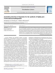
Figure 1. Complexation <strong>of</strong> Fe 3+ with <strong>DOPA</strong>-<strong>PEG</strong> 1. Aliquots <strong>of</strong> 25mM FeCl 3<br />
solution were sequentially added to a cuvette containing 1ml <strong>of</strong> <strong>DOPA</strong>-<strong>PEG</strong> 1<br />
(15mg/ml) in BIS-TRIS buffer: A) 0, B) 10, C) 20, D) 30, E) 40, F) 50, G) 60,<br />
H) 70, I) 80 µl FeCl 3 . Each solution was equilibrated for 30 minutes prior to<br />
measurement.<br />
We also investigated the possibility <strong>of</strong> thermally triggering<br />
complexation by stimuli-induced release <strong>of</strong> Fe 3+ from lipid vesicles. Our<br />
approach to accomplish this mirrors our earlier work in which calcium-loaded<br />
lipid vesicles were used to form calcium phosphate mineral <strong>and</strong> polymer<br />
hydrogels. 8-10 In our previous systems, liposomes were designed to release<br />
entrapped ions when heated from ambient to body temperature, a result <strong>of</strong> an<br />
increased lipid bilayer permeability at the lipid chain melting temperature. In<br />
this work, we have entrapped a 25mM FeCl 3 solution within liposomes, <strong>and</strong><br />
suspended these liposomes in Fe 3+ -free buffer. The resulting liposome<br />
suspension had a light orange color which reflects the color <strong>of</strong> the entrapped<br />
FeCl 3 solution.<br />
When Fe-liposomes were combined with a clear solution <strong>of</strong> 1 at 20ºC,<br />
the suspension maintained a faint orange color for at least several hours when<br />
maintained at 20ºC. As shown in Figure 2, little or no color change occurred<br />
when the suspension was heated to 30ºC, however when heated to 37ºC a<br />
purple complex with a peak maximum at 578nm rapidly formed during the<br />
first few minutes. This behavior can be explained by the thermally triggered<br />
release <strong>of</strong> Fe 3+ from liposomes, resulting in the complexation <strong>of</strong> Fe 3+ with<br />
<strong>DOPA</strong>-<strong>PEG</strong>. The peak maximum shifted to 594nm <strong>and</strong> increased significantly<br />
in intensity during the first 30 minutes at 37ºC.<br />
Absorbance<br />
0.6<br />
0.5<br />
0.4<br />
0.3<br />
0.2<br />
0.1<br />
0.0<br />
400 500 600 700 800<br />
Wavelength (nm)<br />
Figure 2. Thermally triggered complexation <strong>of</strong> Fe 3+ with <strong>DOPA</strong>-<strong>PEG</strong> 1.<br />
136µl <strong>of</strong> Fe-liposomes were combined with 1ml <strong>DOPA</strong>-<strong>PEG</strong> 1 in BIS-TRIS<br />
buffer. From bottom to top: 30 minutes at 20ºC, 30 minutes at 30ºC, <strong>and</strong> 0, 5,<br />
10, 15, 20, 25, <strong>and</strong> 30 minutes at 37ºC.<br />
Conclusions<br />
Linear <strong>and</strong> branched <strong>DOPA</strong>-<strong>PEG</strong> <strong>conjugates</strong> with acid, amine, <strong>and</strong> Bocprotected<br />
endgroups were synthesized. The linear <strong>DOPA</strong>-<strong>PEG</strong> with acidic<br />
endgroups was found to form complexes with free Fe 3+ as determined by<br />
spectrophotometric analysis. Similar complexes between Fe 3+ <strong>and</strong> <strong>DOPA</strong>-<br />
<strong>PEG</strong> were rapidly formed when a <strong>DOPA</strong>-<strong>PEG</strong> solution containing Feliposomes<br />
was heated to 37ºC. Ongoing experiments are aimed at elaborating<br />
the nature <strong>of</strong> the complexes formed (i.e. bis versus tris) <strong>and</strong> whether the<br />
formation <strong>of</strong> intermolecular complexes between <strong>PEG</strong>-bound <strong>DOPA</strong><br />
endgroups can result in physical gelation.<br />
Acknowledgement. This research was supported by the Whitaker<br />
Foundation <strong>and</strong> by NIH grant R01 DE 13030.<br />
References<br />
1) Waite, J.H. Int. J. Adhesion Adhesives 1987, 7, 9.<br />
2) Deming, T.J. Curr. Op. Chem. Biol. 1999, 3, 100.<br />
3) Avdeef, A.; et al. J. Am. Chem. Soc. 1978, 100, 5362.<br />
4) Taylor, S.W.; Luther, G.W.; Waite, J.H. Inorg. Chem. 1994, 33, 5819.<br />
5) Taylor, S.W.; et al. Inorg. Chem. 1996, 35, 7572.<br />
6) Yu, M.; Deming, T.J. Macromolecules 1998, 31, 4739.<br />
7) Yu, M.; Hwang, J.; Deming, T.J. J. Am. Chem. Soc. 1999, 121:5825.<br />
8) Messersmith, P.B.; Starke, S. Chemistry <strong>of</strong> Materials 1998, 10, 117.<br />
9) Murphy, W.; Messersmith, P.B. Polyhedron, in press.<br />
10) Cui, H.; Messersmith, P.B. in Tailored Polymeric Materials for<br />
Controlled Delivery Systems, I. McCullough <strong>and</strong> S. Shalaby, eds., ACS<br />
Symposium Series 709, 1998, pp. 203-211.<br />
Polymer Preprints 2000, 41(1), 990