Characterization of English ivy (Hedera helix) adhesion force and ...
Characterization of English ivy (Hedera helix) adhesion force and ...
Characterization of English ivy (Hedera helix) adhesion force and ...

Create successful ePaper yourself
Turn your PDF publications into a flip-book with our unique Google optimized e-Paper software.
J Nanopart Res (2011) 13:1029–1037<br />
DOI 10.1007/s11051-010-0091-3<br />
RESEARCH PAPER<br />
<strong>Characterization</strong> <strong>of</strong> <strong>English</strong> <strong>ivy</strong> (<strong>Hedera</strong> <strong>helix</strong>) <strong>adhesion</strong><br />
<strong>force</strong> <strong>and</strong> imaging using atomic <strong>force</strong> microscopy<br />
Lijin Xia • Scott C. Lenaghan • Mingjun Zhang •<br />
Yu Wu • Xiaopeng Zhao • Jason N. Burris •<br />
C. Neal Stewart Jr.<br />
Received: 20 January 2010 / Accepted: 14 September 2010 / Published online: 30 September 2010<br />
Ó Springer Science+Business Media B.V. 2010<br />
Abstract <strong>English</strong> <strong>ivy</strong> (<strong>Hedera</strong> <strong>helix</strong>) is well known<br />
for its ability to climb onto <strong>and</strong> strongly adhere to a<br />
variety <strong>of</strong> solid substrates. It has been discovered that<br />
the <strong>ivy</strong> aerial rootlet secretes an adhesive composed<br />
<strong>of</strong> polysaccharide <strong>and</strong> spherical nanoparticles. This<br />
study aims to characterize the mechanical properties<br />
<strong>of</strong> the nanocomposite adhesive using atomic <strong>force</strong><br />
microscopy (AFM). The adhesive was first imaged by<br />
AFM to visualize the nanocomposite. Mechanical<br />
properties were then determined at various time<br />
points, from secretion to hardening. The experimental<br />
results indicate that the <strong>ivy</strong> adhesive exhibited strong<br />
<strong>adhesion</strong> strength <strong>and</strong> high elasticity. There was a<br />
decrease in adhesive <strong>force</strong> over time, from 298 to<br />
202 nN during the 24-h study. Accompanying with it<br />
were the limited changes in extension length <strong>and</strong><br />
Young’s modulus. The limited curing process <strong>of</strong> the<br />
<strong>ivy</strong> adhesive helps fill gaps in the attaching surface,<br />
leading to more intimate contact <strong>and</strong> increased van<br />
der Waals interactions with the surface. However,<br />
L. Xia S. C. Lenaghan M. Zhang (&) <br />
Y. Wu X. Zhao<br />
Department <strong>of</strong> Mechanical, Aerospace <strong>and</strong> Biomedical<br />
Engineering, University <strong>of</strong> Tennessee, Knoxville,<br />
TN 37996, USA<br />
e-mail: mjzhang@utk.edu<br />
J. N. Burris C. Neal Stewart Jr.<br />
Department <strong>of</strong> Plant Sciences, University <strong>of</strong> Tennessee,<br />
Knoxville, TN 37996, USA<br />
study based on a mechanical model indicated that van<br />
der Waals <strong>force</strong> alone is not significant enough to<br />
account for all <strong>of</strong> the measured <strong>force</strong>. Other chemical<br />
interactions <strong>and</strong> cross linking likely contribute to the<br />
strong <strong>adhesion</strong> strength <strong>of</strong> <strong>ivy</strong>.<br />
Keywords Ivy Nanoparticle Adhesion <strong>force</strong> <br />
Atomic <strong>force</strong> microscopy Nanobiotechnology<br />
Introduction<br />
<strong>Hedera</strong>, or <strong>ivy</strong>, is a genus containing approximately<br />
16 species <strong>of</strong> climbing or ground-creeping evergreen<br />
plants (Jennifer <strong>and</strong> Jun 2003). On suitable surfaces,<br />
such as trees <strong>and</strong> rock faces, <strong>ivy</strong> has the ability to<br />
climb 25–30 m above the ground <strong>and</strong> hold fast to<br />
vertical surfaces. This unique ability to climb has<br />
drawn great interest from scientists as early as the<br />
1800s. Charles Darwin first observed that the aerial<br />
rootlets produced on creeping <strong>and</strong> climbing stems<br />
‘‘secreted a little yellowish matter’’ upon first contact<br />
with an attaching surface (Darwin 1876). Little<br />
progress has been made in determining the structure<br />
<strong>and</strong> composition <strong>of</strong> this yellowish adhesive since the<br />
initial observation from Darwin. In the mid 1970s,<br />
Endress <strong>and</strong> Thomson investigated the adhesive <strong>of</strong><br />
Boston <strong>ivy</strong> (Parthenocissus tricuspidata), using<br />
transmission electron microscopy (TEM) (Endress<br />
<strong>and</strong> Thomson 1976, 1977). Their studies provided<br />
123
1030 J Nanopart Res (2011) 13:1029–1037<br />
in-depth details about the structure <strong>of</strong> attaching<br />
Boston <strong>ivy</strong> tendrils, but limited information about<br />
the nature <strong>of</strong> the secreted adhesive. Other studies<br />
have focused on the morphology <strong>of</strong> the rootlets using<br />
light <strong>and</strong> scanning electron microscopy, but have not<br />
provided information on the structure <strong>and</strong> composition<br />
<strong>of</strong> the adhesive (Moens 1956; Melzer et al.<br />
2010). In 2008, Zhang et al. conducted one <strong>of</strong> the first<br />
studies focused on identifying the structural components<br />
<strong>of</strong> the adhesive secreted from naturally growing<br />
<strong>ivy</strong> rootlets. Using atomic <strong>force</strong> microscopy (AFM),<br />
they observed a high abundance <strong>of</strong> uniform nanoparticles<br />
deposited onto the attaching surface. These<br />
nanoparticles were about 70 nm in diameter, <strong>and</strong><br />
were observed on a variety <strong>of</strong> surfaces including<br />
mica, silicon wafer, <strong>and</strong> glass (Zhang et al. 2008).<br />
From the discovery, it is hypothesized that nanoparticles<br />
are the key component that generates the strong<br />
<strong>adhesion</strong> used by <strong>ivy</strong> to climb vertical surfaces.<br />
Intense research has been conducted over the last<br />
decade on the contribution <strong>of</strong> nanostructures to<br />
surface <strong>adhesion</strong>. One <strong>of</strong> the most celebrated examples<br />
is the discovery <strong>of</strong> nan<strong>of</strong>ibers on the gecko<br />
footpads. The existence <strong>of</strong> these abundant <strong>and</strong><br />
deformable nan<strong>of</strong>ibers maximizes the intimate contact<br />
area between the toes <strong>of</strong> the gecko <strong>and</strong> the<br />
climbing surfaces. This intimate contact facilitates<br />
inter-molecular interactions through van der Waals<br />
<strong>force</strong> (Autumn et al. 2002; Peattie <strong>and</strong> Full 2007).<br />
Similar attachment mechanisms have been observed<br />
in flies, beetles, <strong>and</strong> lizards (Steinbrecher et al. 2010).<br />
Although these studies have focused exclusively on<br />
dry <strong>adhesion</strong> <strong>and</strong> on fibrillar structures from the<br />
micro- to nano-scale, shape-insensitive optimal <strong>adhesion</strong><br />
has been studied in mathematic models (Gao <strong>and</strong><br />
Yao 2004). From these models, it was predicted that<br />
for nano-sized structures, shape is not as important as<br />
the size <strong>of</strong> the structure in relation to generating<br />
adhesive strength. Following this principle, we expect<br />
that nanoparticles <strong>of</strong>fer the same potential for surface<br />
<strong>adhesion</strong> due to their size scale.<br />
A theoretical model was recently developed by our<br />
group to evaluate the impact <strong>of</strong> van der Waals<br />
interactions between <strong>ivy</strong> nanoparticles <strong>and</strong> solid<br />
substrates. From the model, it was confirmed that<br />
van der Waals <strong>force</strong> alone was significant to account<br />
for the strength <strong>of</strong> attachment <strong>of</strong> <strong>ivy</strong> (Wu et al. 2010).<br />
Experimentally, various nanoparticles have been<br />
tested to enhance adhesive strength or as filler for<br />
polymeric adhesives (Zhai et al. 2006; Corkery et al.<br />
2008). Although it is not clear whether the nanoparticles<br />
contribute directly or indirectly to the strength<br />
<strong>of</strong> <strong>adhesion</strong>, a recent study has provided firm<br />
evidence <strong>of</strong> the <strong>adhesion</strong> ability <strong>of</strong> polystyrene<br />
nanoparticles (Xing et al. 2010).<br />
The existence <strong>of</strong> adhesive nanoparticles will<br />
prompt their potential applications in a variety <strong>of</strong><br />
fields. Nanoparticles have already been explored as<br />
fillers in the glue industry. Adherent nanoparticles<br />
also hold a significant promise to future research in<br />
drug delivery. Biodegradable nanoparticles provide<br />
controllable, sustained drug delivery in vitro (Panyam<br />
<strong>and</strong> Labhasetwar 2003). In vivo studies, however,<br />
have shown that nanoparticles are not effective at<br />
adhering to vascular walls under shear stress. Adherent<br />
nanoparticles may overcome this deficiency,<br />
allowing the lingering <strong>of</strong> nanoparticles in activated<br />
or inflammatory endothelial cells under fluid shear<br />
stress thus improving the cell uptake <strong>of</strong> drug-carrying<br />
nanoparticles. An additional advantage <strong>of</strong> <strong>ivy</strong> nanoparticles<br />
is their natural origin <strong>and</strong> biocompatibility<br />
which provide them extra potential use in medical<br />
<strong>and</strong> bioengineering fields. Ivy nanoparticles have<br />
recently been proposed to be a prospective c<strong>and</strong>idate<br />
to replace metal-based nanoparticles for sunscreens<br />
(Xia et al. 2010). Biocompatibility combined with<br />
strong <strong>adhesion</strong> ability will make <strong>ivy</strong> nanoparticles<br />
an ideal active component in the design <strong>of</strong> bioglue,<br />
surgical suture, or other wound healing materials.<br />
Before taking significant steps in the direction <strong>of</strong><br />
proposed applications, it is essential to first underst<strong>and</strong><br />
the strength <strong>of</strong> the adhesive, as well as the<br />
principles involved in generating the attachment<br />
strength. In an effort to underst<strong>and</strong> how nanoparticles<br />
contribute to the attachment strength, accurate measurement<br />
<strong>of</strong> the adhesive <strong>force</strong> at the proper scale was<br />
necessary. AFM is a fundamental tool used to<br />
determine nano-scale <strong>force</strong>s, <strong>and</strong> is accurate even<br />
when measuring intermolecular <strong>force</strong>s in piconewtons<br />
(Aoki et al. 1997). AFM can also be used<br />
to monitor the fundamental mechanical properties <strong>of</strong><br />
materials such as adhesive strength, compressibility,<br />
<strong>and</strong> elasticity (Binnig et al. 1986; Muller <strong>and</strong> Dufrene<br />
2008; Noy 2006; Xing et al. 2010). Small noncovalent<br />
interactions, such as van der Waals <strong>force</strong>,<br />
hydrophobic <strong>and</strong> electrostatic interactions, <strong>and</strong> protein<br />
folding can also be determined using AFM<br />
(Matthias et al. 1997; Châtellier et al. 1998). As such,<br />
123
J Nanopart Res (2011) 13:1029–1037 1031<br />
we have applied this AFM-based approach to examine<br />
the nano-scale material properties <strong>of</strong> the nanocomposite<br />
adhesive secreted from <strong>English</strong> <strong>ivy</strong>.<br />
In this study, a method was first developed for<br />
growing <strong>English</strong> <strong>ivy</strong> rootlets in tissue culture, to<br />
eliminate concern over contamination <strong>and</strong> to control<br />
the growth stages <strong>of</strong> <strong>ivy</strong> rootlets. The adhesive <strong>of</strong><br />
tissue-cultured rootlets was then compared to naturally<br />
grown rootlets for their nanostructures. Both<br />
were found to contain nanoparticles with similar<br />
shape <strong>and</strong> size. After confirming the existence <strong>of</strong><br />
nanocomposite adhesive, its strength was further<br />
determined using <strong>force</strong> versus distance curves generated<br />
by AFM during the 24-h study. At the same<br />
time, the elasticity <strong>and</strong> extension length <strong>of</strong> the<br />
secreted adhesive were monitored, to collect useful<br />
data for analyzing <strong>and</strong> underst<strong>and</strong>ing the adhesive<br />
hardening process.<br />
Materials <strong>and</strong> methods:<br />
Sample preparation<br />
Juvenile <strong>Hedera</strong> <strong>helix</strong> shoots, cut from a natural<br />
source on the University <strong>of</strong> Tennessee, Knoxville<br />
campus, were grown in a greenhouse under controlled<br />
temperature, humidity, <strong>and</strong> light availability. Leaves<br />
were removed from the shoots, <strong>and</strong> shoots were<br />
trimmed to 6 cm. Shoots were then sterilized using<br />
1.23% sodium hypochlorite [20% (v/v) commercial<br />
bleach] plus 0.05% Tween 20, shaken at 200 rpm for<br />
20 min, <strong>and</strong> subsequently washed with sterile water<br />
three times. Sterile shoots were then placed upright<br />
into Magenta GA7 boxes containing MS medium<br />
when they were grown at 24 °C at 16:8-h photoperiod<br />
under 82 lmol/m 2 /s 1 irradiance (Murashige <strong>and</strong> Skoog<br />
1962). Aerial roots were produced after ca. 2 days <strong>and</strong><br />
were allowed to grow for an additional 2 days in MS<br />
media. Aerial roots were excised from source plants<br />
when they were approximately 3 cm in length. These<br />
were transferred to Petri dishes containing MS medium<br />
until analysis was performed (between 3 days <strong>and</strong><br />
2 weeks). In preparation for adhesive analysis, the tip<br />
<strong>of</strong> rootlets was gently cut under a bio-safety cabinet <strong>and</strong><br />
touched on the silicon wafer surface to release the<br />
content. Samples were then either processed immediately,<br />
or allowed to air-dry in a bio-safety cabinet for at<br />
least 5 h.<br />
AFM imaging<br />
The freshly prepared or air-dried <strong>ivy</strong> adhesive<br />
samples were scanned for the existence <strong>of</strong> nanoparticles<br />
using an Agilent 5500 AFM system (Agilent<br />
Technologies, Santa Clara, CA). The samples were<br />
imaged at room temperature (20 °C) using Picoview<br />
TM in AC mode, which minimizes sample<br />
distortion due to mechanical interactions between the<br />
AFM tip <strong>and</strong> the surface. To further optimize imaging,<br />
both the integral <strong>and</strong> proportional gains were adjusted<br />
to avoid excessive loading <strong>force</strong> applied to the<br />
samples. Using this system, three-dimensional imaging<br />
<strong>of</strong> the surface morphology with very high lateral<br />
<strong>and</strong> vertical resolution could be obtained. The probes<br />
used were commercially available silicon probes<br />
TAP300Al Ò (Budget Sensors, S<strong>of</strong>ia, Bulgaria) with<br />
a spring constant <strong>of</strong> 20–75 N/m, a resonant frequency<br />
<strong>of</strong> 300 ± 100 kHz, <strong>and</strong> a radius <strong>of</strong> curvature <strong>of</strong> less<br />
than 10 nm.<br />
Adhesive <strong>force</strong> measurement<br />
To measure the adhesive properties <strong>of</strong> the <strong>ivy</strong><br />
secretion, <strong>force</strong> versus distance curves were generated<br />
using an Agilent 5500 AFM system. The AFM<br />
probe was first calibrated with bare silicon wafer as<br />
a substrate to obtain a deflection sensitivity <strong>of</strong><br />
33.5 nm/V <strong>and</strong> a <strong>force</strong> constant <strong>of</strong> 4.33 N/m for the<br />
TAP150Al-G Ò (Budget Sensors) cantilever probes.<br />
The location <strong>of</strong> the adhesive was verified with a<br />
CCD camera mounted to a 10X objective, <strong>and</strong> the<br />
cantilever was moved over the center <strong>of</strong> the<br />
adhesive. Once the location had been established,<br />
approach <strong>of</strong> the tip was initiated <strong>and</strong> the tip was<br />
allowed to contact the sample surface for 2 s so that<br />
the probe <strong>and</strong> adhesive formed a complex. The tip<br />
was then withdrawn from the adhesive, <strong>and</strong> the<br />
deflection <strong>of</strong> the cantilever was measured. A series<br />
<strong>of</strong> 30 <strong>force</strong> curves exhibiting <strong>adhesion</strong> events were<br />
taken for each time point during the curing process.<br />
The mean <strong>and</strong> st<strong>and</strong>ard deviations were calculated<br />
using Micros<strong>of</strong>t Excel Ò . To calculate Young’s<br />
modulus, the slope <strong>of</strong> the approach line for each<br />
<strong>force</strong> curve was obtained using Picoimage TM <strong>and</strong><br />
was then applied to equation E = (F/dL)*(L/A). In<br />
this equation, E is the Young’s modulus, F/dL is<br />
the slope <strong>of</strong> the approaching curve, L is height <strong>of</strong><br />
123
1032 J Nanopart Res (2011) 13:1029–1037<br />
the material, <strong>and</strong> A is the contact area. The height<br />
<strong>of</strong> the material, L, was determined by imaging the<br />
adhesive <strong>and</strong> obtaining a vertical pr<strong>of</strong>ile from the<br />
topography scan in Picoimage TM . The area<br />
<strong>of</strong> contact, A, was calculated based on the diameter<br />
<strong>of</strong> the tip according to the manufacturer’s<br />
specifications.<br />
Results <strong>and</strong> discussion<br />
Nanoparticles produced from in vitro <strong>ivy</strong> rootlets<br />
Because <strong>of</strong> instrument limitation, a real-time monitoring<br />
<strong>of</strong> the attachment process <strong>of</strong> <strong>ivy</strong> at nano-scale<br />
in the fields becomes difficult. Also, concerns emerge<br />
for the contamination <strong>of</strong> environmental nanoparticles<br />
in the open air <strong>and</strong> for the growth stage difference <strong>of</strong><br />
<strong>ivy</strong> rootlets in natural conditions. Thus, a method was<br />
first developed to grow <strong>English</strong> <strong>ivy</strong> rootlets in vitro.<br />
This method allowed us to have an opportunity to<br />
investigate the adhesives properties in different<br />
stages. For topographical characterization <strong>of</strong> the<br />
adhesives from cultured <strong>ivy</strong> rootlets, the rootlets<br />
were washed with 20-nm-filtered water <strong>and</strong> cut at the<br />
tips to release adhesive. Freshly prepared samples<br />
were extremely s<strong>of</strong>t <strong>and</strong> prone to smearing by the<br />
AFM tip during the scanning. After 5 h <strong>of</strong> settlement,<br />
however, the adhesive could be clearly imaged. As<br />
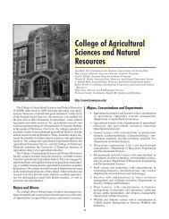
shown in Fig. 1, the nanocomposite was mainly<br />
composed <strong>of</strong> nanoparticles 60–85 nm in diameter.<br />
The average diameter for the nanoparticles was<br />
70 ± 6.5 nm, which was comparable to the previous<br />
report (Zhang et al. 2008).<br />
This observation confirmed the previous report <strong>of</strong><br />
nanoparticles in <strong>ivy</strong> adhesive. Nanoparticles embedded<br />
in a polymeric matrix are not unique to <strong>ivy</strong><br />
adhesive. The existence <strong>of</strong> similar types <strong>of</strong> nanocomposites<br />
has been confirmed in the secretions <strong>of</strong> a<br />
variety <strong>of</strong> marine species, including polychaetes,<br />
mussels, barnacles, <strong>and</strong> sea stars (Stevens et al. 2007;<br />
Zhang et al. 2009; Berglin <strong>and</strong> Gatenholm 2003). The<br />
size <strong>of</strong> spherical particles in these adhesives is in the<br />
same range as those found in the <strong>ivy</strong> secretions,<br />
60 nm for mussel, 80 nm for barnacles, <strong>and</strong><br />
50–100 nm for polychaetes <strong>and</strong> sea stars (Hennebert<br />
et al. 2008). The fact that these nanoparticles can<br />
be observed in a variety <strong>of</strong> species indicates a conserved<br />
approach to generating adhesive materials.<br />
Fig. 1 Nanoparticles released from the tip <strong>of</strong> in vitro grown<br />
<strong>ivy</strong> rootlets. AFM tapping-mode images <strong>of</strong> content from the<br />
tips <strong>of</strong> cultured <strong>ivy</strong> rootlets after 5 h <strong>of</strong> settlement.<br />
a 3.3 9 3.3 nm scanning window. b 1.0 9 1.0 nm scanning<br />
window<br />
Most reported marine adhesives consist <strong>of</strong> protein<br />
complexes dispersed in a polysaccharide matrix<br />
(Hennebert et al. 2008), probably due to the multifunctionality<br />
<strong>and</strong> diversity <strong>of</strong> protein. A study done<br />
by Callow et al. showed that algal spores become less<br />
sensitive to detachment after treatment with proteolytic<br />
enzymes (Callow et al. 2000), indicating the key<br />
role <strong>of</strong> proteins in attachment. Some proteins in<br />
adhesives have important biochemical characteristics,<br />
such as being rich in polar <strong>and</strong> charged residues,<br />
which presents a significant number <strong>of</strong> functional<br />
123
J Nanopart Res (2011) 13:1029–1037 1033<br />
groups for cross linking <strong>and</strong> <strong>adhesion</strong>. A recently<br />
identified barnacle cement protein is a surfacecoupling<br />
protein (Mrcp-19 k) which could efficiently<br />
adsorb to surfaces with various characteristics,<br />
regardless <strong>of</strong> charge <strong>and</strong> hydrophobicity (Urushida<br />
et al. 2007).<br />
High <strong>adhesion</strong> strength <strong>of</strong> <strong>ivy</strong> nanoparticles<br />
As observed in the above biological adhesives, the<br />
existence <strong>of</strong> a large amount <strong>of</strong> proteins in the form <strong>of</strong><br />
nanoparticles implies that they play an important role<br />
in attachment. However, direct evidence for the<br />
<strong>adhesion</strong> ability <strong>of</strong> these nanoparticle-dominated<br />
adhesives has not been obtained. After confirmation<br />
<strong>of</strong> the existence <strong>of</strong> nanoparticles from cultured <strong>ivy</strong><br />
rootlets, its <strong>adhesion</strong> <strong>force</strong> was studied for the first<br />
time using AFM. After calibration <strong>and</strong> determination<br />
<strong>of</strong> the deflection sensitivity <strong>and</strong> <strong>force</strong> constant <strong>of</strong> the<br />
AFM tip, <strong>force</strong> versus distance curves were performed<br />
to measure the pull-<strong>of</strong>f <strong>force</strong> generated from<br />
the interaction between the tip <strong>and</strong> the adhesive.<br />
Thirty successful approach-retract curves <strong>of</strong> freshly<br />
prepared <strong>ivy</strong> adhesive samples were conducted to<br />
determine the optimal adhesive <strong>force</strong>. Out <strong>of</strong> these<br />
curves, the values <strong>of</strong> pull-<strong>of</strong>f <strong>force</strong> ranged from 274<br />
to 307 nN, with an average value <strong>of</strong> 298 ± 8.34 nN.<br />
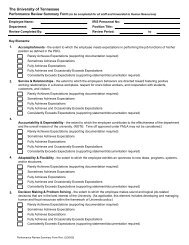
A typical <strong>force</strong> curve exhibited a single pull-<strong>of</strong>f event<br />
during the measurements (Fig. 2a), even though sawtoothed<br />
curves were observed occasionally (Fig. 2b).<br />
The saw-toothed pull-<strong>of</strong>f is typical <strong>of</strong> polymer-based<br />
materials, <strong>and</strong> indicated that the tip was interacting<br />
with multiple components <strong>of</strong> the nanocomposite<br />
adhesive.<br />
The nano-scale <strong>adhesion</strong> <strong>force</strong> for freshly released<br />
<strong>ivy</strong> adhesive averaged 298 nN, which is four times<br />
greater than what could be measured for the adhesive<br />
from algal spores using AFM (Callow et al. 2000).<br />
However, the readings are close to a recent study <strong>of</strong><br />
<strong>adhesion</strong> <strong>force</strong> between a polystyrene nanoparticle<br />
<strong>and</strong> a nan<strong>of</strong>iber in the range <strong>of</strong> 159.5–384.9 nN (Xing<br />
et al. 2010). Also, to ensure that the measured <strong>force</strong><br />
was not due to the tip contamination, the tip was<br />
frequently moved to a bare silicon surface to calibrate<br />
the pull-<strong>of</strong>f <strong>force</strong>. The <strong>force</strong> measured on the bare<br />
silicon wafer surface was around 30 nN, which is<br />
comparable to previous reports (Grinevich et al.<br />
1999).<br />
Fig. 2 A typical approach-retract cycle illustrates the <strong>adhesion</strong><br />
pr<strong>of</strong>ile <strong>and</strong> other mechanical properties <strong>of</strong> <strong>ivy</strong> nanoparticles<br />
examined under AFM in a. Saw-toothed curves were occasionally<br />
observed in b<br />
High elasticity <strong>of</strong> <strong>ivy</strong> nanoparticles<br />
The addition <strong>of</strong> filler materials to adhesives normally<br />
strengthens the adhesive, but reduces the tack which is<br />
a required character for permanent attachment. The<br />
addition <strong>of</strong> a nanomaterial, however, showed different<br />
behaviors, exhibiting an increase in both strength <strong>and</strong><br />
tack (Wang et al. 2006), which might explain the<br />
advantage <strong>of</strong> nanoparticles for strong attachment.<br />
Tackiness theory predicts that as the elastic modulus<br />
<strong>of</strong> the coating increases, the tack energy decreases<br />
(Gay <strong>and</strong> Leibler 1999). The different tack performance<br />
from nanomaterial fillers might be due to their<br />
unusual interactions with polymeric matrix, which<br />
increases the elastic modulus <strong>of</strong> the composite adhesive.<br />
For this purpose, we examined the elasticity <strong>of</strong><br />
the <strong>ivy</strong> adhesive. In this study, the slope <strong>of</strong> the<br />
approach curve was used to calculate Young’s modulus.<br />
The obtained Young’s modulus through this<br />
method ranged from 1.035 to 1.297 GPa for the <strong>ivy</strong><br />
adhesive, <strong>and</strong> was much greater than what was<br />
calculated for algal spores. Young’s modulus <strong>of</strong> the<br />
dried adhesive was comparable to a hard material such<br />
as rubber, nylon, <strong>and</strong> some plastics. High elasticity <strong>of</strong><br />
the <strong>ivy</strong> nanocomposite could be attributed in part to<br />
the Hall–Petch effect, which predicts that as grain size<br />
123
1034 J Nanopart Res (2011) 13:1029–1037<br />
decreases into the nanometer range, stiffness increases<br />
due to increased trapping <strong>of</strong> dislocations at interfaces<br />
(Waychunas <strong>and</strong> Zhang 2008).<br />
Time-based loss <strong>of</strong> <strong>adhesion</strong><br />
The time from initial secretion <strong>of</strong> the adhesive to<br />
permanent attachment is a slow process that involves<br />
corresponding changes in the adhesive. These<br />
changes could be due to water evaporation after<br />
secretion, or chemical changes due to polymerization.<br />
Ultimately the changes will be reflected in the<br />
hardening <strong>of</strong> the adhesive, which are believed to<br />
correspond to the formation <strong>of</strong> strong permanent<br />
attachment. To follow the change in adhesive properties<br />
over time, <strong>force</strong> versus distance curves were<br />
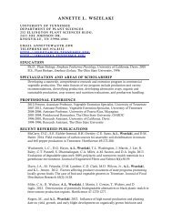
taken over the course <strong>of</strong> 24 h. The time-course <strong>of</strong><br />
<strong>adhesion</strong> revealed a slow reduction in the first 8.5 h<br />
from 298 to 202 nN, followed by a constant level after<br />
24 h (Fig. 3). A 30.2% decrease <strong>of</strong> <strong>adhesion</strong> <strong>force</strong> was<br />
observed over a curing period <strong>of</strong> 8.5 h. This change<br />
was not as great when compared to a 65% decrease<br />
observed during the first hour <strong>of</strong> settlement in the<br />
adhesive secreted from Enteromorpha, an algae<br />
(Callow et al. 2000).<br />
One explanation for the decreased adhesive <strong>force</strong><br />
was the hardening <strong>of</strong> the limited amount <strong>of</strong> polymer<br />
matrix material in the <strong>ivy</strong> adhesive. As observed with<br />
many polymer glues, the hydrated form has a high<br />
adhesive strength, whereas the dried form lacks any<br />
<strong>adhesion</strong>. This is due to a change in conformation <strong>of</strong><br />
Fig. 3 Change <strong>of</strong> <strong>adhesion</strong> <strong>force</strong> within 24 h. The <strong>force</strong> was<br />
measured at various time points after deposition. Each point<br />
represents an average <strong>of</strong> 30 retraction curves ±95% confidence<br />
limits<br />
the material <strong>and</strong> the gradual formation <strong>of</strong> a solid upon<br />
drying. Another explanation includes the loss <strong>of</strong><br />
capillary <strong>force</strong>s due to the decreased water content<br />
during the drying process. Capillary <strong>force</strong>s play a<br />
significant role in the adhesive <strong>force</strong> <strong>of</strong> the gecko, <strong>and</strong><br />
decreased relative humidity has been related to<br />
decreased <strong>adhesion</strong> <strong>force</strong> (Sun et al. 2005). The<br />
freshly released adhesive contains a significant percentage<br />
<strong>of</strong> water which is lost as the sample dries. The<br />
capillary <strong>force</strong>s associated with water in the hydrated<br />
state would become smaller during the drying process,<br />
leading to the decreased <strong>adhesion</strong> <strong>force</strong>.<br />
Limited change in extension length<br />
<strong>and</strong> Young’s modulus<br />
The decreased <strong>force</strong> over time can be due to the<br />
existence <strong>of</strong> polymeric matrix <strong>and</strong> be related to<br />
changes <strong>of</strong> elasticity <strong>and</strong> extension length as observed<br />
in previous studies (Callow et al. 2000), which is also<br />
a typical response for liquid adhesives during the<br />
hardening process. Because <strong>of</strong> the existence <strong>of</strong> a small<br />
amount <strong>of</strong> polymeric matrix in <strong>ivy</strong> adhesive, it is<br />
expected similar changes exist during the hardening<br />
process <strong>of</strong> the adhesive. For this purpose, elasticity<br />
<strong>and</strong> extension length for the <strong>ivy</strong> adhesive were also<br />
investigated over the 24-h time-course. From Figs. 4<br />
<strong>and</strong> 5, we concluded that the curing pr<strong>of</strong>iles <strong>of</strong> the <strong>ivy</strong><br />
adhesive were consistent with the pattern observed<br />
from the adhesive <strong>of</strong> the algae Enteromorpha (Callow<br />
et al. 2000), but at a much lower degree. The values<br />
for extension length (interpreted in Fig. 2a) show a<br />
limited decrease over 24 h, indicating a minimal<br />
hardening over the time-course. Correspondingly,<br />
Young’s modulus increased by 20.2% over 6 h, much<br />
less than normally found in liquid or s<strong>of</strong>t polymer<br />
adhesives (Callow et al. 2000). This observed difference<br />
in extension length <strong>and</strong> Young’s modulus change<br />
over time was most likely due to the low percentage <strong>of</strong><br />
polymeric matrix. The existence <strong>of</strong> a large amount <strong>of</strong><br />
nanoparticles, on the other h<strong>and</strong>, does not change in<br />
elasticity <strong>and</strong> extension length during the hardening<br />
process. The fact that polymeric adhesives, such as<br />
tape, are s<strong>of</strong>t <strong>and</strong> deformable allows for intimate<br />
contact over a relatively large surface area making<br />
these adhesives optimal to explore the sub-atomic<br />
distance <strong>of</strong> van der Waals <strong>force</strong> (Gay <strong>and</strong> Leibler<br />
1999). The strong curing process in normal liquid<br />
adhesives works well when contact surfaces are<br />
123
J Nanopart Res (2011) 13:1029–1037 1035<br />
Fig. 4 Increased Young’s modulus value for <strong>ivy</strong> nanoparticles.<br />
Each point represents an average <strong>of</strong> 30 approach<br />
curves ±95% confidence limits<br />
Fig. 5 Decreased extension length for <strong>ivy</strong> nanoparticles. Each<br />
point represents an average <strong>of</strong> 30 retraction curves ±95%<br />
confidence limits<br />
smooth. However, as in the case <strong>of</strong> <strong>ivy</strong>, the attaching<br />
surfaces for these plants are extremely hard, rough,<br />
<strong>and</strong> irregularly shaped. The strong curing process in<br />
the contact areas will lead to the notably decreased<br />
volume thus the decreased contact area <strong>of</strong> the rootlets<br />
with attaching surfaces. The limited curing process in<br />
<strong>ivy</strong> adhesive thus works to maintain the maximal<br />
contact areas through nanoparticle part, <strong>and</strong> facilitate<br />
intimate contact <strong>and</strong> van der Waals interactions<br />
through deformable matrix part.<br />
Mechanics model <strong>of</strong> <strong>ivy</strong> nanoparticle <strong>adhesion</strong><br />
As studied in marine adhesives, different mechanisms<br />
might exist for strong attachment <strong>of</strong> nanoparticle<br />
components. Because <strong>of</strong> its strong attachment to<br />
surfaces <strong>of</strong> different physical <strong>and</strong> chemical properties,<br />
van der Waals <strong>force</strong> is <strong>of</strong> priority to be investigated. A<br />
recent study proposed three mechanical models to<br />
explain the strong <strong>adhesion</strong> <strong>of</strong> <strong>ivy</strong> nanoparticles to a<br />
rigid substrate based on van der Waals <strong>force</strong> (Wu et al.<br />
2010). The selection <strong>of</strong> a specific model depends on<br />
the contact angle <strong>and</strong> mechanical properties, especially<br />
Young’s modulus, <strong>of</strong> the nanoparticles. Based<br />
on the Young’s modulus obtained from this study, we<br />
have chosen the Johnson–Kendall–Robert (JKR)<br />
model. JKR theory was created to characterize the<br />
adhesive contact between elastic spheres based on van<br />
der Waals <strong>force</strong>, <strong>and</strong> is applicable to large, s<strong>of</strong>t, <strong>and</strong><br />
compliant materials with high surface energy (Johnson<br />
et al. 1971). The theory can be applied to various<br />
conditions occurring between the <strong>ivy</strong> nanoparticles<br />
<strong>and</strong> substrate surfaces. Equation (1), based on JKR<br />
theory, describes the method to calculate the pull-<strong>of</strong>f<br />
<strong>force</strong> <strong>of</strong> an elastic sphere in contact with a rigid<br />
substrate; essentially a model for the contact between<br />
an <strong>ivy</strong> nanoparticle <strong>and</strong> a solid surface.<br />
pffiffiffiffiffiffiffiffiffiffiffiffiffiffiffiffiffiffiffiffiffiffiffiffiffiffiffiffiffiffi<br />
F p ¼ 8pE xR 3 sin 3 a<br />
ð1Þ<br />
In Eq. (1), E is the effective Young’s modulus,<br />
R is the radius <strong>of</strong> the particles, <strong>and</strong> a is the contact<br />
angle. x is the work <strong>of</strong> <strong>adhesion</strong>, <strong>and</strong> can be<br />
calculated as:<br />
x ¼ c 1 þ c 2 c 12 ð2Þ<br />
where c 1 <strong>and</strong> c 2 represent the surface energy <strong>of</strong> the<br />
two surfaces, <strong>and</strong> c 12 is the interface energy between<br />
the two materials. The value <strong>of</strong> x is set here as<br />
0.025 J/m 2 as previously discussed (Wu et al. 2010).<br />
Based on these equations, the <strong>adhesion</strong> <strong>force</strong> for<br />
spherical nanoparticles with different effective<br />
Young’s moduli was calculated at two different<br />
contact angles <strong>of</strong> 30° <strong>and</strong> 45°. As shown in Fig. 6, a<br />
larger contact angle translates to a larger contact area,<br />
which provides a stronger <strong>adhesion</strong> <strong>force</strong> for each<br />
individual nanoparticle. Similarly, for material with a<br />
larger Young’s modulus, there will be a stronger van<br />
der Waals <strong>force</strong> formed between nanoparticles <strong>and</strong><br />
the surface. From the calculated value <strong>of</strong> Young’s<br />
modulus for <strong>ivy</strong> nanoparticles, between 1.035 <strong>and</strong><br />
1.297 nN/nm 2 (GPa), this model indicates that van<br />
der Waals <strong>force</strong> alone could provide <strong>force</strong>s in the<br />
range <strong>of</strong> 55 to 110 nN, depending on actual value for<br />
Young’s modulus <strong>and</strong> the contact angle <strong>of</strong> these<br />
nanoparticles to the surface.<br />
123
1036 J Nanopart Res (2011) 13:1029–1037<br />
Fig. 6 Curves from the mechanics model <strong>of</strong> <strong>adhesion</strong> <strong>force</strong><br />
between nanoparticles <strong>and</strong> the substrate for materials with<br />
different Young’s modulus <strong>and</strong> at two contact angle <strong>of</strong> 30°<br />
(solid line) <strong>and</strong> 45° (dashed line)<br />
The exact chemical composition <strong>of</strong> the <strong>ivy</strong> nanoparticles<br />
is currently under investigation, <strong>and</strong> the<br />
mechanism that contributes to the strong <strong>adhesion</strong><br />
will be more accurately interpreted <strong>and</strong> investigated<br />
upon collecting this data. However, calculations<br />
based on JKR theory indicated that van der Waals<br />
<strong>force</strong> alone would provide *100 nN <strong>of</strong> <strong>force</strong> for the<br />
nanoparticles, which is 1/3 <strong>of</strong> what was measured<br />
using AFM. This difference indicates that chemical<br />
or mechanical interactions other than van der Waals<br />
<strong>force</strong> are involved in <strong>ivy</strong> <strong>adhesion</strong>. These possible<br />
interactions include capillary <strong>force</strong>s, hydrogen bonding,<br />
electronic <strong>force</strong>s, or even cross linking <strong>of</strong> the<br />
matrix with attaching surfaces (Israelachvili 1992).<br />
Conclusions<br />
This study used AFM to characterize the structural<br />
<strong>and</strong> mechanical properties <strong>of</strong> the nanocomposite<br />
adhesive from cultured <strong>ivy</strong> rootlets. The adhesive<br />
from cultured rootlets was examined <strong>and</strong> found to<br />
maintain the similar structure <strong>of</strong> nanocomposite as<br />
the naturally grown <strong>ivy</strong> rootlets. The nanocomposite<br />
adhesive was then examined for <strong>adhesion</strong> strength<br />
using AFM. The average <strong>force</strong> for the fresh adhesive<br />
was found to be 298 nN. This <strong>force</strong> was comparable<br />
to other reports <strong>of</strong> nanoparticle–nan<strong>of</strong>iber <strong>adhesion</strong>,<br />
but four times larger than the <strong>adhesion</strong> strength <strong>of</strong><br />
algal adhesive. The mechanical properties <strong>of</strong> the <strong>ivy</strong><br />
adhesive were also investigated, <strong>and</strong> a large value <strong>of</strong><br />
Young’s modulus was observed for the nanocomposite<br />
adhesive, explaining the strong tackiness <strong>of</strong> the<br />
<strong>ivy</strong> adhesive. In addition, the curing process <strong>of</strong> the<br />
<strong>ivy</strong> adhesive was tracked over 24-h. The limited<br />
curing process <strong>of</strong> the <strong>ivy</strong> adhesive helps fill gaps in<br />
the attaching surface, leading to more intimate<br />
contact <strong>and</strong> increased van der Waals interactions<br />
with the surface. A theoretical study based on van der<br />
Waals <strong>force</strong> <strong>of</strong> <strong>ivy</strong> nanoparticles was performed, <strong>and</strong><br />
it was found that other <strong>force</strong>s exist that contribute to<br />
the strong attachment <strong>of</strong> <strong>ivy</strong> rootlets except van der<br />
Waals <strong>force</strong>. This study has provided first-h<strong>and</strong><br />
experimental data to display <strong>and</strong> analyze one <strong>of</strong> the<br />
strongest adhesives in nature. Future study will focus<br />
on underst<strong>and</strong>ing interactions <strong>of</strong> <strong>ivy</strong> nanoparticles<br />
each other <strong>and</strong> with the adhesive matrix using<br />
functionalized AFM tips.<br />
Acknowledgments This research is sponsored by the US<br />
Army Research Office, Life Sciences Division, Biochemistry<br />
Program under the contract W911NF-10-1-0114. We would<br />
like to thank Susan Brocker for her assistance with the tissue<br />
culture work. Burris <strong>and</strong> Stewart’s work are partially funded by<br />
the Tennessee Agricultural Experiment Station.<br />
References<br />
Aoki T, Hiroshima M, Kitamura K, Tokunaga M, Yanagida T<br />
(1997) Non-contact scanning probe microscopy with subpiconewton<br />
<strong>force</strong> sensitivity. Ultramicroscopy 70(1–2):<br />
45–55. doi:10.1016/S0304-3991(97)00069-7<br />
Autumn K, Sitti M, Liang YA, Peattie AM, Hansen WR,<br />
Sponberg S, Kenny TW, Fearing R, Israelachvili JN, Full<br />
RJ (2002) Evidence for van der Waals <strong>adhesion</strong> in gecko<br />
setae. Proc Natl Acad Sci USA 99(19):12252–12256. doi:<br />
10.1073/pnas.192252799<br />
Berglin M, Gatenholm P (2003) The barnacle adhesive plaque:<br />
morphological <strong>and</strong> chemical differences as a response to<br />
substrate properties. Colloids Surf B 28(2–3):107–117.<br />
doi:10.1016/S0927-7765(02)00149-2<br />
Binnig G, Quate CF, Gerber C (1986) Atomic <strong>force</strong> microscope.<br />
Phys Rev Lett 56(9):930–933. doi:10.1103/Phys<br />
RevLett.56.930<br />
Callow JA, Stanley MS, Wetherbee R, Callow ME (2000)<br />
Cellular <strong>and</strong> molecular approaches to underst<strong>and</strong>ing primary<br />
<strong>adhesion</strong> in enteromorpha: an overview. Bi<strong>of</strong>ouling<br />
16(2):141–150. doi:10.1080/08927010009378439<br />
Châtellier X et al (1998) Detachment <strong>of</strong> a single polyelectrolyte<br />
chain adsorbed on a charged surface. Europhys Lett<br />
41(3):303. doi:10.1209/epl/i1998-00147-6<br />
Corkery RW, Fleischer C, Daly RC (2008) Polymeric adhesive<br />
including nanoparticle filler. US Patent WO/2008/124389,<br />
16 Oct 2008<br />
Darwin CR (1876) The movements <strong>and</strong> habits <strong>of</strong> climbing<br />
plants. D. Appleton <strong>and</strong> company, New York<br />
Endress AG, Thomson WW (1976) Ultrastructural <strong>and</strong> cytochemical<br />
studies on the developing adhesive disc <strong>of</strong><br />
123
J Nanopart Res (2011) 13:1029–1037 1037<br />
Boston <strong>ivy</strong> tendrils. Protoplasma 88(2):315–331. doi:<br />
10.1007/BF01283255<br />
Endress AG, Thomson WW (1977) Adhesion <strong>of</strong> the Boston <strong>ivy</strong><br />
tendril. Can J Bot 55(8):918–924. doi:10.1139/b77-112<br />
Gao H, Yao H (2004) Shape insensitive optimal <strong>adhesion</strong> <strong>of</strong><br />
nanoscale fibrillar structures. Proc Natl Acad Sci USA<br />
101(21):7851–7856. doi:10.1073/pnas.0400757101<br />
Gay C, Leibler L (1999) Theory <strong>of</strong> tackiness. Phys Rev Lett<br />
82(5):936. doi:10.1103/PhysRevLett.82.936<br />
Grinevich O, Mejiritski A, Neckers DC (1999) AFM <strong>force</strong>–<br />
distance curve methods for measuring the kinetics <strong>of</strong> silicon<br />
chemical etching <strong>and</strong> reactions between silylating<br />
agents <strong>and</strong> a silicon surface. Langmuir 15(6):2077–2079.<br />
doi:10.1021/la981107r<br />
Hennebert E, Viville P, Lazzaroni R, Flammang P (2008)<br />
Micro- <strong>and</strong> nanostructure <strong>of</strong> the adhesive material secreted<br />
by the tube feet <strong>of</strong> the sea star asterias rubens. J Struct<br />
Biol 164(1):108–118. doi:10.1016/j.jsb.2008.06.007<br />
Israelachvili JN (1992) Intermolecular <strong>and</strong> surface <strong>force</strong>s.<br />
Second edition: with applications to colloidal <strong>and</strong> biological<br />
systems (colloid science) edn. Academic Press,<br />
New York<br />
Jennifer A, Jun W (2003) Evolution <strong>of</strong> hedera (the <strong>ivy</strong> genus,<br />
araliaceae): insights from chloroplast DNA data. Int J<br />
Plant Sci 164(4):593–602. doi:10.2307/3691873<br />
Johnson KL, Kendall K, Roberts AD (1971) Surface energy<br />
<strong>and</strong> the contact <strong>of</strong> elastic solids. Proc R Soc London A<br />
324(1):301–313. doi:10.1098/rspa.1971.0141<br />
Matthias R, Filipp O, Berthold H, Hermann EG (1997) Single<br />
molecule <strong>force</strong> spectroscopy on polysaccharides by<br />
atomic <strong>force</strong> microscopy. Science 275(5):1295–1297. doi:<br />
10.2307/2892391<br />
Melzer B, Steinbrecher T, Seidel R, Kraft O, Schwaiger R,<br />
Speck T (2010) The attachment strategy <strong>of</strong> <strong>English</strong> <strong>ivy</strong>: a<br />
complex mechanism acting on several hierarchical levels.<br />
J R Soc Interface 7(50):1383–1389. doi:10.1098/rsif.<br />
2010.0140<br />
Moens P (1956) Ontogenese des vrilles et differenciation des<br />
ampoules adhesives chez quelques vegetaux (ampelopsis,<br />
bignonia, glaziovia). Cellule 57:369–401<br />
Muller DJ, Dufrene YF (2008) Atomic <strong>force</strong> microscopy as a<br />
multifunctional molecular toolbox in nanobiotechnology.<br />
Nat Nanotechnol 3(5):261–269. doi:10.1038/nnano.<br />
2008.100<br />
Murashige T, Skoog F (1962) A revised medium for rapid<br />
growth <strong>and</strong> bio assays with tobacco tissue cultures.<br />
Physiol Plant 15(3):473–497. doi:10.1111/j.1399-3054.<br />
1962.tb08052.x<br />
Noy A (2006) Chemical <strong>force</strong> microscopy <strong>of</strong> chemical <strong>and</strong><br />
biological interactions. Surf Interface Anal 38(11):1429–<br />
1441. doi:10.1002/sia.2374<br />
Panyam J, Labhasetwar V (2003) Biodegradable nanoparticles<br />
for drug <strong>and</strong> gene delivery to cells <strong>and</strong> tissue. Adv Drug<br />
Deliv Rev 55(3):329–347. doi:10.1016/S0169-409X(02)<br />
00228-4<br />
Peattie AM, Full RJ (2007) Phylogenetic analysis <strong>of</strong> the scaling<br />
<strong>of</strong> wet <strong>and</strong> dry biological fibrillar adhesives. Proc Natl<br />
Acad Sci USA 104(47):18595–18600. doi:10.1073/pnas.<br />
0707591104<br />
Steinbrecher T, Danninger E, Harder D, Speck T, Kraft O,<br />
Schwaiger R (2010) Quantifying the attachment strength<br />
<strong>of</strong> climbing plants: a new approach. Acta Biomater<br />
6(4):1497–1504. doi:10.1016/j.actbio.2009.10.003<br />
Stevens MJ, Steren RE, Hlady V, Stewart RJ (2007) Multiscale<br />
structure <strong>of</strong> the underwater adhesive <strong>of</strong> phragmatopoma<br />
californica: a nanostructured latex with a steep microporosity<br />
gradient. Langmuir 23(9):5045–5049. doi:10.1021/<br />
la063765e<br />
Sun W, Neuzil P, Kust<strong>and</strong>i TS, Oh S, Samper VD (2005) The<br />
nature <strong>of</strong> the gecko lizard adhesive <strong>force</strong>. Biophys J<br />
89(2):L14–L17. doi:10.1529/biophysj.105.065268<br />
Urushida Y, Nakano M, Matsuda S, Inoue N, Kanai S,<br />
Kitamura N, Nishino T, Kamino K (2007) Identification<br />
<strong>and</strong> functional characterization <strong>of</strong> a novel barnacle cement<br />
protein. FEBS J 274(16):4336–4346. doi:10.1111/j.1742-<br />
4658.2007.05965.x<br />
Wang T, Lei C-H, Dalton AB, Creton C, Lin Y, Fern<strong>and</strong>o<br />
KAS, Sun Y-P, Manea M, Asua JM, Keddie JL (2006)<br />
Waterborne, nanocomposite pressure-sensitive adhesives<br />
with high tack energy, optical transparency, <strong>and</strong> electrical<br />
conductivity. Adv Mater 18(20):2730–2734. doi:10.1002/<br />
adma.200601335<br />
Waychunas GA, Zhang H (2008) Structure, chemistry, <strong>and</strong><br />
properties <strong>of</strong> mineral nanoparticles. Elements 4(6):<br />
381–387. doi:10.2113/gselements.4.6.381<br />
Wu Y, Zhao X, Zhang M (2010) Adhesion mechanics <strong>of</strong> <strong>ivy</strong><br />
nanoparticles. J Colloid Interface Sci 344(2):533–540.<br />
doi:10.1016/j.jcis.2009.12.041<br />
Xia L, Lenaghan S, Zhang M, Zhang Z, Li Q (2010) Naturally<br />
occurring nanoparticles from <strong>English</strong> <strong>ivy</strong>: an alternative to<br />
metal-based nanoparticles for UV protection. J Nanobiotechnol<br />
8(1):12. doi:10.1186/1477-3155-8-12<br />
Xing M, Zhong W, Xu X, Thomson D (2010) Adhesion <strong>force</strong><br />
studies <strong>of</strong> nan<strong>of</strong>ibers <strong>and</strong> nanoparticles. Langmuir 26(14):<br />
11809–11814. doi:10.1021/la100443d<br />
Zhai L, Ling G, Li J, Wang Y (2006) The effect <strong>of</strong> nanoparticles<br />
on the <strong>adhesion</strong> <strong>of</strong> epoxy adhesive. Mater Lett<br />
60(25–26):3031–3033. doi:10.1016/j.matlet.2006.02.038<br />
Zhang M, Liu M, Prest H, Fischer S (2008) Nanoparticles<br />
secreted from <strong>ivy</strong> rootlets for surface climbing. Nano Lett<br />
8(5):1277–1280. doi:10.1021/nl0725704<br />
Zhang M, Liu M, Bewick S, Suo Z (2009) Nanoparticles to<br />
increase adhesive properties <strong>of</strong> biologically secreted<br />
materials for surface affixing. J Biomed Nanotechnol<br />
5:294–299. doi:10.1166/jbn.2009.1034<br />
123