The Centropyxis aerophila Complex (Protozoa: Testacea)
The Centropyxis aerophila Complex (Protozoa: Testacea)
The Centropyxis aerophila Complex (Protozoa: Testacea)

Create successful ePaper yourself
Turn your PDF publications into a flip-book with our unique Google optimized e-Paper software.
Acta Protozool. (2000) 39: 257 - 273<br />
<strong>The</strong> <strong>Centropyxis</strong> <strong>aerophila</strong> <strong>Complex</strong> (<strong>Protozoa</strong>: <strong>Testacea</strong>)<br />
Wilhelm FOISSNER 1 and Galina A. KORGANOVA 2<br />
1<br />
Universität Salzburg, Institut für Zoologie, Salzburg, Austria; 2 Russian Academy of Sciences, Institute of Evolutionary<br />
Morphology and Ecology of Animals, Moscow, Russia<br />
Summary. We show that the varieties <strong>Centropyxis</strong> <strong>aerophila</strong> <strong>aerophila</strong>, C. <strong>aerophila</strong> sphagnicola, and C. <strong>aerophila</strong> sylvatica, described<br />
by Deflandre in 1929 and later recorded by many workers worldwide, cannot be distinguished with the features provided in the original<br />
descriptions, and even not with refined morphometrical data. Very likely, this applies also to most varieties and forms described later. Some<br />
of these taxa are obviously extremes of variability clines, while others are distinct but morphometrically inseparable (sibling) species or<br />
subspecies, as indicated by concrete morphological traits and/or different ecologies. A representative example is the variety sylvatica, which<br />
is a distinct species because it has a unique inner pseudostome (ventral lip perforation) produced by material agglutinated on the dorsal<br />
and lateral shell wall. This peculiar feature, first recognised by Bonnet and Thomas (1955), is shown by clear light and scanning electron<br />
micrographs in the present paper. However, the inner pseudostome is recognisable only on refined investigation. Thus, we suggest to lump<br />
taxa, which are morphologically and/or morphometrically difficult to distinguish, into “complexes”, as it has been done in sibling ciliate<br />
species. This would make testacean alpha-taxonomy more reliable and elegant, especially for field ecologists. Nomenclaturally, the varieties<br />
described by Deflandre (1929) are subspecies because he did not unambiguously indicate that the names were proposed for infrasubspecific<br />
entities.<br />
Key words: <strong>Centropyxis</strong> spp., <strong>Centropyxis</strong> sylvatica, <strong>Centropyxis</strong> <strong>aerophila</strong> sphagnicola, morphology, morphometry, nomenclature, sibling<br />
species, varieties.<br />
INTRODUCTION<br />
Testate amoebae are common in terrestrial habitats<br />
and some authors consider them as the most important<br />
soil protozoa for their large biomass and production<br />
reaching that of earthworms (Lousier and Parkinson<br />
Address for correspondence: Wilhelm Foissner, Universität<br />
Salzburg, Institut für Zoologie, Hellbrunnerstrasse 34, A-5020<br />
Salzburg, Austria; Fax: +43 (0) 662 8044-5698<br />
1984; Foissner 1987, 1999; Schönborn 1992b). Accordingly,<br />
they are valuable bioindicators in a variety of<br />
terrestrial (Foissner 1987, 1999) and limnetic (Schönborn<br />
1973) habitats. However, testacean ecology has been<br />
notoriously plagued by nomenclatural and taxonomical<br />
problems because many taxa are superficially described<br />
and taxonomy is almost entirely based on shell morphologies<br />
(Medioli and Scott 1985, Foissner and Korganova<br />
1995). Recent studies show that shell features are<br />
valuable, if supported by sufficient morphometric data<br />
and used cautiously. Nonetheless, there is accumulating
258 W. Foissner and G. Korganova<br />
evidence that many of the subspecies, varieties, and<br />
forms described fall into the range of natural variability<br />
of a species (Wanner 1991, Schönborn 1992a, Wanner<br />
and Meisterfeld 1994, Bobrov et al. 1995, Foissner and<br />
Korganova 1995).<br />
So far, several hundred testacean taxa have been<br />
recorded from terrestrial habitats worldwide, and some<br />
of these are very likely restricted to such biotopes<br />
and/or certain biogeographical regions (Bonnet 1975,<br />
Chardez and Lambert 1981, Foissner 1987). One of the<br />
most abundant and frequent species, contained in most<br />
species lists worldwide, is <strong>Centropyxis</strong> <strong>aerophila</strong><br />
Deflandre, 1929 and its varieties, C. <strong>aerophila</strong> sylvatica<br />
Deflandre, 1929 and C. <strong>aerophila</strong> sphagnicola<br />
Deflandre, 1929. <strong>The</strong> variety sylvatica was later classified<br />
as a distinct species by Bonnet and Thomas (1955,<br />
1960). <strong>The</strong>y observed that it has an enlarged ventral<br />
pseudostome lip extending to the shell’s dorsal wall,<br />
dividing it in an anterior and posterior compartment<br />
connected by a roundish opening in the lip through which<br />
the pseudopods can extend. <strong>The</strong> observations of Bonnet<br />
and Thomas (1955, 1960) were confirmed by Lüftenegger<br />
et al. (1988) and Rauenbusch (1987). Thus, if classical<br />
morphological traits are used, C. sylvatica must be<br />
considered as a distinct species, although the lip perforation<br />
is often difficult to recognise. In the fifties and<br />
sixties further varieties were described (see Discussion).<br />
Problems in separating the varieties of C. <strong>aerophila</strong><br />
were mentioned by many authors, for instance, Jung<br />
(1936), Schönborn (1966, 1975), and Chardez (1979).<br />
<strong>The</strong>y became obvious also during our work, and several<br />
redescribers could not find better features for distinguishing<br />
these varieties, but usually emphasised their<br />
high similarity (Schönborn 1966, 1975; Rauenbusch 1987;<br />
Lüftenegger et al. 1988). On the other hand, the three<br />
taxa were mentioned in hundreds of ecological and<br />
faunistic studies, including our own (Foissner and Peer<br />
1985, Korganova 1988, Todorov 1993, Aescht<br />
and Foissner 1994), and ecologists even provided<br />
different autecologies for C. <strong>aerophila</strong> <strong>aerophila</strong> and<br />
C. <strong>aerophila</strong> sphagnicola (Bonnet 1989). Finally, an<br />
examination of the original descriptions revealed that<br />
they contain very few, if any, features justifying the<br />
establishment of distinct taxa.<br />
<strong>The</strong> purpose of our study was: (i) to investigate<br />
whether three taxa can be distinguished with the features<br />
provided by Deflandre (1929); (ii) to investigate<br />
how previous authors separated Deflandre’s taxa; (iii) to<br />
confirm C. <strong>aerophila</strong> var. sylvatica as a distinct species<br />
with the features provided by Bonnet and Thomas (1955,<br />
1960); and (iv) to suggest a practicable solution of the<br />
problem acceptable for both taxonomists and ecologists.<br />
MATERIALS AND METHODS<br />
Material<br />
<strong>The</strong> specimens were isolated from the upper litter and soil layer<br />
of an about 110 years old secondary spruce forest (mixed with some<br />
Corylus and Tilia; ground covered mainly by Carex spp.) circa 50 km<br />
out of Moscow, that is, in the territory of the biogeocoenological<br />
experimental station “Malinki” of the Institute of Ecology and Evolution<br />
of the Russian Academy of Sciences. Soil was a “dern-podsol”,<br />
that is, a podsol with a rather distinct, about 10 cm deep, greyish<br />
humus layer having 7% total organic matter and pH 5.2. It was<br />
covered by an up to 6 cm thick litter layer, depending on microhabitat<br />
and season. <strong>The</strong> sample was taken in August 1999, when soil water<br />
content was circa 40%, from the “F-horizon”, that is, the upper 2 cm<br />
of the dern-podsol and the overlying 2cm of the rather strongly<br />
decomposed litter. <strong>The</strong> sample, which consisted of 5 subsamples<br />
taken from an area of about 25 m 2 , was air-dried for four weeks and<br />
then stored in a paper bag.<br />
Isolation of taxa and sampling protocol<br />
In October 1999, the air-dried sample was rewetted with distilled<br />
water and gently shaken some minutes to separate shells from soil<br />
particles. <strong>Centropyxis</strong> <strong>aerophila</strong>-like shells were selected from the<br />
soil suspension with a micropipette under a dissecting microscope<br />
(x70), using “classical” features, such as shape, shell structure, size,<br />
and length:width ratio. Fortunately, the sample contained few other<br />
<strong>Centropyxis</strong> species, which could be easily distinguished by size<br />
(C. orbicularis) or size and shape (C. constricta). <strong>The</strong>re was no<br />
selection for full (alive), empty (dead), and cystic specimens. <strong>The</strong><br />
shells were put into glycerol drops to make them clearer and easier to<br />
handle while measuring. <strong>The</strong> following sample program was performed<br />
to meet the objectives mentioned in the introduction: (a) 127<br />
specimens were taken at random for basic statistics and considerations;<br />
(b) Selected specimens, that is, shells identified with<br />
Deflandre’s (1929) features either as C. <strong>aerophila</strong> <strong>aerophila</strong>,<br />
C. <strong>aerophila</strong> sphagnicola or C. <strong>aerophila</strong> sylvatica. We did not look<br />
for the perforation in the ventral pseudostome lip, which unequivocally<br />
separates C. sylvatica from C. <strong>aerophila</strong> <strong>aerophila</strong> and<br />
C. <strong>aerophila</strong> sphagnicola (Bonnet and Thomas 1955) in selecting<br />
these tests. 30 specimens from each “species” were collected and<br />
measured. All specimens (about 50%), which could not unequivocally<br />
be assigned to one of the three taxa, were discarded. Selected<br />
specimens were also used for scanning electron microscopy, using the<br />
technique described by Schönborn et al. (1983); (c) With respect to<br />
Deflandre’s remark on C. <strong>aerophila</strong> var. sylvatica “Cette varieté differe<br />
de l’espèce type par ses dimensions généralement supérieurs” and<br />
because the inner pseudostome is difficult to recognise, we made the<br />
following experiment: 30 large (length ≥ 85 µm) C. <strong>aerophila</strong> sylvatica-
<strong>The</strong> <strong>Centropyxis</strong> <strong>aerophila</strong> complex 259<br />
like looking shells were compared with 30 small (length 60-75 µm)<br />
shells more similar to either C. <strong>aerophila</strong> <strong>aerophila</strong> or C. <strong>aerophila</strong><br />
sphagnicola.<br />
Measurements and statistics<br />
Six classical features were measured in series (a) and (b), namely<br />
shell height (character 1 in Figure 3); shell length (character 2 in<br />
Figure 2); maximum shell width (3); “abdomen length” (4), that is, the<br />
distance between posterior margin of the pseudostome and posterior<br />
shell end; long axis of pseudostome (5), and short axis of pseudostome<br />
(6). Furthermore, ratios between characters 2 and 4 and 3 and 4 were<br />
calculated because they provide some overall measure for shell shape.<br />
All measurements were made in glycerinated specimens at middle<br />
magnification (x400), where a micrometer unit was 5 µm; accordingly,<br />
values were usually rounded up in 5 µm steps.<br />
Statistics and diagrams were prepared according to textbooks<br />
using the computer program BIOMstat, version 3.3. As most variables<br />
were not normally distributed, the parameter-free analysis of<br />
variance (ANOVA) of Kruskal-Wallis was applied. To increase n<br />
(sample number) in some tests, we put together measurements from<br />
series (a) and (b).<br />
Figures and terminology<br />
Line drawings were taken from the literature because our investigations<br />
did not show significant new details. All micrographs were<br />
taken from the Moscow material under bright field illumination or in<br />
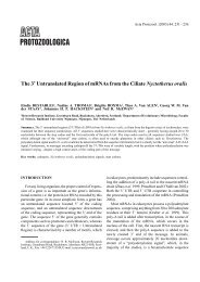
Figs. 1-3. Terminology and morphometrics (numbers 1 - 6 in Figs. 2 and 3) used in this study.
260 W. Foissner and G. Korganova<br />
the scanning electron microscope. Specimens usually must be strongly<br />
tilted and very carefully orientated to show the inner pseudostome of<br />
C. sylvatica.<br />
Terminology of testate amoebae is rather confused and many<br />
terms, for instance, the so-called visor, vestibulum, and pseudostome<br />
are used in different ways by different or even the same authors<br />
(Bonnet 1964, 1975). Thus, we use “simple” terms, shown in Figures<br />
1 and 3, for the scope of the present paper.<br />
Permanent slides<br />
<strong>The</strong> specimens used, and several others, were embedded in a drop<br />
of glycerol sealed by a rather thick ring of artificial resin (Histofluid,<br />
Merck Company, Germany) to prevent shells from desiccation and<br />
distortion by the cover glass pressure. All slides and the<br />
SEM-preparations have been stored in the Oberösterreichische<br />
Landesmuseum in Linz (LI).<br />
RESULTS<br />
Brief description of the taxa involved<br />
To understand our argumentation it is crucial to know<br />
the original description and status of the taxa involved.<br />
Thus, we provide the original descriptions (translated<br />
from French) and supplement them with recent literature<br />
data and/or our observations from the Moscow material.<br />
<strong>Centropyxis</strong> <strong>aerophila</strong> Deflandre, 1929 (Figs. 4-14,<br />
28-33; Tables 1, 2)<br />
“Test small, abdomen globular, dorsal wall strongly<br />
flattened towards pseudostome. Ventral outline oval with<br />
circular or slightly elliptical abdomen, sides not or only<br />
slightly converging towards pseudostome having semicircular<br />
outline. Sides only slightly curved, often almost<br />
straight. In ventral view, abdomen seemingly separated<br />
from pseudostomal area, looking like being attached to<br />
the abdomen. Pseudostomal region more transparent<br />
than abdominal region in resin preparations.<br />
Pseudostome usually semicircular with straight, occasionally<br />
slightly concave (Figs. 7, 10) posterior margin.<br />
Abdomen distinctly inflated in lateral view, but steeply<br />
flattens towards pseudostome, whose anterior margin is<br />
more or less distinctly turned inside.<br />
Test entirely chitinous, finely and irregularly punctuated<br />
or with rather distinct flakes, carries sometimes<br />
brown or dark organic debris and small quartz grains,<br />
colourless or yellowish, occasionally rather dark brownyellow.<br />
At certain sites, shell often appears entirely<br />
covered with foreign particles.<br />
Pseudopods and nucleus not yet observed. Dimensions<br />
(number of specimens measured not given): overall<br />
size 53 - 85 x 42 - 66 µm, shell height about 2/3 of length;<br />
pseudostome 21 - 28 x 15 - 21 µm”.<br />
<strong>The</strong>re are several notes and brief redescriptions<br />
available in the recent literature, adding significantly to<br />
the morphology of the shell and its inhabitant, but leaving<br />
untouched the basic features described by Deflandre<br />
(1929). Our selected material from Russia also matches<br />
Deflandre’s description (Figs. 28-33; Table 2). No cement<br />
structures are recognisable in the scanning electron<br />
microscope.<br />
Lobose pseudopods and their movements, as well as<br />
two contractile vacuoles, were described in detail by<br />
Bartoš (1954) and Bonnet (1961). Ogden and Hedley<br />
(1980) provided some helpful scanning electron micrographs<br />
of the shell, which is usually more chitinous in<br />
freshwater than terrestrial habitats, where it is often<br />
entirely covered by sand grains (Fig. 33). Measurements,<br />
but no detailed morphometrics, were given by<br />
various authors (Decloitre 1954, 1956; Laminger 1972;<br />
Ogden and Hedley 1980; Rauenbusch 1987), broadening,<br />
however, like our detailed data (Table 2), Deflandre’s<br />
limits only slightly: 47 - 93 (length) x 32-77 (width) µm;<br />
pseudostome: 19-34 (long axis) x 10-22 (short axis) µm.<br />
Schönborn (1966) found very small specimens (about<br />
30 x 30 µm) in the sediment of small Tundra lakes in<br />
Lapland.<br />
<strong>Centropyxis</strong> <strong>aerophila</strong> var. sphagnicola Deflandre,<br />
1929 (Figs. 15-20, 34-40, 58; Table 2)<br />
“Test very similar to that of type species. In ventral<br />
view, it differs from C. <strong>aerophila</strong> <strong>aerophila</strong> and<br />
C. <strong>aerophila</strong> sylvatica in being more frequently circular.<br />
Pseudostome very eccentric, its outline forms two<br />
more or less convex arcs, ventral lip extends deeply into<br />
shell leaving free only a narrow slit between lip end and<br />
dorsal shell wall (Rauenbusch 1987). In lateral view less<br />
inflated than type and less flattened towards pseudostome.<br />
Wall chitinous, flaky, more frequently incrusted with<br />
foreign particles, small irregular platelets, or quartz grains<br />
mostly at anterior margin of pseudostome. Dimensions<br />
(number of specimens measured not given): diameter<br />
49-66 µm, shell height 1/2 - 3/5 of diameter; pseudostome<br />
(long axis) 25-37 µm”.<br />
<strong>The</strong>re are several notes and brief redescriptions<br />
available in the recent literature, adding significantly to<br />
the morphology of the shell and its inhabitant, but leaving
<strong>The</strong> <strong>Centropyxis</strong> <strong>aerophila</strong> complex 261<br />
Table 1. Morphometric data on <strong>Centropyxis</strong> <strong>aerophila</strong>. Upper line: 127 randomly selected specimens (including varieties and C. sylvatica)<br />
identified with the features given by Deflandre (1929). Lower line: the 127 specimens mentioned above and the 90 specimens from<br />
Table 2 pooled<br />
Characteristics 1 x M SD SE CV Min Max n<br />
Shell, height (1) 42.3 43.0 7.8 0.7 18.5 15.0 65.0 127<br />
43.5 44.0 8.3 0.6 19.0 15.0 65.0 217<br />
Shell, length (2) 70.8 70.0 10.2 0.9 14.4 51.0 110.0 127<br />
71.2 70.0 9.7 0.7 13.6 51.0 110.0 217<br />
Shell, width (3) 66.1 65.0 9.9 0.9 14.9 47.0 95.0 127<br />
66.6 65.0 10.2 0.7 15.3 47.0 95.0 217<br />
Abdomen, length (4) 45.0 45.0 6.6 0.6 14.7 30.0 65.0 127<br />
45.4 45.0 6.5 0.4 14.3 30.0 65.0 217<br />
Pseudostome, long axis (5) 29.6 30.0 5.6 0.5 18.8 16.0 47.0 127<br />
29.6 30.0 5.6 0.4 19.0 16.0 47.0 217<br />
Pseudostome, short axis (6) 16.3 15.0 3.8 0.3 23.5 10.0 27.0 127<br />
16.2 15.0 3.8 0.3 23.4 10.0 27.0 217<br />
Shell length:abdomen length, ratio 1.6 1.6 0.1 0.0 7.1 1.3 1.9 127<br />
1.6 1.6 0.1 0.0 6.5 1.3 1.9 217<br />
Shell width:abdomen length, ratio 1.5 1.5 0.2 0.0 11.5 1.0 2.0 127<br />
1.5 1.5 0.2 0.0 11.0 1.0 2.0 217<br />
1<br />
Numbers in parenthesis designate features as shown in Figs. 2 and 3. Measurements in µm. CV- coefficient of variation in %; M - median;<br />
Max - maximum; Min - minimum; n - number of specimens investigated; SD - standard deviation; SE - standard error of mean; x- arithmetic<br />
mean<br />
Table 2. Morphometric data on 30 “ selected specimen ” each (see Materials and Methods section) of <strong>Centropyxis</strong> <strong>aerophila</strong> <strong>aerophila</strong> (CAA),<br />
C. <strong>aerophila</strong> sphagnicola (CAS), and C. <strong>aerophila</strong> sylvatica (CS), identified with the features given by Deflandre (1929)<br />
Characteristics 1 Species x M SD SE CV Min Max n<br />
Shell, height (1) CAA 36.6 36.0 5.7 1.0 15.6 25.0 45.0 30<br />
CAS 44.9 45.0 3.8 0.7 8.4 37.0 51.0 30<br />
CS 54.1 55.0 4.5 0.8 8.3 45.0 62.0 30<br />
Shell, length (2) CAA 66.6 65.0 4.5 0.8 6.8 55.0 80.0 30<br />
CAS 66.2 65.0 4.5 0.8 6.7 60.0 75.0 30<br />
CS 82.8 82.0 4.4 0.8 5.3 75.0 90.0 30<br />
Shell, width (3) CAA 56.4 55.0 4.5 0.8 8.1 50.0 70.0 30<br />
CAS 65.6 65.0 5.1 0.9 7.8 57.0 76.0 30<br />
CS 79.5 79.5 4.8 0.9 6.0 74.0 90.0 30<br />
Abdomen, length (4) CAA 42.2 42.0 3.9 0.7 9.3 35.0 50.0 30<br />
CAS 42.8 41.5 4.1 0.8 9.7 35.0 54.0 30<br />
CS 53.2 52.0 3.4 0.6 6.5 50.0 60.0 30<br />
Pseudostome, long axis (5) CAA 24.9 25.0 3.3 0.6 13.2 20.0 30.0 30<br />
CAS 28.9 26.5 4.6 0.8 15.7 25.0 37.0 30<br />
CS 35.2 35.0 3.6 0.7 10.3 27.0 42.0 30<br />
Pseudostome, short axis (6) CAA 14.3 15.0 2.6 0.5 18.3 10.0 20.0 30<br />
CAS 13.7 14.5 2.4 0.4 17.4 10.0 19.0 30<br />
CS 19.8 20.0 2.6 0.5 13.2 16.0 25.0 30<br />
Shell length:abdomen length, ratio CAA 1.6 1.6 0.1 0.0 6.5 1.4 1.9 30<br />
CAS 1.6 1.6 0.1 0.0 4.5 1.4 1.7 30<br />
CS 1.6 1.5 0.1 0.0 5.0 1.5 1.8 30<br />
Shell width:abdomen length, ratio CAA 1.3 1.3 0.1 0.0 11.0 1.1 1.6 30<br />
CAS 1.5 1.5 0.1 0.0 7.9 1.3 1.9 30<br />
CS 1.5 1.5 0.1 0.0 7.2 1.3 1.7 30<br />
1<br />
Numbers in parenthesis designate features as shown in Figs. 2 and 3. Measurements in µm. CV - coefficient of variation in %; M - median;<br />
Max - maximum; Min - minimum; n - number of specimens investigated; SD - standard deviation; SE - standard error of mean; x- arithmetic<br />
mean
262 W. Foissner and G. Korganova<br />
untouched the basic features described by Deflandre<br />
(1929). Our selected material from Russia also matches<br />
Deflandre’s description (Figs. 34-40; Table 2). No cement<br />
structures are recognisable in the scanning electron<br />
microscope.<br />
Lobose pseudopodia, which form rapidly and eruptively,<br />
were described by Bonnet (1963), who provided<br />
also detailed ecological data (Bonnet 1989). Detailed<br />
morphometrics and helpful scanning electron micrographs<br />
were published by Lüftenegger et al. (1988) and<br />
Rauenbusch (1987). Scattered measurements were provided<br />
by other authors (Laminger 1972, Chardez 1979),<br />
broadening, however, Deflandre’s limits only slightly:<br />
shell diameter 47-75 µm, shell height 30-45 µm;<br />
pseudostome 18-44 x 9-23 µm. Decloitre (1956) and<br />
Rosa (1971) observed very small specimens with a<br />
length of 30-36 µm.<br />
<strong>Centropyxis</strong> <strong>aerophila</strong> var. sylvatica Deflandre, 1929<br />
(Figs. 21-27, 41-61; Table 2)<br />
“This variety differs from the type species in that it is<br />
usually larger and more robust and the anterior<br />
pseudostome margin has a well-recognisable double<br />
contour. 1 <strong>The</strong> pseudostome is elliptical, more often it is<br />
semicircular (again, this is unclear and thus we provide<br />
the original text: “la bouche elle-même est elliptique plus<br />
souvent que semicirculaire”). In ventral view, the shell is<br />
usually exactly (“régulière”) elliptical or subcircular. <strong>The</strong><br />
sides are more strongly curved than in the type.<br />
<strong>The</strong> shell is chitinous as in the type; usually, it carries<br />
a certain amount of small, angular quartz grains towards<br />
the posterior end. It appears more punctuated and flaky<br />
than the type and is reinforced by fine, transparent,<br />
irregular platelets and very small quartz grains. Dimensions<br />
(number of specimens measured not given):<br />
68-102 x 63-85 µm; pseudostome 32-53 x 17-30 µm”.<br />
<strong>The</strong>re are several notes and redescriptions available<br />
in the recent literature, significantly modifying Deflandre’s<br />
original description. Most important is the observation by<br />
Bonnet and Thomas (1955) that the ventral pseudostome<br />
lip [called “membrane ventrale” by Bonnet and Thomas<br />
(1955) and “diaphragma” by Lüftenegger et al. (1988)<br />
and Rauenbusch (1987)] extends to the dorsal shell wall,<br />
dividing the test in a small “pseudostomal chamber” and<br />
a large “abdominal chamber” connected with each other<br />
by a more or less large opening (lip perforation) through<br />
which the pseudopods can extend. <strong>The</strong> observations of<br />
Bonnet and Thomas (1955) were confirmed by<br />
Lüftenegger et al. (1988) and Rauenbusch (1987). We<br />
put much effort in observing and documenting these<br />
details in our material and thus can, for the first time,<br />
clearly show the lip perforation which, in fact, is an inner<br />
pseudostome (Figs. 41-61).<br />
<strong>The</strong> 30 large shells, which should have been<br />
C. <strong>aerophila</strong> sylvatica according to Deflandre’s criteria<br />
(see “isolation of taxa and sampling protocol”), were<br />
very difficult to investigate because they were composed<br />
of rather large quartz grains making them opaque<br />
and refractive. Thus, nothing definite could be seen; only<br />
in five to six out of the 30 specimens there was a bright<br />
spot, possibly the lip perforation, at the right place. In<br />
contrast, we could see the lip perforation in 22 out of the<br />
30 small, transparent shells, when they were optimally<br />
orientated, that is, viewed frontally; if debris adhered to<br />
the pseudostome, visibility of the lip perforation decreased.<br />
<strong>The</strong> transparent specimens showed that the<br />
site where the lip internally abuts to the dorsal shell wall<br />
is often marked by an inconspicuous groove (Figs. 52,<br />
61), less distinct than those shown by Lüftenegger et al.<br />
(1988).<br />
<strong>The</strong> lip perforation has a mean size of 17 x 11 µm<br />
(10-25 x 8-15 µm, n 22) in specimens with a pseudostome<br />
size of 28 x 15 µm (20-38 x 10-25 µm). Thus, it is<br />
elliptical and rather large; the margin is uneven. In the<br />
population investigated by Bonnet and Thomas (1955),<br />
the perforation is roundish with a diameter of only<br />
5-8 µm. In the specimens studied by Lüftenegger et al.<br />
(1988), the lip perforation is very similar to our material<br />
having a mean size of 18 µm. In Rauenbusch’s (1987)<br />
specimens the perforation is sickle-shaped (unfortunately,<br />
the scanning micrographs are too dark to be<br />
entirely convincing). Thus, there is considerable variability<br />
in the size and shape of the lip perforation; however,<br />
some of the variability might be caused by the observation<br />
problems described above.<br />
In the scanning electron microscope, the inner<br />
pseudostome (lip perforation) was better recognisable in<br />
the large than in the small specimens (in contrast to the<br />
light microscope, see above). <strong>The</strong> scanning electron<br />
microscope investigations suggest that the lip perforation<br />
1<br />
“... vers la bouche dont le bord extérieur montre un double contour tres net”. Very likely, Deflandre (1929) describes a dorsal pseudostome<br />
lip as shown in Figure 3. However, such a lip has not yet been convincingly shown and we could not find it in our material.
<strong>The</strong> <strong>Centropyxis</strong> <strong>aerophila</strong> complex 263<br />
Figs. 4-27. Ventral (4, 5, 7, 9 - 18, 21, 23, 25 - 27), lateral (6, 8, 19, 20, 22), and frontal (24) views of shells of the <strong>Centropyxis</strong> <strong>aerophila</strong> complex<br />
in the light microscope (bright field illumination). All figures are reproductions (Xerox copies) from the original literature cited below.<br />
4- 14 - <strong>Centropyxis</strong> <strong>aerophila</strong> <strong>aerophila</strong>, size 53 - 85 x 42 - 66 µm (from Deflandre 1929); 15-20 - <strong>Centropyxis</strong> <strong>aerophila</strong> sphagnicola, size<br />
49 - 66 x 25 - 37 µm (from Deflandre 1929); 21-27 - <strong>Centropyxis</strong> sylvatica, size 65 - 105 x 60 - 87 µm (21, 22, from Bonnet and Thomas 1960;<br />
23, 24, from Bonnet and Thomas 1955; 25 - 27, from Deflandre 1929). Arrowheads in Figs. 22 and 24 mark inner pseudostome, the main<br />
species character.
264 W. Foissner and G. Korganova<br />
Figs. 28-33. <strong>Centropyxis</strong> <strong>aerophila</strong> <strong>aerophila</strong>, selected specimens<br />
identified with the characteristics given by Deflandre (1929), in the<br />
light (28) and scanning electron microscope (29 - 33). Note lack of an<br />
inner pseudostome; thus, the xenosomes of the dorsal shell wall are<br />
recognisable. Test shape varies from broadly elliptical to almost<br />
circular. 28 - ventral view, bright field, length 63 µm; 29, 30 - ventral<br />
views, 80 µm and 75 µm; 31, 32 - ventral view of same specimen at<br />
low and high magnification, length 72 µm. Arrowheads mark inner<br />
margin of ventral pseudostome lip, which extends to near dorsal shell<br />
wall; 33 - lateral view showing shell to be composed of quartz grains,<br />
length 60 µm.
<strong>The</strong> <strong>Centropyxis</strong> <strong>aerophila</strong> complex 265<br />
Figs. 34-40. <strong>Centropyxis</strong> <strong>aerophila</strong> sphagnicola, selected specimens identified with the characteristics given by Deflandre (1929), in the light<br />
(34 - 36) and scanning electron microscope (37 - 40). Note lack of an inner pseudostome; thus, the xenosomes of the dorsal shell wall are<br />
recognisable. <strong>The</strong> shells are circular or slightly broader than long and have agglutinated small quartz grains mainly in posterior portion.<br />
34-36 - same specimen in ventral and frontal views, size 68 x 68 x 46 µm. Arrowheads mark ventral pseudostome lip; 37, 38 - ventral view of<br />
same specimen at low and high magnification, length 61 µm. Arrowheads mark inner margin of ventral pseudostome lip, which extends to near<br />
dorsal shell wall; 39 - ventral view, length 63 µm; 40 - frontolateral view, length 58 µm.
266 W. Foissner and G. Korganova<br />
Figs. 41-47. <strong>Centropyxis</strong> sylvatica, selected small (“transparent”) specimens identified with the characteristics given by Deflandre (1929), in<br />
the light (41-43) and scanning electron microscope (44-47). Note that specimens are circular or slightly broader than long and have agglutinated<br />
small quartz grains mainly in posterior portion. 41-43 - same specimen in ventral view and two frontal focal plans showing lower and upper<br />
margin of inner pseudostome (arrowheads), size 68 x 74 x 53 µm; 44-47 - ventral views at low and high magnification showing the outer (arrow)<br />
and inner (arrowheads) pseudostome, length 61 µm and 66 µm. <strong>The</strong> inner pseudostome is formed by agglutinated material on the dorsal and<br />
lateral shell wall
<strong>The</strong> <strong>Centropyxis</strong> <strong>aerophila</strong> complex 267<br />
Figs. 48-55. <strong>Centropyxis</strong> sylvatica, selected small (“transparent”) specimens (48-53) and a large opaque specimen (54, 55) identified with the<br />
characteristics given by Deflandre (1929), in the light (48-53) and scanning electron microscope (54, 55). Arrow in Figs. 51 and 52 marks minute<br />
groove, where the inner pseudostome abuts to the dorsal shell wall. 48-51 - same specimen (size 68 x 74 x 46 µm) in ventral view, where the<br />
inner pseudostome is not recognisable; in oblique ventral view, where the pseudostome becomes minute; in frontal view, where the broadly<br />
elliptical inner pseudostome is well recognisable (arrowheads); and in lateral view, where the lip perforation (inner pseudostome, arrowheads)<br />
is difficult to recognise; 52, 53 - lateral and frontal view of another specimen showing the inner pseudostome (arrowheads); 54, 55 - ventral<br />
view of same specimen at low and high magnification showing impressively the outer (arrow) and inner (arrowheads) pseudostome, length<br />
94 µm. <strong>The</strong> inner pseudostome is obviously made of small xenosomes attached to the dorsal wall of the shell and the lateral walls of the outer<br />
pseudostome
268 W. Foissner and G. Korganova<br />
Figs. 56-61. <strong>Centropyxis</strong> sylvatica, selected small (“transparent”) specimens (58-60) and large opaque specimens (56, 57, 61) identified with<br />
the characteristics given by Deflandre (1929), in the light (58, 60) and scanning electron microscope (56, 57, 59, 61). 56, 57 - ventral view of<br />
same specimen (length 87 µm) at low and high magnification showing the outer (arrow) and inner (arrowheads) pseudostome; 58, 60 - ventral<br />
views of C. sylvatica (CS) and C. <strong>aerophila</strong> sphagnicola (CAS), size of specimen shown in Fig. 60, 61 x 65 x 43 µm; 59, 61 - lateral views<br />
showing shells to be composed mainly of quartz grains, length 73 µm and 88 µm. Arrow marks minute depression, where the inner<br />
pseudostome attaches to the dorsal shell wall (cp. Figs. 51, 52)
<strong>The</strong> <strong>Centropyxis</strong> <strong>aerophila</strong> complex 269<br />
is mainly brought about by accumulation of agglutinated<br />
material on the dorsal and lateral shell wall. Thus, the<br />
ventral lip is possibly not “perforated” in the strict sense<br />
of the word; this is also indicated by its general appearance,<br />
which is as in the other varieties (Figs. 32, 38). In<br />
the scanning electron microscope, a lip perforation was<br />
seen in several small (transparent; Figs. 41-47, 48-53)<br />
and large (“typical”) C. sylvatica specimens (Figs. 54,<br />
55, 56, 57), showing that the species cannot be recognised<br />
by size.<br />
Lobose pseudopods and their movements, as well as<br />
two contractile vacuoles, were described by Bonnet<br />
(1961) in C. sylvatica var. minor. Rauenbusch (1987)<br />
and Lüftenegger et al. (1988) provided some helpful<br />
scanning electron micrographs showing that shell wall<br />
structure highly depends on the substrate the organisms<br />
live. Lüftenegger et al. (1988) provided also detailed<br />
morphometrics showing that pseudostome features are<br />
more variable than the length and width of the shell.<br />
Scattered measurements were given by other authors<br />
(Bonnet and Thomas 1955, Rosa 1971), broadening,<br />
however, Deflandre’s limits only slightly: 56-113 (length)<br />
x 47-100 (width) x 45-68 (height) µm; pseudostome<br />
23-55 (long axis) x 20-32 (short axis) µm.<br />
Morphometry<br />
Basic statistics show that most variables have usual<br />
coefficients of variation and the number of specimens<br />
investigated is sufficient because mean and median<br />
hardly change if 127 or 217 specimens are analysed<br />
(Table 1). Of course, variation is distinctly lower in the<br />
selected specimens (Table 2). Only a few of the variables<br />
measured are normally distributed, viz. shell width,<br />
ratio shell length: abdomen length, and ratio shell width:<br />
abdomen length. All other features are slightly skewed to<br />
the left.<br />
Analysis of variance: If the 30 selected specimens of<br />
each are compared (all intermediate shells removed, see<br />
Method section!), all variables tested (length, width…)<br />
are significantly different (p ≤ 0.001), except the ratio<br />
shell length: abdomen length, that is, the three taxa can<br />
be clearly distinguished. If the 127 randomly chosen<br />
specimens are compared with the 30 selected specimens<br />
of either C. <strong>aerophila</strong> <strong>aerophila</strong> or C. <strong>aerophila</strong><br />
sylvatica, highly significant differences (p ≤ 0.001 to<br />
p ≤ 0.05) still occur in most variables, except for the<br />
ratios; in contrast, only shell height, shell length, short<br />
pseudostome axis, and the length: width ratio are different<br />
(p ≤ 0.05) in C. <strong>aerophila</strong> sphagnicola, indicating<br />
that this variety is intermediate between the two others.<br />
Finally, when the 30 selected specimens of each are<br />
pooled (= 90 specimens) and compared with the 127<br />
randomly chosen specimens, all variables become indistinguishable<br />
(p ≥ 0.05).<br />
Frequency distributions and relationships between<br />
variables (only some representative examples each are<br />
shown, Figs. 62a-f): Frequency distributions show curves<br />
with a single peak (Fig. 62d). Likewise, rather homogenous<br />
clusters are formed in the randomly chosen<br />
specimens, when variables are plotted against each<br />
other (Figs. 62a-c). In contrast, two distinct clusters are<br />
usually formed, if the selected specimens are plotted<br />
(Figs. 62e, f): one contains C. <strong>aerophila</strong> <strong>aerophila</strong> and<br />
C. <strong>aerophila</strong> sphagnicola, the other C. <strong>aerophila</strong><br />
sylvatica.<br />
DISCUSSION<br />
Morphometry<br />
Although species cannot be proven mathematically,<br />
some basic statistics are often useful to distinguish them<br />
more properly. As concerns the present material, neither<br />
the coefficients of variation (Table 1) nor frequency<br />
distributions (Fig. 62d) and relationships between the<br />
variables tested (Figs. 62a - c) give any indication that<br />
the randomly chosen specimens consist of more than<br />
one species. Variation coefficients (Tables 1, 2) are of<br />
the same order of magnitude as in multicellular organisms<br />
(Mayr 1975) and other protozoans, e.g. ciliates<br />
(Foissner 1984, 1993), and as in testate amoebae in<br />
general (Lüftenegger et al. 1988, Wanner 1991, Foissner<br />
and Korganova 1995). Only the pseudostome variables<br />
have coefficients higher than 20%; however, this seems<br />
to be a general feature of testacean shells (Lüftenegger<br />
et al. 1988, Wanner 1991).<br />
However, the situation changes drastically in the<br />
selected material, that is, when all intermediate specimens,<br />
which could not unequivocally assigned to one of<br />
Deflandre’s varieties, are excluded (Table 2). <strong>The</strong>n,<br />
three taxa can be distinguished by analysis of variance<br />
and at least two in scatter diagrams (Figs. 62e, f), just as<br />
different species were compared (Lüftenegger et al.<br />
1988). <strong>Centropyxis</strong> <strong>aerophila</strong> sylvatica is separated by<br />
its larger dimensions, while C. <strong>aerophila</strong> <strong>aerophila</strong> and<br />
C. <strong>aerophila</strong> sphagnicola are distinguished mainly by<br />
the shell proportions (Table 2): the former is broadly<br />
elliptical (66.6 x 56.4 µm), the latter almost circular<br />
(66.2 x 65.6 µm); furthermore, the long pseudostome<br />
axis is distinctly larger in C. <strong>aerophila</strong> sphagnicola
270 W. Foissner and G. Korganova<br />
Figs. 62a - f. Representative examples from the measurements (for details, see chapter on morphometry). 62a - c - when all (217) specimens<br />
of <strong>Centropyxis</strong> <strong>aerophila</strong> <strong>aerophila</strong>, C. <strong>aerophila</strong> sphagnicola and C. sylvatica are pooled and the main characteristics plotted, rather<br />
homogenous clouds of dots are formed, indicating that the taxa are morphometrically inseparable; 62d - likewise, frequency distributions of the<br />
217 specimens do not provide any indication that several taxa are mixed; 62e, f - when the 30 selected (all intermediates discarded, see Materials<br />
and Methods) specimens each are plotted, at least two distinct clouds are formed, one contains C. sylvatica (*), the other C. <strong>aerophila</strong><br />
<strong>aerophila</strong> (•) and C. <strong>aerophila</strong> sphagnicola (♦). We hypothesise that most authors distinguished Deflandres varieties by a similar (mental)<br />
selection process
<strong>The</strong> <strong>Centropyxis</strong> <strong>aerophila</strong> complex 271<br />
than in C. <strong>aerophila</strong> <strong>aerophila</strong> (28.9 vs. 24.9 µm).<br />
However, such result is expected and can be obtained<br />
with most living things, even with man, when intermediate<br />
specimens are removed. In fact, the experiment was<br />
performed specifically with the goal of obtaining information<br />
as to how previous authors probably distinguished<br />
Deflandre’s varieties (see next but one chapter).<br />
<strong>Centropyxis</strong> sylvatica is a distinct morphospecies<br />
We agree with Bonnet and Thomas (1955) that the lip<br />
perforation of C. sylvatica is difficult to observe and<br />
usually recognisable only in optimally orientated and<br />
transparent specimens. Indeed, when looking at an<br />
unorientated shell assemblage one gets doubts whether<br />
the lip perforation exists at all! And this doubt is strengthened<br />
by the fact that, contrary to Deflandre’s (1929)<br />
claim, C. sylvatica is obviously not larger than<br />
C. <strong>aerophila</strong> <strong>aerophila</strong> and C. <strong>aerophila</strong> sphagnicola<br />
(Figs. 62a - c), this being also evident from the size<br />
(69-72 x 67-73 µm) of the specimens studied by Bonnet<br />
and Thomas (1955). Nonetheless, the lip perforation<br />
exists and distinguishes C. sylvatica from the other<br />
varieties (Figs. 41 - 61). This conclusion is supported by<br />
the investigations of Lüftenegger et al. (1988) and<br />
Rauenbusch (1987). Furthermore, the lip perforation has<br />
been clearly shown, i.e. by scanning electron microscopy,<br />
in a larger, related species, <strong>Centropyxis</strong> matthesi<br />
Rauenbusch, 1987; and a similar, probably homologous<br />
opening occurs in Paracentropyxis mimetica Bonnet,<br />
1960. Thus, we agree with Bonnet and Thomas (1955,<br />
1960) that C. <strong>aerophila</strong> var. sylvatica should obtain<br />
species rank. It is well defined by its extraordinary lip<br />
perforation, forming a second, inner pseudostome.<br />
Much more difficult is the question whether<br />
C. sylvatica is a member of the genus <strong>Centropyxis</strong> at<br />
all! <strong>The</strong>re are at least two other, well-defined species<br />
with a distinct lip perforation, viz., <strong>Centropyxis</strong> matthesi<br />
and C. deflandriana. We cannot exclude that these<br />
three taxa represent a specific (homologous) evolutionary<br />
line different from that of <strong>Centropyxis</strong>, that is, it<br />
could happen that the similarities in shape and size of<br />
C. <strong>aerophila</strong> and C. sylvatica are an analogy. <strong>The</strong><br />
solution of this question will require genetic and molecular<br />
methods.<br />
How did previous authors distinguish Deflandre’s<br />
<strong>Centropyxis</strong> <strong>aerophila</strong> varieties<br />
As mentioned in the introduction, Deflandre’s varieties<br />
of C. <strong>aerophila</strong> have been reported by many<br />
testacean researchers from terrestrial and freshwater<br />
habitats worldwide, usually even from the same sample.<br />
Obviously, all used Deflandre’s traits to distinguish the<br />
varieties, at least none mentioned to have applied other<br />
and/or additional features. Furthermore, none mentioned<br />
having looked for the lip perforation, even after the<br />
pioneering paper of Bonnet and Thomas (1955), although<br />
it is the sole feature unequivocally separating<br />
C. <strong>aerophila</strong> sylvatica from C. <strong>aerophila</strong> <strong>aerophila</strong><br />
and C. <strong>aerophila</strong> sphagnicola.<br />
We showed that it is impossible to distinguish three<br />
varieties in C. <strong>aerophila</strong> with the morphological and<br />
morphometrical features given by Deflandre (1929); and<br />
our data give no indication that other, as yet undescribed<br />
reliable characteristics exist (Figs. 62a-d; Table 1). How<br />
then, could so many authors distinguish Deflandre’s<br />
varieties, although some mentioned problems (Jung 1936;<br />
Schönborn 1966, 1975; Chardez 1979) In our opinion it<br />
is because they considered mainly the extremes of a<br />
variability cline, which fit to Deflandre’s descriptions,<br />
obviously assigning intermediate specimens more or less<br />
arbitrarily to one of the varieties. We could also distinguish<br />
three taxa when we sorted out all intermediates,<br />
that is, about half of the shells (Figs. 62e, f; Table 2).<br />
A practicable solution for the problem:<br />
the “<strong>Centropyxis</strong> <strong>aerophila</strong> complex”<br />
<strong>The</strong> species problem in general and of testate amoebae<br />
in particular has been extensively investigated and<br />
discussed recently (Schönborn and Peschke 1988, Medioli<br />
et al. 1990, Schönborn 1992a, Foissner and Korganova<br />
1995, Bobrov et al. 1995, Wanner 1999). <strong>The</strong>se studies<br />
showed the lack of a simple answer and emphasised the<br />
need for thorough species descriptions and avoidance of<br />
infrasubspecific taxa, unless they can be proven by<br />
reliable morphological and/or morphometrical features.<br />
<strong>The</strong>y also showed that testacean shells are not extraordinarily<br />
variable, in contrast to the widespread believe,<br />
because the variation coefficients are hardly greater<br />
than in other organisms. This is emphasised by the<br />
present results (Tables 1, 2).<br />
We showed that it is impossible to distinguish varieties<br />
in C. <strong>aerophila</strong> with the features used by Deflandre<br />
(1929). <strong>The</strong> situation became even worse when the<br />
varieties described later were taken into account<br />
(for reviews, see Decloitre 1978, 1979).<br />
Basically, there are two ways to solve the problem.<br />
First, one might recognise only a single species, namely,<br />
<strong>Centropyxis</strong> <strong>aerophila</strong>, and classify all varieties and<br />
forms as falling into the species natural range of variability.<br />
Actually, this has been done by the describers of the
272 W. Foissner and G. Korganova<br />
varieties and forms. This, however, resulted in an extraordinary<br />
variability of a single species casting doubts<br />
on the decision because such a broad range indicates the<br />
inclusion of several species or, at least, several cryptic<br />
(sibling) species (Mayr 1975), which is emphasised by<br />
the varying proportions the varieties were found in field<br />
samples (Chardez 1979, Bonnet 1989, Aescht and<br />
Foissner 1994). On the other hand, a reliable separation<br />
and identification of the varieties is often impossible and<br />
will be difficult also with modern molecular techniques<br />
(Wanner et al.1997), if they exist at all. Accordingly, we<br />
prefer the second way at the present state of knowledge,<br />
that is, to unite all taxa in a “<strong>Centropyxis</strong> <strong>aerophila</strong><br />
complex”, as has been done in several “difficult” (cryptic)<br />
ciliate species [Paramecium aurelia, Tetrahymena<br />
pyriformis, Sterkiella histriomuscorum; see Foissner<br />
and Berger (1999) for a brief review]. This will at least<br />
relieve ecologists and practitioners of work which can<br />
hardly be done during field investigation, e.g., counting<br />
work. <strong>The</strong> theoretical background of this suggestion is<br />
the assumption that the phenospectrum <strong>aerophila</strong>sphagnicola<br />
is an adaptive polymorphism to meet wetter<br />
(<strong>aerophila</strong>) and drier (sphagnicola) environmental<br />
conditions by selective shifting of certain genes.<br />
We suggest that C. sylvatica is also included in the<br />
C. <strong>aerophila</strong> complex, although it is a distinct species,<br />
because its overall appearance is so similar and the<br />
species character, the lip perforation, is too difficult to<br />
recognise in routine counting work. Certainly, taxonomists<br />
and biogeographers (faunists) must look for this<br />
feature to get a reliable species list. Very likely, further<br />
taxa need to be put into the complex, for instance,<br />
C. cassis and C. constricta. However, these and other<br />
candidates, especially the subspecies and varieties of<br />
C. <strong>aerophila</strong> <strong>aerophila</strong> and C. <strong>aerophila</strong> sphagnicola<br />
(for reviews, see Decloitre 1978, 1979), are still poorly<br />
known and thus a final decision must await further<br />
investigations.<br />
Nomenclature<br />
<strong>The</strong>re is great uncertainty whether Deflandre’s<br />
“varieties” should be considered as infrasubspecific taxa<br />
or as subspecies. Indeed, many authors cited the varieties<br />
like subspecies or species, but none formally raised<br />
C. <strong>aerophila</strong> sphagnicola to subspecies or species<br />
rank. Fortunately, the matter can be unambiguously<br />
decided by the International Code of Zoological Nomenclature<br />
(1999), article 45.6. Deflandre (1929) described<br />
C. <strong>aerophila</strong> sphagnicola and C. <strong>aerophila</strong> sylvatica<br />
as “var. n.”; however, his work does not unambiguously<br />
reveal that the names were proposed for<br />
infrasubspecific entities, which are not regulated by the<br />
code, and accordingly sphagnicola and sylvatica have<br />
subspecific rank from their original publication. Thus,<br />
Deflandre (1929) is author of the subspecies C. <strong>aerophila</strong><br />
sylvatica, raised to species rank by Bonnet and Thomas<br />
(1955). Accordingly, the species formally must be cited<br />
as “<strong>Centropyxis</strong> sylvatica Deflandre, 1929”. <strong>The</strong> same<br />
applies to C. <strong>aerophila</strong> var. sphagnicola, if it is not<br />
considered as synonym of C. <strong>aerophila</strong>, and to most<br />
other varieties and forms described before 1961 (see<br />
previous chapter). All varieties and forms described<br />
after 1961 have infrasubspecific rank, unless they were<br />
adopted as valid name of a species or subspecies before<br />
1985 (e.g. C. <strong>aerophila</strong> var. globulosa Bonnet and<br />
Thomas, 1955).<br />
Acknowledgements. Dr. Galina A. Korganova was supported by<br />
a grant of the Wilhelm and Ilse Foissner Stiftung. <strong>The</strong> technical<br />
assistance of Dr. H. Berger, Dr. E. Herzog, Dr. B. Moser, and<br />
Mag. E. Strobl is greatly acknowledged.<br />
REFERENCES<br />
Aescht E., Foissner W. (1994) Effects of organically enriched magnesite<br />
fertilizers on the testate amoebae of a spruce forest. Europ.<br />
J. Soil Biol. 30: 79-92<br />
Bartoš E. (1954) Koreòonožce Radu <strong>Testacea</strong>. Vydavatel’Stvo<br />
Slovenskej Akadémie Vied, Bratislava (in Czech)<br />
Bobrov A. A., Yazvenko S. B., Warner B. G. (1995) Taxonomic and<br />
ecological implications of shell morphology of three testaceans<br />
(<strong>Protozoa</strong>: Rhizopoda) in Russia and Canada. Arch. Protistenkd.<br />
145: 119-126<br />
Bonnet L. (1960) Thécamoebiens des sols d’Angola (1). Publ. cult.<br />
Co. Diam. Ang. (Lisboa) 51: 79-86<br />
Bonnet L. (1961) L’émission pseudopodique, chez les thécamoebiens<br />
endogés (I). Bull. Soc. zool. Fr. 86: 17-28<br />
Bonnet L. (1963) L’émission pseudopodique chez les thécamoebiens<br />
endogés (II). Bull. Soc. zool. Fr. 88: 57-63<br />
Bonnet L. (1964) Le peuplement thécamoebien des sols. Revue Ecol.<br />
Biol. Sol 2: 123-408<br />
Bonnet L. (1975) Types morphologiques, écologie et évolution de la<br />
thèque chez les thécamoebiens. Protistologica 11: 363-378<br />
Bonnet L. (1989) Données écologiques sur quelques Centropyxidae<br />
(thécamoebiens) des sols. Bull. Soc. Hist. nat. Toulouse 125:<br />
7-16<br />
Bonnet L., Thomas R. (1955) Étude sur les thécamoebiens du sol.<br />
Bull. Soc. Hist. nat. Toulouse 90: 411-428<br />
Bonnet L., Thomas R. (1960) Thécamoebiens du sol. Vie Milieu<br />
11(Suppl.): 1-103<br />
Chardez D. (1979) Variations chez <strong>Centropyxis</strong> <strong>aerophila</strong> sphagnicola<br />
Deflandre. Revue verviét. Hist. nat. 36 (Nos 1 à 3): 4 pages<br />
without pagination<br />
Chardez D., Lambert J. (1981) Thécamoebiens indicateurs biologiques<br />
(<strong>Protozoa</strong> Rhizopoda <strong>Testacea</strong>). Bull. Rech. Agron. Gembloux<br />
16: 181-203<br />
Decloitre L. (1954) Biométrie et thécamoebiens d’A. O. F. Bull. Inst.<br />
fr. Afr. noire 16: 834-847<br />
Decloitre L. (1956) Les Thécamoebiens del’Eqe (Groenland). Hermann<br />
& Cie, Paris
<strong>The</strong> <strong>Centropyxis</strong> <strong>aerophila</strong> complex 273<br />
Decloitre L. (1978) Le genre <strong>Centropyxis</strong> I. Compléments à jour au<br />
31. décembre 1974 de la monographie du genre parue en 1929.<br />
Arch. Protistenkd. 120: 63-85<br />
Decloitre L. (1979) Le genre <strong>Centropyxis</strong> II. Compléments à jour au<br />
31. décembre 1974 de la monographie du genre parue en 1929.<br />
Arch. Protistenkd. 121: 162-192<br />
Deflandre G. (1929) Le genre <strong>Centropyxis</strong> Stein. Arch. Protistenkd.<br />
67: 322-375<br />
Foissner W. (1984) Infraciliatur, Silberliniensystem und Biometrie<br />
einiger neuer und wenig bekannter terrestrischer, limnischer und<br />
mariner Ciliaten (<strong>Protozoa</strong>: Ciliophora) aus den Klassen<br />
Kinetofragminophora, Colpodea und Polyhymenophora. Stapfia<br />
(Linz) 12: 1-165<br />
Foissner W. (1987) Soil protozoa: fundamental problems, ecological<br />
significance, adaptations in ciliates and testaceans, bioindicators,<br />
and guide to the literature. Progr. Protistol. 2: 69-212<br />
Foissner W. (1993) Colpodea (Ciliophora). Protozoenfauna 4/1:<br />
I-X, 1-798<br />
Foissner W. (1999) Soil protozoa as bioindicators: pros and cons,<br />
methods, diversity, representative examples. Agric. Ecosyst.<br />
Environ. 74: 95-112<br />
Foissner W., Berger H. (1999) Identification and ontogenesis of the<br />
nomen nudum hypotrichs (<strong>Protozoa</strong>: Ciliophora) Oxytricha nova<br />
(= Sterkiella nova sp. n.) and O. trifallax (= S. histriomuscorum).<br />
Acta Protozool. 38: 215-248<br />
Foissner W., Korganova G. A. (1995) Redescription of three testate<br />
amoebae (<strong>Protozoa</strong>, Rhizopoda) from a Caucasian soil: <strong>Centropyxis</strong><br />
plagiostoma Bonnet & Thomas, Cyclopyxis kahli (Deflandre) and<br />
C. intermedia Kufferath. Arch. Protistenkd. 146: 13-28<br />
Foissner W., Peer T. (1985) Protozoologische Untersuchungen an<br />
Almböden im Gasteiner Tal (Zentralalpen, Österreich). I.<br />
Charakteristik der Taxotope, Faunistik und Autökologie der<br />
<strong>Testacea</strong> und Ciliophora. Veröff. Österr. MaB-Programms 9:<br />
27-50<br />
Jung W. (1936) <strong>The</strong>kamoeben eines Eggegebirgsmoores und zweier<br />
Moore im Hohen Venn. Annls Protist. 5: 83-123<br />
International Commission on Zoological Nomenclature (1999) International<br />
Code of Zoological Nomenclature. Tipografia La<br />
Garangola, Padova<br />
Korganova G. A. (1988) Testate amoebae (<strong>Protozoa</strong>) in minor<br />
tropical islands of Oceania. Pedobiologia 32: 117-127<br />
Laminger H. (1972) Ein Beitrag zur Kenntnis der Hochgebirgs-<br />
Testaceen Österreichs. Arch. Protistenkd. 114: 101-151<br />
Lousier J. D., Parkinson D (1984) Annual population dynamics and<br />
production ecology of <strong>Testacea</strong> (<strong>Protozoa</strong>, Rhizopoda) in an<br />
aspen woodland soil. Soil Biol. Biochem. 16: 103-114<br />
Lüftenegger G., Petz W., Berger H., Foissner W., Adam H. (1988)<br />
Morphologic and biometric characterization of twenty-four soil<br />
testate amoebae (<strong>Protozoa</strong>, Rhizopoda). Arch. Protistenkd. 136:<br />
153-189<br />
Mayr E. (1975) Grundlagen der zoologischen Systematik. Parey,<br />
Hamburg and Berlin<br />
Medioli F. S., Scott D. B. (1985) Designation of types, for one genus<br />
and nine species of arcellaceans (thecamoebians), with additional<br />
original reference material for four other species. J. Foram. Res.<br />
15: 24-37<br />
Medioli F. S., Scott D. B., Collins E. S., McCarthy F. M. G. (1990)<br />
Fossil thecamoebiens: present status and prospects for the<br />
future. In: Paleoecology, Biostratigraphy, Paleoceanography and<br />
Taxonomy of Agglutinated Foraminifera, (Ed. C. Hemleben et al.).<br />
Dordrecht, Netherlands, 813-839<br />
Ogden C. G., Hedley R. H. (1980) An Atlas of Freshwater Testate<br />
Amoebae. British Museum (Natural History), Oxford University<br />
Press, Oxford<br />
Rauenbusch K. (1987) Biologie und Feinstruktur (REM-<br />
Untersuchungen) terrestrischer Testaceen in Waldböden<br />
(Rhizopoda, <strong>Protozoa</strong>). Arch. Protistenkd. 134: 191-294<br />
Rosa K. (1971) Mikroedafon der Waldböden in ÈSSR. Edafon,<br />
year 1971: 1-49 (in Czech)<br />
Schönborn W. (1966) Untersuchungen über die Testaceen Schwedisch-<br />
Lapplands. Ein Beitrag zur Systematik und Ökologie der beschalten<br />
Rhizopoden. Limnologica (Berlin) 4: 517-559<br />
Schönborn W. (1973) Paläolimnologische Studien an Testaceen aus<br />
Bohrkernen des Latnjajaure (Abisko-Gebiet; Schwedisch-<br />
Lappland). Hydrobiologia 42: 63-75<br />
Schönborn W. (1975) Studien über die Testaceenbesiedlung der Seen<br />
und Tümpel des Abisko-Gebietes (Schwedisch-Lappland).<br />
Hydrobiologia 46: 115-139<br />
Schönborn W. (1992a) Adaptive polymorphism in soil-inhabiting<br />
testate amoebae (Rhizopoda): its importance for delimination and<br />
evolution of asexual species. Arch. Protistenkd. 142: 139-155<br />
Schönborn W. (1992b) Comparative studies on the production<br />
biology of protozoan communities in freshwater and soil ecosystems.<br />
Arch. Protistenkd. 141: 187-214<br />
Schönborn W., Peschke T. (1988) Biometric studies on species, races,<br />
ecophenotypes and individual variations of soil-inhabiting <strong>Testacea</strong><br />
(<strong>Protozoa</strong>: Rhizopoda), including Trigonopyxis minuta n. sp. and<br />
Corythion asperulum n. sp. Arch. Protistenkd. 136: 345-363<br />
Schönborn W., Foissner W., Meisterfeld R. (1983) Lichtund<br />
rasterelektronenmikroskopische Untersuchungen zur<br />
Schalenmorphologie und Rassenbildung bodenbewohnender<br />
Testaceen (<strong>Protozoa</strong>: Rhizopoda) sowie Vorschläge zur<br />
biometrischen Charakterisierung von Testaceen-Schalen.<br />
Protistologica 19: 553-566<br />
Todorov M. T. (1993) Testate amoebae (<strong>Protozoa</strong>, Rhizopoda) in<br />
soils of Vitosa mountain (Bulgaria). Acta zool. Bulg. 46: 16-23<br />
Wanner M. (1991) Zur Morphologie von <strong>The</strong>kamöben (<strong>Protozoa</strong>:<br />
Rhizopoda) in süddeutschen Wäldern. Arch. Protistenkd. 140:<br />
45-66<br />
Wanner M. (1999) A review on the variability of testate amoebae:<br />
methodological approaches, environmental influences and taxonomical<br />
implications. Acta Protozool. 38: 15-29<br />
Wanner M., Meisterfeld R. (1994) Effects of some environmental<br />
factors on the shell morphology of testate amoebae (Rhizopoda,<br />
<strong>Protozoa</strong>). Europ. J. Protistol. 30: 191-195<br />
Wanner M., Nähring J. M., Fischer R. (1997) Molecular identification<br />
of clones of testate amoebae using single nuclei for PCR<br />
amplification. Europ. J. Protistol. 33: 192-199<br />
Received on 14th March, 2000; accepted on 15th May, 2000