Isolation and identification of bacteria and fungi from ... - ictp
Isolation and identification of bacteria and fungi from ... - ictp
Isolation and identification of bacteria and fungi from ... - ictp
- No tags were found...

You also want an ePaper? Increase the reach of your titles
YUMPU automatically turns print PDFs into web optimized ePapers that Google loves.
60<br />
ARTICLE IN PRESS<br />
C. Abrusci et al. / International Biodeterioration & Biodegradation 56 (2005) 58–68<br />
2. Materials <strong>and</strong> methods<br />
2.1. Cinematographic film samples<br />
Film samples <strong>from</strong> collections at Barcelona, Madrid<br />
<strong>and</strong> Gran Canaria archives were supplied by Filmoteca<br />
Espan˜ ola.The sampling <strong>and</strong> transport <strong>of</strong> the samples<br />
were carried out under aseptic conditions.The films<br />
supplied appeared to be in good condition.<br />
2.2. <strong>Isolation</strong> <strong>of</strong> microorganisms <strong>from</strong> cinematographic<br />
films<br />
The isolation procedure for all samples was performed<br />
in a laminar-flow chamber <strong>and</strong> manual operations<br />
were carried out using sterile disposable gloves.<br />
From each cinematographic sample, two or three pieces<br />
were placed on trypticase-soya-agar (TSA), for heterotrophic<br />
<strong>bacteria</strong>, <strong>and</strong> Sabouraud-glucose-agar (SAB)<br />
supplemented with choramphenicol for <strong>fungi</strong>.Petri<br />
dishes were incubated at 30 1C for 1–2 weeks.Cultures<br />
were observed daily under a stereoscopic microscope to<br />
check the presence <strong>of</strong> <strong>bacteria</strong>l colonies <strong>and</strong>/or fungal<br />
mycelium (Fig.1).<br />
2.3. Characterization <strong>of</strong> <strong>bacteria</strong><br />
Each <strong>bacteria</strong>l strain isolated <strong>from</strong> the film samples<br />
was cultured on TSA either at 30 or 37 1C for 48 h.<br />
Replicates were made <strong>and</strong> preserved at 4 1C or 80 1C<br />
using TSB liquid medium supplemented with glycerol.<br />
Gram- <strong>and</strong> simple staining were performed using<br />
exponential TSA cultures, <strong>and</strong> oxidase <strong>and</strong> catalase tests<br />
made.Acid production <strong>from</strong> glucose <strong>and</strong> sucrose on OF<br />
basal medium, growth on Simmons citrate agar, <strong>and</strong><br />
starch <strong>and</strong> gelatin hydrolysis, were tested.In all Grampositive<br />
(<strong>and</strong> some Gram-negative strains), the ability to<br />
form spores was tested by examining 24-, 48- <strong>and</strong> 72-h,<br />
<strong>and</strong> 1-week cultures on TSA, as well as the Schaeffer-<br />
Fulton specific stain (Forbes et al., 2002).Morphological<br />
characters were examined under differential interference<br />
contrast (Nomarski) <strong>and</strong> phase contrast using a<br />
Zeiss Laboval 4 optical microscope.<br />
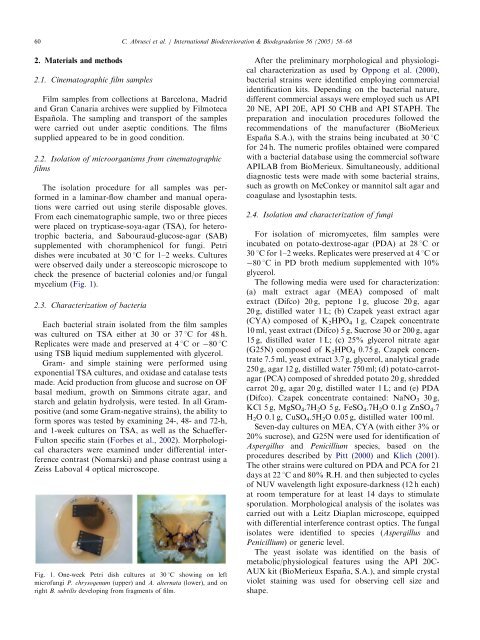
Fig.1.One-week Petri dish cultures at 30 1C showing on left<br />
micr<strong>of</strong>ungi P. chrysogenum (upper) <strong>and</strong> A. alternata (lower), <strong>and</strong> on<br />
right B. subtilis developing <strong>from</strong> fragments <strong>of</strong> film.<br />
After the preliminary morphological <strong>and</strong> physiological<br />
characterization as used by Oppong et al.(2000),<br />
<strong>bacteria</strong>l strains were identified employing commercial<br />
<strong>identification</strong> kits.Depending on the <strong>bacteria</strong>l nature,<br />
different commercial assays were employed such us API<br />
20 NE, API 20E, API 50 CHB <strong>and</strong> API STAPH.The<br />
preparation <strong>and</strong> inoculation procedures followed the<br />
recommendations <strong>of</strong> the manufacturer (BioMerieux<br />
Espan˜ a S.A.), with the strains being incubated at 30 1C<br />
for 24 h.The numeric pr<strong>of</strong>iles obtained were compared<br />
with a <strong>bacteria</strong>l database using the commercial s<strong>of</strong>tware<br />
APILAB <strong>from</strong> BioMerieux.Simultaneously, additional<br />
diagnostic tests were made with some <strong>bacteria</strong>l strains,<br />
such as growth on McConkey or mannitol salt agar <strong>and</strong><br />
coagulase <strong>and</strong> lysostaphin tests.<br />
2.4. <strong>Isolation</strong> <strong>and</strong> characterization <strong>of</strong> <strong>fungi</strong><br />
For isolation <strong>of</strong> micromycetes, film samples were<br />
incubated on potato-dextrose-agar (PDA) at 28 1C or<br />
30 1C for 1–2 weeks.Replicates were preserved at 4 1Cor<br />
80 1C in PD broth medium supplemented with 10%<br />
glycerol.<br />
The following media were used for characterization:<br />
(a) malt extract agar (MEA) composed <strong>of</strong> malt<br />
extract (Difco) 20 g, peptone 1 g, glucose 20 g, agar<br />
20 g, distilled water 1 L; (b) Czapek yeast extract agar<br />
(CYA) composed <strong>of</strong> K 2 HPO 4 1 g, Czapek concentrate<br />
10 ml, yeast extract (Difco) 5 g, Sucrose 30 or 200 g, agar<br />
15 g, distilled water 1 L; (c) 25% glycerol nitrate agar<br />
(G25N) composed <strong>of</strong> K 2 HPO 4 0.75 g, Czapek concentrate<br />
7.5 ml, yeast extract 3.7 g, glycerol, analytical grade<br />
250 g, agar 12 g, distilled water 750 ml; (d) potato-carrotagar<br />
(PCA) composed <strong>of</strong> shredded potato 20 g, shredded<br />
carrot 20 g, agar 20 g, distilled water 1 L; <strong>and</strong> (e) PDA<br />
(Difco).Czapek concentrate contained: NaNO 3 30 g,<br />
KCl 5 g, MgSO 4 .7H 2 O 5 g, FeSO 4 .7H 2 O 0.1 g ZnSO 4 .7<br />
H 2 O 0.1 g, CuSO 4 .5H 2 O 0.05 g, distilled water 100 ml.<br />
Seven-day cultures on MEA, CYA (with either 3% or<br />
20% sucrose), <strong>and</strong> G25N were used for <strong>identification</strong> <strong>of</strong><br />
Aspergillus <strong>and</strong> Penicillium species, based on the<br />
procedures described by Pitt (2000) <strong>and</strong> Klich (2001).<br />
The other strains were cultured on PDA <strong>and</strong> PCA for 21<br />
days at 22 1C <strong>and</strong> 80% R.H. <strong>and</strong> then subjected to cycles<br />
<strong>of</strong> NUV wavelength light exposure-darkness (12 h each)<br />
at room temperature for at least 14 days to stimulate<br />
sporulation.Morphological analysis <strong>of</strong> the isolates was<br />
carried out with a Leitz Diaplan microscope, equipped<br />
with differential interference contrast optics.The fungal<br />
isolates were identified to species (Aspergillus <strong>and</strong><br />
Penicillium) or generic level.<br />
The yeast isolate was identified on the basis <strong>of</strong><br />
metabolic/physiological features using the API 20C-<br />
AUX kit (BioMerieux Espan˜ a, S.A.), <strong>and</strong> simple crystal<br />
violet staining was used for observing cell size <strong>and</strong><br />
shape.