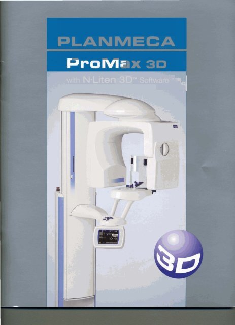
Planmeca Promax 3D with N-Liten 3D Software
Planmeca Promax 3D with N-Liten 3D Software
Planmeca Promax 3D with N-Liten 3D Software
- No tags were found...

Create successful ePaper yourself
Turn your PDF publications into a flip-book with our unique Google optimized e-Paper software.
PLANMECA<br />
- m<br />
rn<br />
L)X <strong>3D</strong><br />
1<br />
<strong>with</strong> Na<strong>Liten</strong> <strong>3D</strong>Tm S~ftware~--%~~
& all yaur diagnostic needs.<br />
*Cephalometric imaging only available when Cephalometric unit is purchased.
Any Existing PLANMECA ProMax Can<br />
be Upgraded to 30<br />
r<br />
'hanks to the original, technologically advanced design, any PLANMECA<br />
. ProMax can be upgraded to a <strong>3D</strong> Cone Beam Volumetric<br />
Tomography (CBVT) unit.<br />
1<br />
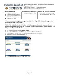
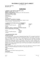
Rendering <strong>Software</strong><br />
N-<strong>Liten</strong> <strong>3D</strong> software's design includes the<br />
following main modules: DBM (Database<br />
Management); DVR (Digital Volume<br />
Rendering); Dynamic Lightbox; and<br />
Report Writer.<br />
Within each ofthese modules are the functions<br />
and features that will allow you tocreate pseudo<br />
pans; cross-sectional slices; trace nerves for<br />
implants; view and render three-dimensional<br />
volumes; and so much more. N-<strong>Liten</strong> <strong>3D</strong> will<br />
supply you <strong>with</strong> unlimited knowledge at<br />
your fingertips, and simple secure methods of<br />
archiving; sharing; printing; or retrieving your<br />
patient information. N-<strong>Liten</strong> <strong>3D</strong> software has<br />
r remote PACS Server sending capabilities and is<br />
fully DlCOM compatible.<br />
N-<strong>Liten</strong> <strong>3D</strong> is so advanced, there is no way to list all of its<br />
features in a brochure, however, hereare someofthe other<br />
tools included in the N-<strong>Liten</strong> 30 software package:Zooming;<br />
Panning;Windowing; Rotating; 1nverting;Text 0verlay;Volume<br />
of Interest Overlay; Ru1er;Tapeline; Angle; Profile; Area; Region of<br />
Interest; Notes; Mask; Arch; Segment; Color; and Opacity.<br />
u<br />
I<br />
With its simplistic design, advanced features, and useful tools, N-<strong>Liten</strong><br />
<strong>3D</strong> will definitely put the knowledge you need for your patients and your 1<br />
practice at your fingertips.<br />
Images may concatn optional items not IthAluded<br />
in standmd Uelhrwv.Rlghtrfor changes reserved.
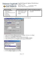
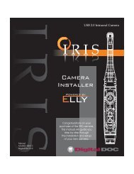
Diagnostic Specific Image Sizes<br />
With PLANMECA ProMax<strong>3D</strong>CBVT,studyvolumesizesareselectable<br />
to meet diagnostic needs <strong>with</strong>out excess radiation outside the<br />
region of interest (ROI).<br />
The80x80 mm imagesize isoptimum for most diagnosticapplications<br />
requiring wholedentition, mandibleand maxilla in the same study<br />
volume.The 80 x50 mm volume can be used for single views of the<br />
jaw mandible or maxilla, which lowers the radiation by almost 40%.<br />
The small 40 x 50 mm volume is intended for molar area studies or<br />
for planning third molar extractions.<br />
30 rendition Impaction analysis<br />
Implant Placement<br />
PLANMECA ProMax <strong>3D</strong> CBVT produces high resolution volumetric<br />
studiesofthe mandible and maxilla for analyzing the bone structure<br />
available, the location of the mandibular canal, and the correct<br />
4<br />
Nerve mapping<br />
lzzez!<br />
position for the implant. Pre-surgical planning will reach a new<br />
level of precision, as the proposed site is visible in all three imaging<br />
planes: sagittal, axial, and coronal.<br />
TMJ view<br />
Maxillofacial Surgery<br />
Third molars, maxillarycuspids, supernumeraryteeth,and impactions<br />
challenge the clinician to identify the tooth's orientation. PLANMECA<br />
ProMax <strong>3D</strong> CBVT makes all angles and orientations easily visible.
El<br />
Close-up view of condyle<br />
I<br />
I<br />
Cephalometricimagesarethefull<br />
I<br />
the<br />
Orthodontic Planning and<br />
TUI Analysis<br />
PLANMECA ProMax <strong>3D</strong> CBVT studies supported <strong>with</strong> digital<br />
visualization ofall classesoforthodontic<br />
malocclusion and planning, saving timeand reducing patient radiation<br />
dose. Unlike traditional orthodontic analyses, PLANMECA <strong>3D</strong> provides<br />
orthodontist <strong>with</strong> image data in the correct anatomic 1:1 ratio<br />
<strong>with</strong> no need to correct for geometric magnification.<br />
-! 4~ Cross-sectional slice<br />
Pseudo panoramic<br />
I 1<br />
PLANMECA ProMax <strong>3D</strong> CBVT also provides high-resolution TMJ<br />
studies for true and accurate evaluations of the joint arthritides,<br />
condylar morphology and the condyle-fossa relationship.<br />
PLANMECA ProMax <strong>3D</strong> CBVT complies <strong>with</strong> a multitude of<br />
diagnostic requirements: endodontic, periodontic, orthodontic,<br />
and implantology.<br />
WR (Digital volume renderings <strong>with</strong> axial & pseudo pans'<br />
With its high resolution (3lp/mm) and reconstruction technology,<br />
PLANMECA ProMax <strong>3D</strong> CBVTwill establish the new standard for <strong>3D</strong><br />
dental radiology.
Features<br />
& Benefits<br />
I Panoramic<br />
The reconstructed image volume<br />
consists of more than 120<br />
million voxels. These voxels are<br />
isotropic, which enables accurate<br />
1 :I measurements and ensures<br />
geometric relations throughout<br />
PLANMECA ProMax <strong>3D</strong> CBVT<br />
utilizes new Cone Beamvolumetric<br />
Tomography (CBVT) technology. It<br />
takes the whole volume needed<br />
in a single half circle scan, as<br />
opposed to medical CT's that take<br />
multipleaxial slices in multiple full<br />
circle scans.<br />
The direct deposit Csl on a<br />
semiconductor flat panel produces<br />
accurate, distortion-free images<br />
for <strong>3D</strong> reconstruction. Image<br />
intensifier sensors useold vacuum<br />
tube technology and multi-step<br />
focusing, whereas flat panels<br />
use single-step image readout5<br />
<strong>with</strong>out geometric distortion, los=<br />
of sensitivity, and the subsequent<br />
need for frequent calibration.<br />
I Lateral Ceph<br />
Rotational Sinus<br />
I<br />
the image.<br />
High Resolution<br />
The extremely small voxel size,<br />
160 I.lm, provides a detailed high<br />
resolution 3 Ip/mm image <strong>with</strong>out<br />
artifacts.<br />
PLANMECA ProMax's patented<br />
SCARA robotic arm ensures<br />
accurate and reliable image<br />
volume positioning. All controls<br />
are made on a full-color graphic<br />
user interface in the language of<br />
your choice.<br />
Pulsed Exposure:<br />
More Accuracy,<br />
Smaller Dose<br />
Unique 30<br />
Reconstruction<br />
PLANMECA's proprietary 30<br />
reconstruction<br />
algorithm<br />
converts the original 2D<br />
transillumination images to <strong>3D</strong><br />
volume study and is the core<br />
component for high-quality <strong>3D</strong><br />
imaging. It handles high contrast<br />
objects like amalgam fillings in a<br />
special way in order to produce<br />
undisturbed study views.<br />
During the scan, each image is :<br />
made using a short X-ray pulse<br />
instead of continuous radiation.<br />
The total scanning time is 16-18<br />
seconds, but theactual exposure ,<br />
time is only 6 seconds. This<br />
technique reduces the patient ,<br />
dose and forms a stroboscopic 1<br />
X-ray effect which together <strong>with</strong><br />
theshortened rotation scan (only ;<br />
I<br />
194 degrees), virtually eliminates i<br />
artifacts, contributing to the ~<br />
outstanding image quality.<br />
Images may contain optional items not inc.----<br />
... -.-.~dard delivery. Rights for changes reserved.
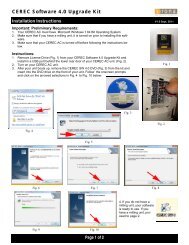
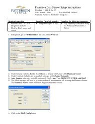
Data Sewer Description<br />
Stores all data. Romexis stores data to MS<br />
SQL Express or Solid. N-<strong>Liten</strong> <strong>3D</strong>M stores<br />
data to XML. The Data Server should have<br />
large hard disk space, hardware redundancy,<br />
and a logical backup solution.<br />
Hardware Requirements<br />
Processor: 2Ghz Duo Core<br />
System Memory: 2 GB<br />
Hard Disk Space: Multiple large hard disks.<br />
Storage size depends on usage. Each <strong>3D</strong> data<br />
set is approximately 250 MB. Each hard disk<br />
size should be a minimum of 500 GB.<br />
<strong>Software</strong> Requirements<br />
Windows XP or 2003 Server OS, .NET 2.0,<br />
and IIS must be installed on server.<br />
Network Server<br />
Hardware<br />
Diagram<br />
& System<br />
Requirements<br />
Image Acquieltion Workstation<br />
Romexis acquires images from the<br />
Recon- struction PC and utilizes<br />
N-Uten <strong>3D</strong>m for advanced image<br />
processing.<br />
Hardware Requirements<br />
Processor: 2Ghz Duo Core<br />
System Memory: 2 GB<br />
Video Card: 256 MB, 32-bR color<br />
Hard Disk Space: 10 GB free space<br />
Monitor: 1600x1200 mlution,<br />
32-bit color<br />
Network: 2 network interface cards<br />
+m- -<br />
-<br />
lmage Acquisition Workstation<br />
ProMax <strong>3D</strong> images are<br />
taken similar to a pan.<br />
The ProMax has the<br />
ability to capture pans,<br />
Cephs, and <strong>3D</strong> CBVT<br />
Reconstructs all raw data and<br />
transfers it to the Romexis<br />
workstation. All calibration files<br />
and software needed to capture<br />
CBVT images are preloaded<br />
by PLANMECA.<br />
Reconstruction PC
PLANMECA USA, INC. 100 N. Gary Ave., Suite A, Roselle, IL 601 72,<br />
Telephone: 630.529.2300 Website: \nnnrw.planmeca.com E-mail: sales@planmecausa.com