A Conversation with Gordon Christensen - New York State Dental ...
A Conversation with Gordon Christensen - New York State Dental ...
A Conversation with Gordon Christensen - New York State Dental ...
- No tags were found...

You also want an ePaper? Increase the reach of your titles
YUMPU automatically turns print PDFs into web optimized ePapers that Google loves.
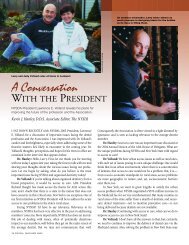
NYSDJ Editor Elliott Moskowitz, right, talks about the latest in dental techniques and trends <strong>with</strong> international expert <strong>Gordon</strong> <strong>Christensen</strong>.<br />
A <strong>Conversation</strong> <strong>with</strong> <strong>Gordon</strong> <strong>Christensen</strong><br />
Renowned dental expert shares his thoughts on variety of topics of interest to today’s professional.<br />
Elliott M. Moskowitz, D.D.S., M.Sd<br />
Editor, The <strong>New</strong> <strong>York</strong> <strong>State</strong> <strong>Dental</strong> Journal<br />
GORDON CHRISTENSEN is among the most sought after speakers<br />
in dentistry today. His comprehensive experience <strong>with</strong> materials,<br />
laboratory procedures, clinical practice, and long-term follow-up<br />
and research also make him an ideal colleague to interview.<br />
I had the opportunity to “catch up” <strong>with</strong> Dr. <strong>Christensen</strong> at the<br />
2004 Greater <strong>New</strong> <strong>York</strong> <strong>Dental</strong> Meeting, where he is an annual featured<br />
clinician. His perspectives on many different topics in dentistry<br />
are presented in this interview.<br />
I would like to thank the following NYSDA members for serving<br />
as expert consultants for this interview project: Van Thompson,<br />
Diane Rekow, Warren Scherer, Steven Mondre, Farhad Vahidi,<br />
Charles Kaner and John Cavallaro Jr.<br />
Dr. Moskowitz: A lot of talk has been directed in the area of moving<br />
dentistry to more of a medical model. For example, in <strong>New</strong> <strong>York</strong><br />
<strong>State</strong>, we are now mandating a PGY-1 (additional year of residency<br />
training after dental school) as a requirement for licensure. Where<br />
do you see dentistry heading in the next five years in this respect<br />
Dr. <strong>Christensen</strong>: I see a continued movement to the medical<br />
model. However, that may or may not be good, since many current<br />
graduates are relatively unprepared at graduation. My personal<br />
preference would be, as in numerous other countries, a dentist has<br />
an M.D. degree, and dentistry is similar to any other specialty of<br />
medicine. However, that is not about to happen in the U.S.<br />
Dr. Moskowitz: Prevention has always been a priority of the<br />
dental profession. Should the preventive programs supported by<br />
third-party systems include caries risk assessment and payment<br />
for this diagnostic procedure<br />
Dr.<strong>Christensen</strong>: Risk assessment is the current popular academic<br />
phrase.Yes,I feel this service should qualify for third-party payment.<br />
Dr. Moskowitz: When there is radiographic evidence of proximal<br />
caries penetrating to the DEJ, should it be eradicated and a<br />
restoration placed<br />
Dr. <strong>Christensen</strong>: Yes.At this time remineralization concepts are<br />
too slow and unpredictable to stop such lesions.Additionally, dentists<br />
are not informed about remineralization concepts and techniques.<br />
Dr.Moskowitz: What is the recommended cavity design for an<br />
initial Cl II cavity design when the caries is limited to the first third<br />
of dentin<br />
Dr. <strong>Christensen</strong>: I assume no caries on the occlusal. The prep<br />
should be a minimal extension slot either from the occlusal or,<br />
preferably, from the facial, preserving the marginal ridge.<br />
Caries Detection<br />
Dr. Moskowitz: Given the limited detection sensitivity of direct<br />
vision and radiographs for occlusal caries, how should a device<br />
such as laser fluorescence (Diagnodent) be incorporated to detect<br />
and diagnose occlusal caries<br />
Dr. <strong>Christensen</strong>: The Diagnodent has performed well in<br />
Clinical Research Associates studies. However, it still requires judgment<br />
to determine if the higher Diagnodent scoring teeth should be<br />
restored. Restoring a tooth <strong>with</strong> a level 20 on the Diagnodent is<br />
14 NYSDJ • APRIL 2005
probably appropriate on a high-caries-risk 16-year-old, but contraindicated<br />
on a caries-stable 50-year-old.<br />
Dr. Moskowitz: Should dentists consider differentiation<br />
between infected and affected dentin in cavity preparation for<br />
resin-based composite restorations<br />
Dr. <strong>Christensen</strong>: Yes, but this is very difficult. I suggest using<br />
dyes or the Diagnodent on partially excavated lesions.<br />
Dr. Moskowitz: Should dentin bonding agent be applied as<br />
part of occlusal sealant placement procedures<br />
Dr.<strong>Christensen</strong>: No, unless the lesion has progressed into dentin.<br />
Dr. Moskowitz: Do you see any more advances in bonding to<br />
dentin, or have we reached the limits of the materials<br />
Dr. <strong>Christensen</strong>: Dentin bonding looks good during “in vitro”<br />
studies. However, observation in clinical examples shows release of<br />
some dentin-bonded restorations over a period of service. There is<br />
still a long way to go to reach perfection for dentin bonding.<br />
Currently, I suggest mechanical retention in most dentin-bonded<br />
situations, if it is at all possible. The forces of mastication soon<br />
depreciate dentin bonds, as manifested by the frequent release of<br />
veneers placed nearly totally on dentin. But similar veneers are<br />
almost impossible to remove on properly acid-etched enamel.<br />
Reversible Pulpitis<br />
Dr. Moskowitz: What is the recommended procedure for indirect<br />
and direct pulp capping and restoration for a tooth diagnosed as<br />
having a reversible pulpitis associated <strong>with</strong> a deep occlusal carious<br />
lesion<br />
Dr. <strong>Christensen</strong>: Highly controversial! Calcium hydroxide still<br />
works well, as evidenced by relatively recent studies. Bonding<br />
agents also have positive research support as pulp capping materials.<br />
At this moment, I prefer resin-modified glass ionomer—<br />
Vitrebond (3M ESPE), or Fuji Lining Cement (GC), which are, in fact,<br />
self-etching/bonding materials filled <strong>with</strong> glass ionomer particles.<br />
Dr. Moskowitz: Are fiber-reinforced resin-based composite<br />
posts recommended for core resistance and retention in anterior<br />
and posterior teeth, in preference to a prefabricated or cast post<br />
Dr. <strong>Christensen</strong>: Prefabricated posts and cores are state-ofthe-art,<br />
and fiber-reinforced resin-based composite posts are highly<br />
popular. Our research shows the composite posts used <strong>with</strong><br />
build-up composite to be quite adequate for most situations.<br />
Dr. Moskowitz: The cast post/core has always been the gold<br />
standard. Is this standard of care shifting,and,ifso,what is the<br />
standard of care for posts and cores<br />
Dr. <strong>Christensen</strong>: Custom cast posts and cores are now of historical<br />
interest in the mainstream of dental practice. Pre-fabricated<br />
titanium alloy posts or fiber-reinforced resin-based composite are<br />
state-of-the-art. In spite of historically oriented criticism, research<br />
supports the use of prefabricated posts.<br />
Dr. Moskowitz: When a patient asks,“Will the composite resin<br />
last as long as the amalgam restoration,” what is your answer<br />
Dr. <strong>Christensen</strong>: Well-done composite restorations in smallto-moderate-sized<br />
Class II restorations compete very well <strong>with</strong><br />
amalgam in our long-term studies. Poorly done composites are<br />
total failures.<br />
Custom cast posts and cores are now of historical<br />
interest in the mainstream of dental practice.<br />
Pre-fabricated titanium alloy posts or fiberreinforced<br />
resin-based composite are state-ofthe-art.<br />
In spite of historically oriented criticism,<br />
research supports the use of prefabricated posts.<br />
The Future for Amalgams<br />
Dr. Moskowitz: Is amalgam dead<br />
Dr. <strong>Christensen</strong>: No, nor will it be in the near future. However,<br />
many patients will not accept amalgam now because of esthetic<br />
concerns or alleged health challenges.<br />
Dr.Moskowitz: In the area of tooth bleaching, many and more<br />
systems have evolved that do not carry the ADA Seal of Approval.<br />
Do you have a concern about over-the-counter bleaching delivery<br />
systems as compared to those that are dentist-delivered<br />
Dr. <strong>Christensen</strong>: Bleaching teeth is considered to be a “cosmetic”<br />
procedure by the FDA. As such, the ADA approval is not as<br />
significant as it might be for a dental restorative material.<br />
Dr. Moskowitz: Many dentists are now using either systemic or<br />
local delivery systems, for example, Periostat or Arestin. Do you see<br />
the general dentist more and more assuming the role of a periodontist,<br />
given the fact that less surgery may be implemented on a patient<br />
Dr. <strong>Christensen</strong>: I welcome general dentists becoming<br />
involved <strong>with</strong> these and other chemicals, but most general dentists<br />
will not treat periodontal disease. Periodontal disease is now very<br />
dominant in adults and needs treatment from general dentists or<br />
anyone else who will treat it.<br />
Dr. Moskowitz: In this world of resin cements, is there a place<br />
for zinc phosphate cement, and, if so, what is its place<br />
Dr. <strong>Christensen</strong>: Zinc phosphate still works. I have 45-year<br />
zinc phosphate restorations in my own mouth. This statement certainly<br />
undermines the “microleakage theory,” since zinc phosphate<br />
has total microleakage. Zinc phosphate is still a viable cement for<br />
dentists who want to use it, but resin-modified glass ionomer is the<br />
cement of choice today.<br />
Cement of Choice<br />
Dr. Moskowitz: What is your cement of choice for the single ceramo-metal<br />
crown, and does this cement choice differ for large<br />
cases, like full arch splinted crown and bridge restorations<br />
Dr. <strong>Christensen</strong>: I prefer RelyX, Luting Cement Plus or Fuji<br />
Cem, which are resin-modified glass ionomers.Although becoming<br />
more popular, resin cement is not cariostatic.<br />
Dr. Moskowitz: There seems to be increased patient demand<br />
for porcelain laminates. What are your current feelings about<br />
tooth preparations for laminates <strong>with</strong> respect to breaking interproximal<br />
contacts<br />
Dr. <strong>Christensen</strong>: In my opinion, ceramic veneers should be<br />
NYSDJ • APRIL 2005 15
In my opinion, ceramic veneers should be thin<br />
and conservative. When possible, I do not<br />
recommend totally breaking the contact areas.<br />
I feel strongly that the current trend toward<br />
grossly cut, deep “veneers” is not necessary<br />
and frequently represents overt overtreatment.<br />
thin and conservative. When possible, I do not recommend totally<br />
breaking the contact areas. I feel strongly that the current trend<br />
toward grossly cut, deep “veneers” is not necessary and frequently<br />
represents overt overtreatment.<br />
Dr.Moskowitz: What about an incisal coverage If incisal coverage<br />
is to be achieved,how far onto the lingual surface should it extend<br />
Dr. <strong>Christensen</strong>: I prefer that incisal coverage extend over the<br />
incisal edge but not into the occlusal centric stop area.<br />
Dr. Moskowitz: Temporization, in general, is an important<br />
aspect of restorative dentistry. What is your preferred method of<br />
temporization for laminate cases<br />
Dr. <strong>Christensen</strong>: Temporary restorations for veneers I prefer<br />
microfill handmade or matrix formed provisionals, bonded <strong>with</strong><br />
resin cement to a 2 mm circle of acid-etched enamel in the center<br />
of the facial surface of the tooth preparation.<br />
Laminate Material<br />
Dr.Moskowitz: Should the amount of coverage influence the material<br />
or the technology from which laminates are made That is to<br />
say, should it be a pressed ceramic, that is, Empress for one vs. feldspathic<br />
laminates for another situation<br />
Dr. <strong>Christensen</strong>: In the case of thin laminates, I prefer fired<br />
ceramic. If the tooth preparations are deeper, pressed ceramic is<br />
preferable.<br />
Dr. Moskowitz: Does the esthetic need relate to the choice of<br />
material in the fabrication of veneers<br />
Dr.<strong>Christensen</strong>: Yes. Significant malpositioning or dark colors<br />
demand deeper preps and probably pressed ceramic veneers or<br />
crown preps and crown restorations.<br />
Dr. Moskowitz: Is there an optimum thickness for veneers<br />
Dr. <strong>Christensen</strong>: I prefer up to 0.75 mm, fading to almost<br />
nothing at the gingival margins.<br />
Dr.Moskowitz: What is your preference as to the bonding system<br />
for veneers Is there any advantage to self-cure or dual-cure<br />
resins for bonding veneers Incidentally, we hear these restorations<br />
referred to as veneers, laminates, laminate-veneers, etc. This might<br />
be confusing to the public and to the profession.<br />
Dr. <strong>Christensen</strong>: Light-cure cement is best for veneers<br />
since dual-cure and auto-cure have observable color darkening<br />
over a period of months. By the way, such restorations are<br />
veneers or laminates. Calling them by the commonly heard<br />
name laminate veneers is similar to saying I want a car-auto-<br />
mobile. Laminate and veneer are synonymous.<br />
Dr. Moskowitz: What is your preferred sequence for bonding<br />
of laminates of the six upper anterior teeth<br />
Dr. <strong>Christensen</strong>: I prefer one at a time, rapidly moving from<br />
one side of the arch to the other, <strong>with</strong> initial finishing (one minute)<br />
of each before progressing to the next one.<br />
Corporate-Sponsored Research<br />
Dr. Moskowitz: There seems to be a great deal of corporate-sponsored<br />
research in dentistry. Do you see any potential problems <strong>with</strong><br />
this type of corporate research support <strong>with</strong> respect to conflicts of<br />
interest and inherent biases<br />
Dr.<strong>Christensen</strong>: Yes! You cannot believe much of it. That’s why<br />
we initiated Clinical Research Associates (CRA). The amount of<br />
misinformation in the dental “literature” today is appalling. And<br />
that comment also relates to so called “peer-reviewed” but corporate-sponsored<br />
“research.”<br />
Dr. Moskowitz: It has been suggested that, perhaps, the<br />
American <strong>Dental</strong> Association could play an important role in the<br />
oversight of corporate- or privately funded research. Do you favor<br />
such an expanded role of the ADA in this particular area<br />
Dr. <strong>Christensen</strong>: I love the ADA, but NO! The ADA must ask<br />
for money from manufacturers for many significant projects. I<br />
am currently on an ADA committee to raise one billion dollars<br />
for dental education, some of which will come from dental manufacturers.<br />
Such financial demands by our organization and the<br />
resultant donations from manufacturers cannot help but foster<br />
favoritism.<br />
Dr.Moskowitz: Do you think many dental products and materials<br />
are being marketed prematurely Should not some of these<br />
materials or products undergo greater scrutiny before coming to<br />
the dental marketplace<br />
Dr. <strong>Christensen</strong>: Yes! However, every company must decide<br />
when adequate research has been done to make the product marketable.<br />
If they wait too long another company has taken the market.<br />
It is a delicate balance.<br />
Dr. Moskowitz: What kind of cement do you recommend for<br />
all-ceramic crowns<br />
Dr. <strong>Christensen</strong>: Strong ones (Lava, Cercon) can have any<br />
cement. I prefer resin-modified glass ionomer. Weaker ones<br />
(Empress) should have resin cement.<br />
Dr. Moskowitz: Do you prefer zirconia all-ceramic crowns to<br />
alumina all-ceramic crowns, and, if so, why<br />
Dr. <strong>Christensen</strong>: Yes. Zirconia-based crowns are significantly<br />
stronger than alumina-based crowns.<br />
Dr. Moskowitz: Can you comment on the all-ceramic zirconia<br />
posterior three-unit bridges<br />
Dr. <strong>Christensen</strong>: They have proven themselves in up to seven<br />
years of research (Cercon) in Switzerland. They appear to be very<br />
promising, but long-term research is needed.<br />
Dr. Moskowitz: Do you expect metal-free ceramic restoration<br />
to be the state-of-the-art bridge in the near future<br />
Dr. <strong>Christensen</strong>: Yes. I estimate about five years for their popularization.<br />
16 NYSDJ • APRIL 2005
<strong>Dental</strong> Labs<br />
Dr. Moskowitz: Do you think the increased use of CAD-CAM<br />
restorations will have an impact on the role of commercial labs<br />
Dr. <strong>Christensen</strong>: In-clinic, CAD-CAM is growing, but it is not<br />
influencing labs much. However, I see CAD-CAM in laboratories<br />
soon dominating the fixed prosthodontic market.<br />
Dr. Moskowitz: <strong>Dental</strong> technology is getting more sophisticated;<br />
however, fewer dental technology programs are being<br />
developed. What is your solution to increasing the number of<br />
qualified technicians in the U.S.<br />
Dr. <strong>Christensen</strong>: We have set up a “task force” to evaluate this<br />
challenge. Organized dentistry and laboratory technology must<br />
wake up and begin aggressive recruitment of young people into<br />
technology. It’s a great field, but it needs immediate attention.<br />
Dr. Moskowitz: Do you feel the length of implants is still as<br />
important as it was deemed years ago, since the success rate of<br />
endopore implants is relatively the same as all of the larger lengths<br />
Dr. <strong>Christensen</strong>: There is a relationship between usable surface<br />
area of an implant and its success.Length can be long or short, but the<br />
surface area that is available for integration is the important factor.<br />
Dr. Moskowitz: What is your view on the immediate loading<br />
of implants<br />
Dr. <strong>Christensen</strong>: In general, I prefer to wait until the implant is<br />
osseointegrated. Most of my failures have been on early-loaded<br />
implants. However, I have had success on early loading of multiple<br />
“mini”implants; and I accomplish this procedure on a routine basis.<br />
Dr. Moskowitz: Is there a place for the immediate placement of<br />
implants into an extraction socket<br />
Dr. <strong>Christensen</strong>: Yes. In many cases I prefer this concept since<br />
the bone does not have time to shrink away during the healing period;<br />
and research has supported this concept.<br />
Dr. Moskowitz: How do you feel about using pre-made, labprocessed<br />
provisional bridges that are placed immediately upon the<br />
insertion of implants and guided by computer-generated surgical<br />
stents made by Materialize<br />
Dr. <strong>Christensen</strong>: Great concept! Needs further research and<br />
development.<br />
Dr.Moskowitz: Thanks for taking the time to share your thoughts<br />
<strong>with</strong> our NYSDJ readers. We have certainly covered a lot of important<br />
current clinical topics in dentistry. We look forward to speaking <strong>with</strong><br />
you again. In closing, do you have any advice for our colleagues<br />
Dr. <strong>Christensen</strong>: The future of dentistry is very bright. The<br />
techniques and new concepts in the profession are impressive.<br />
Managed care is getting under control to some extent, and esthetic<br />
dentistry has had a significant impact that will continue to expand.<br />
I am looking forward to the best time in the history of dentistry.<br />
Thanks for the opportunity to respond to your questions. ■<br />
<strong>Gordon</strong> J. <strong>Christensen</strong>, D.D.S., M.S.D., Ph.D., is founder and director of Practical Clinical Courses,<br />
an international continuing education organization for dental professionals based in Provo, UT.<br />
He is co-founder, <strong>with</strong> his wife, Rella, of the nonprofit Clinical Research Associates. For nearly 30<br />
years, CRA has conducted research in all areas of dentistry and published its findings in the CRA<br />
<strong>New</strong>sletter. Dr. <strong>Christensen</strong> practices in Provo.<br />
NYSDJ • APRIL 2005 17
Lisa A. Efron, Ph.D.; Jeffrey A. Sherman, D.D.S.<br />
Abstract<br />
It has been estimated that between 3% and 7% of children<br />
suffer from attention deficit hyperactivity disorder (ADHD). It<br />
is probable, therefore, that these children will present in any<br />
dental office. ADHD is characterized by inattention, overactivity<br />
and impulsivity. Such symptoms can be quite severe<br />
and often present challenges in the dental setting. ADHD<br />
symptoms are often managed by medication; and it is important<br />
for the clinician to be familiar <strong>with</strong> these medications to<br />
determine when to schedule dental appointments. In dealing<br />
<strong>with</strong> ADHD children, adjunctive behavioral strategies are<br />
often useful. Several basic techniques are discussed here.<br />
ATTENTION DEFICIT HYPERACTIVITY DISORDER (ADHD) is a<br />
chronic disorder characterized by inattention, overactivity and<br />
impulsivity. The specific DSM IV criteria for this disorder are displayed<br />
in Table 1. 1 It has been estimated that approximately 3% to<br />
7% of children have the condition. 2 Many children diagnosed <strong>with</strong><br />
ADHD will continue to experience symptoms of the disorder in<br />
adolescence and adulthood. 3 Therefore, it is probable that these<br />
patients will end up in any dental practice.<br />
There is a subset of children <strong>with</strong> ADHD who have difficulties<br />
sustaining attention and who do not experience any overactivity<br />
and impulsivity symptoms. These patients, categorized as having<br />
the inattentive subtype of ADHD, are not likely to be especially challenging<br />
in the dental setting. However, many children <strong>with</strong> ADHD<br />
are overactive and/or impulsive and are likely to present significant<br />
challenges to the clinician.<br />
Among other things, these children tend to be restless, fidgety<br />
and talkative; and they have difficulty remaining seated. Clearly,<br />
these behaviors will make treatment difficult. Therefore, it is<br />
important for the clinician to inquire about the presence of psychiatric<br />
conditions prior to the initial appointment.<br />
Because of the stigma that is often associated <strong>with</strong> mental<br />
health diagnoses, parents may not readily disclose all pertinent<br />
information to the clinician. Given the base rate of ADHD in the<br />
population, it is important for the clinician to be aware of the diagnostic<br />
criteria for this disorder in case he or she is confronted <strong>with</strong><br />
a patient who is displaying the symptoms.<br />
Medication<br />
Medication is the most effective form of treatment for ADHD, and,<br />
as a result, many children <strong>with</strong> the disorder take medication. Given<br />
that the various medications used to treat ADHD have different<br />
durations of effect, it is important to know which medication your<br />
patient is taking to determine what is the best time of day to schedule<br />
your appointment <strong>with</strong> the ADHD patient.<br />
Most ADHD medications fall into the category of stimulants.<br />
Children are likely to experience the effects of stimulants 30 to 60<br />
minutes after dosing; therefore, in the case of an early morning<br />
appointment, clinicians will want to ensure that sufficient time has<br />
elapsed between dosing and the dental appointment.<br />
18 NYSDJ • APRIL 2005
Some stimulants, such as Ritalin, are shortacting,<br />
and are effective for only three to four<br />
hours. Children generally take short-acting<br />
stimulants two or three times a day (for<br />
example, before school, at lunchtime, after<br />
school). Between doses of short-acting stimulants,<br />
children are not covered by medication<br />
and thus are symptomatic. In addition,<br />
they often become extremely irritable<br />
between doses. Clearly, one would want to<br />
avoid scheduling dental appointments during<br />
these “rebound” periods.<br />
Long-acting stimulants such as Concerta<br />
can last up to 12 hours. Information about<br />
type of medication and dosing pattern also has<br />
implications for late afternoon appointments<br />
as well. For example, a 4 p.m. appointment,<br />
which is a popular time slot for school-age<br />
children, would not be ideal for a child on<br />
short-acting Ritalin. Recently, a non-stimulant<br />
medication,Strattera,has become popular<br />
for the treatment of ADHD. This medication<br />
does not have the short half-life seen among<br />
stimulant preparations and, in fact, is similar<br />
to antidepressant medications in its composition.<br />
As a result, the timing issues that are<br />
so crucial in dealing <strong>with</strong> stimulants are<br />
basically irrelevant <strong>with</strong> Straterra.<br />
Positive Reinforcement<br />
Positive reinforcement, in terms of praise<br />
and small tangible rewards (for example,<br />
stickers, pretend tattoos, baseball cards), can<br />
be of great value in obtaining compliance<br />
from an ADHD child. Children will be more<br />
apt to work to please you if they sense you<br />
feel they are doing a good job. Children<br />
should be reinforced often, <strong>with</strong> younger<br />
children requiring more frequent reinforcement<br />
than older children.<br />
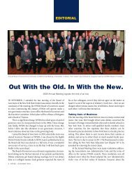
TABLE 1<br />
Diagnostic Criteria for ADHD<br />
A. Either (1) or (2)<br />
(1) Six (or more) of the following symptoms of inattention have persisted for at least 6 months<br />
to a degree that is maladaptive and inconsistent <strong>with</strong> developmental level:<br />
Inattention<br />
● often fails to give close attention to details or makes careless mistakes in schoolwork,<br />
work, or other activities<br />
● often has difficulty sustaining attention in tasks or play activities<br />
● often does not seem to listen when spoken to directly<br />
● often does not follow through on instructions and fails to finish schoolwork, chores,<br />
or duties in the workplace<br />
● often has difficulty organizing tasks and activities<br />
● often avoids, dislikes, or is reluctant to engage in tasks that require sustained mental effort<br />
● often loses things necessary for tasks or activities<br />
● is often distracted by extraneous stimuli<br />
● is often forgetful in daily activities<br />
(2) Six (or more) of the following symptoms of hyperactivity-impulsivity have persisted for at<br />
least 6 months to a degree that is maladaptive and inconsistent <strong>with</strong> developmental level:<br />
Hyperactivity<br />
● often fidgets <strong>with</strong> hands or feet or squirms in seat<br />
● often leaves seat in classroom or in other situations in which remaining<br />
seated is expected<br />
● often runs about or climbs excessively in situations in which it is inappropriate<br />
(in adolescents or adults, may be limited to subjective feelings of restlessness)<br />
● often has difficulty playing or engaging in leisure activities quietly<br />
● is often “on the go” or acts as if “driven by a motor”<br />
● often talks excessively<br />
Impulsivity<br />
● often blurts out answers before questions have been completed<br />
● often has difficulty awaiting turn<br />
● often interrupts or intrudes on others<br />
B. Some hyperactive-impulsive or inattentive symptoms that caused impairment<br />
were present before age 7.<br />
C. Some impairment from the symptoms is present in two or more settings.<br />
D. There must be clear evidence of clinically significant impairment in social,<br />
academic, or occupational functioning.<br />
E. The symptoms do not occur exclusively during the course of a pervasive<br />
developmental disorder, schizophrenia, or other psychotic disorder.<br />
NYSDJ • APRIL 2005 19
In addition to reinforcing unusually positive behaviors, consider reinforcing<br />
the behaviors that most adults would expect from a child and<br />
generally go unnoticed (for example,great job following directions,great<br />
job listening, great job sitting patiently). One can never over-reinforce.<br />
If a child is particularly challenging, the clinician might consider<br />
providing the child <strong>with</strong> a token every few minutes if he or she is on<br />
task (of course, it would be important to explain to the child at the outset<br />
exactly what is expected of him or her).At the end of the appointment,<br />
the child could be given the opportunity to cash in his or her<br />
tokens for a small treat from the dental office or from the parent.<br />
It’s Break Time...Again<br />
Children <strong>with</strong> ADHD tend to do well <strong>with</strong> frequent breaks. These<br />
breaks are likely to be effective even if they are very brief. So, when it<br />
is possible to provide breaks during a procedure, the clinician should<br />
consider doing so. If possible, allow the child to get out of the dental<br />
chair during the break. The clinician may opt to set a timer during<br />
breaks so that the child will know that the break is “officially” over.<br />
Clinicians might consider asking parents to provide a favorite activity<br />
(coloring supplies, book) or preferred toy (Gameboy, toy cars, doll) for<br />
the child to use during the breaks. For guidelines on whether breaks<br />
will be necessary, how often to provide breaks and duration of breaks,<br />
the clinician should consider consulting <strong>with</strong> the child’s parents.<br />
If the clinician has decided to use breaks,he or she should inform<br />
the child at the beginning of the appointment that breaks will be<br />
given, during which the child will be able to play. The clinician can<br />
take this opportunity to let the child know what he or she expects<br />
from the child during the non-break portion of the appointment.<br />
Parents Are Your Best Resource<br />
When in doubt, consult <strong>with</strong> parents. Do not hesitate to let parents<br />
know you are having difficulty managing their children. In all likelihood,<br />
they have heard it before from countless other professionals.<br />
Parents may be able to provide the clinician <strong>with</strong> some simple tips<br />
that will likely work <strong>with</strong> their child. If the clinician feels that he or<br />
she is becoming too involved in behavior management <strong>with</strong> a particular<br />
child, it is reasonable to ask the parent to remain in the treatment<br />
room so that the parent can manage the child’s challenging<br />
behaviors while the clinician is engaged in the dental procedure.<br />
Remember, parents are experts on their children—do not forget<br />
to make use of this very valuable resource. ■<br />
REFERENCES<br />
1. American Psychiatric Association (1994). Diagnostic and Statistical Manual of Mental<br />
Disorders (4th Ed.). Washington, DC.<br />
2. Szatmari P. The epidemiology of attention-deficit hyperactivity disorders. In G. Weiss<br />
(Ed.), Child and Adolescent Psychiatry Clinics of North America: Attention Deficit<br />
Hyperactivity Disorder. Philadelphia: Saunders, 1992:361-371.<br />
3. Applegate B, Lahey BB, Hart EL, et al. Validity of the age-of-onset criterion for ADHD: a<br />
report of the DSM-IV field trials. J Amer. Acad. Child and Adolescent Psychiatry 1997;<br />
36:1211-21.<br />
20 NYSDJ • APRIL 2005
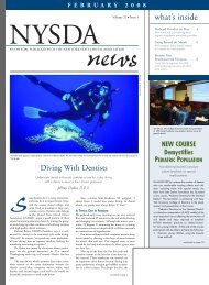
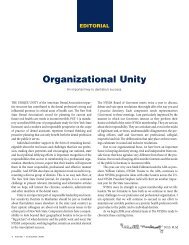
Figure 1. Typical scene of office on third floor of Murrah Federal<br />
Building after the bombing. While parts of two dismembered<br />
victims were found in this area, the chief forensic dentist did<br />
not feel area was safe enough to put members of dental team in<br />
such locales. Rather, trained individuals from D-Mort search and<br />
rescue teams were able to retrieve body parts that allowed for<br />
identification of victims.<br />
R. Thomas Glass, D.D.S., Ph.D.<br />
Abstract<br />
While body identification by dental means has not<br />
changed substantially since 9/11, or even since the bombing<br />
of the Murrah Federal Building in Oklahoma City in<br />
1995, the conditions and potential risks of a bioterrorism<br />
action to the dental personnel is new. The purpose of this<br />
article is to review general forensic dentistry disaster<br />
responses and to address the impact a bioterrorism action<br />
might have on primary, secondary and tertiary dental<br />
responders. It will also examine the triage role that dental<br />
offices might play in the event of such a disaster.<br />
BY THE TIME THE ECHOES of the Murrah Federal Building bombing<br />
on April 19, 1995, had subsided, the Oklahoma dental disaster team<br />
had been activated.As chief forensic dentist for the state and a professor<br />
of pathology/oral pathology who had taught forensic dentistry to<br />
every second-year dental student from the very inception of the dental<br />
program at the University of Oklahoma, I encountered little difficulty<br />
mustering a dental team that was prepared to function.<br />
Word went out through the dental school, and by noon on April<br />
19, more than 150 volunteers had crowded into one of the lecture<br />
rooms. The chief forensic dentist went over the response plan,<br />
dividing the group into site teams, morgue teams (including dental<br />
X-ray teams), ante-mortem data collection teams and data entry<br />
teams. The procedures that were delineated at that first meeting<br />
were the procedures that had been taught in the forensic dentistry<br />
lectures and used daily in the morgue.<br />
The results of this pre-planned program were that at the end of<br />
the 17 days of the resolution, all but three of the 167 victims had<br />
been positively identified. After the building was imploded, these<br />
three bodies were retrieved and identified. Pre-planning and documentation<br />
of the Murrah Federal Building bombing response can<br />
be found in a series of published papers. 1-6<br />
While the culprits in the Murrah Federal Building bombing<br />
were indigenous, the events associated <strong>with</strong> 9/11/2001 were quite<br />
different and presented dental teams <strong>with</strong> additional challenges.<br />
First, the perpetrators of 9/11 were foreign-born terrorists. Second,<br />
they used airplanes, not truck bombs to accomplish their ends.<br />
Third, in the case of the World Trade Center and the Pentagon, there<br />
was not only primary damage (loss of life in the airplane crashes),<br />
but also substantial collateral damage.<br />
Dismemberment was the rule, along <strong>with</strong> high-temperature<br />
fires. All of these factors played a role in the dental teams’ ability to<br />
identify the victims by dental means. In the Murrah Federal Building<br />
bombing, daily inspection of the bombing site by the chief forensic<br />
dentist never found the structural integrity of the building to be such<br />
that would allow for a dental team to be placed there to aid in body<br />
and body part retrieval (Figure 1). The situation was very similar in<br />
the 9/11 sites at the World Trade Center; however, dental team members<br />
did assist at the Pennsylvania site.<br />
NYSDJ • APRIL 2005 21
Lessons Learned<br />
If there is one central after-incident theme, it is that successful<br />
resolution of any disaster depends upon preparedness. For the<br />
dental team, forensic preparedness should be comparable to the<br />
annual or biannual cardiopulmonary resuscitation (CPR) classes<br />
that are mandated by most states. While no one expects that<br />
the next patient, or even a loved one, will need CPR, being skilled<br />
allows the dental professional to respond in an appropriate<br />
manner while awaiting the emergency medical service (EMS) or<br />
fire department.<br />
In deference to CPR training, forensic training is not prescribed<br />
in most states and is not even a part of every dental school<br />
curriculum. And like CPR, if forensic dentistry is not practiced on a<br />
regular basis, when it is needed, it may be found lacking. But who<br />
makes up the forensic dental team and what constitutes forensic<br />
dental preparedness<br />
Experience has shown that the most effective response to any<br />
disaster starts at a local level, because, truly, every disaster is a local<br />
disaster. 7 Even though the federal government, through its Federal<br />
Emergency Management Agency (FEMA), has disaster-mortuary<br />
(D-MORT) teams that include dental personnel that can respond<br />
to any disaster, most of the time it is the local dental team that is<br />
best suited to go to the disaster scene, to work in a familiar morgue<br />
and to talk to the local dentists regarding ante-mortem data. So the<br />
forensic dental team comprises dental professionals in the local<br />
Figure 2. Photograph demonstrates small piece of “field fence” that<br />
can be used by disaster site teams to grid areas of search for body<br />
parts. Actual size of field fence given each searcher is 4 ft x 4 ft.<br />
community who have a grasp of not only forensic dentistry, but<br />
also response.<br />
The composition of the team could be local dentists, dental<br />
hygienists, dental assistants, office personnel and even laboratory<br />
technicians. Each of these people would be expected to perform in<br />
a disaster response, performing the same duties he or she does<br />
every day in the office <strong>with</strong> the objective being to provide the evidence<br />
necessary to make body identifications by dental means.<br />
In terms of the dental forensic team, experience has shown<br />
that having a single chief forensic dentist who makes all of the identifications<br />
is the most reliable way to assure proper victim identification<br />
by dental means. Usually, this person is someone who makes<br />
body identifications routinely for the local medical examiner or<br />
coroner. The rest of the dental disaster team is subdivided into specialty<br />
teams, <strong>with</strong> one member of each specialty team being designated<br />
as the chief of that team.<br />
The first dental team is designated as the disaster site team or<br />
scene team. The function of this team is to go to the scene and<br />
search for body parts, especially those related to the dentition (for<br />
example, skulls, jaws, individual teeth). Prior to being dispatched<br />
to the scene, the chief forensic dentist and the chief of the site team<br />
should visit the scene to consult <strong>with</strong> the incident commander to<br />
determine whether the scene is secure enough to place a dental<br />
team there and whether such a team would be helpful. Of course,<br />
if the dental disaster team has been constituted prior to the incident<br />
and has worked <strong>with</strong> other members of the local disaster<br />
response teams, the incident commander will know the chief<br />
forensic dentist.<br />
If the scene is secured and a dental team is desired, each member<br />
of the site team is given a grid or “field-fence” (four foot square<br />
<strong>with</strong> grids of 6” x 8”) (Figure 2). The scene is divided into sectors,<br />
and each member is asked to systematically search his or her sector<br />
for body parts. When a body part is found, the team member is<br />
instructed to “flag it, bag it, and label it <strong>with</strong> their sector number.”<br />
Operating as we do under threats of terrorism, it is important<br />
that the chief forensic dentist and the site chief determine whether<br />
placing dental personnel at the scene would place these people in<br />
harm’s way. The concern is <strong>with</strong> possible exposure of team mem-<br />
22 NYSDJ • APRIL 2005
Figure 3. Example of Archimedes screw, used in breaking oral<br />
rigor mortis. Point of screw is placed between first and second<br />
bicuspids and turned until rigor is broken. Easiest way to make<br />
such a screw is to use conical-shaped screw found on typical dental<br />
laboratory lathes as prototype. Impression of prototype can be<br />
made and cast in partial denture metal.<br />
bers to biological, chemical or nuclear agents that would make<br />
members sick.<br />
The second dental team works in the morgue doing dental<br />
examinations on the victims. In order to gain access to the dentition,<br />
it may be necessary to either break the postmortem muscle<br />
lock on the jaws (rigor mortis) <strong>with</strong> an Archimedes screw (Figure<br />
3), placed between the bicuspid teeth and turned until the rigors<br />
are broken, or actually gain access by cutting the soft tissues of the<br />
cheeks from the commissures to the mandibular ramus. The decision<br />
on whether to cut or use the Archimedes screw is usually left<br />
to the medical examiner or coroner assigned to each victim.<br />
Given that most disaster resolutions take time, it is critical that<br />
the dental team be given protective clothing, masks and eyewear to<br />
protect its members from exposure to the potential pathogenic<br />
microorganisms so often associated <strong>with</strong> decomposition. Again, as<br />
<strong>with</strong> the site team, it is important that the chief forensic dentist<br />
determine whether the dentition examination poses a biological,<br />
chemical or nuclear threat to team members.<br />
The third dental team is assigned to collect ante-mortem data.<br />
While this team has little fear of biohazard exposure, it must be<br />
concerned <strong>with</strong> HIPAA regulations. Even though medical-legal<br />
facilities are granted certain statutory privileges when it comes to<br />
patient records, care should be taken to protect the privacy of the<br />
patient/victim. This is where dental office personnel are invaluable<br />
in a disaster response, as most are well trained in HIPAA regulations.<br />
Routine dental office procedures can be instituted in the<br />
morgue, especially in the accumulation and handling of antemortem<br />
records. Also, because many offices are now either substantially<br />
computerized or completely “paperless,” the ante-mortem<br />
data can be transported rapidly to the morgue.<br />
The fourth dental team is responsible for placing both the<br />
postmortem data and the ante-mortem data on the computer.<br />
While there are a number of computer programs that are helpful in<br />
sorting data, the computer cannot take the place of the final comparison<br />
of the actual postmortem dental anatomy/X-rays and the<br />
ante-mortem records/X-rays by the chief forensic dentist.<br />
In summary, several additional caveats have been gained from<br />
experience. They are presented here.<br />
●<br />
●<br />
●<br />
●<br />
●<br />
Whenever there is a disaster, everyone wants to help. Often,<br />
people from all over the country will just “show up” at the<br />
morgue wanting to be a part of the dental team. Because these<br />
people have not been through preparedness training, they<br />
may be more of a liability than an asset. Consider thanking<br />
them and sending them on their way.<br />
The best way for forensic dental disaster teams to be an effective<br />
part of the resolution is not only to be prepared, but also to<br />
interact <strong>with</strong> other members of the community disaster<br />
response teams. Such interactions will allow other teams to<br />
know what the dental team can do.<br />
It is important to respond to the disaster as rehearsed. It is a<br />
poor time to change procedures when teams are responding to<br />
a disaster, unless biohazards intervene. A variety of responses<br />
to biohazards should also be practiced prior to the event.<br />
Other disaster agencies should be reminded that rodents in the<br />
wild are calcium deficient and will seek out calcified body<br />
parts. In the same manner, birds will eat the soft tissue and<br />
often will carry off the bony portion of the remains.<br />
Debriefing and stress therapy should be made available and<br />
encouraged for all members of the dental team. In like manner,<br />
dental team members should be compensated for loss of time<br />
from the office. Both the therapy and the office overhead<br />
should be a part of the budget of any disaster response.<br />
<strong>New</strong> Challenges<br />
Among new challenges presented by terrorists is the possibility<br />
they will use chemicals, biological or nuclear agents. For purposes<br />
of this paper, only examples of each will be given and wherever possible,<br />
citations are given to direct the reader to further information.<br />
Finally, just a few comments will be made regarding use of the<br />
dental office as a triage facility for mass disasters.<br />
Chemicals Agents<br />
The most recently used chemical agent is ricin.A quick check of the subject<br />
on the Internet will lead the reader to the Centers for Disease Control<br />
and Prevention’s Web site (http://www.bt.cdc.gov/agent/ricin/facts.asp).<br />
What follows is a synopsis of the information found there:<br />
NYSDJ • APRIL 2005 23
“Ricin is a chemical poison, made from the waste left over from<br />
processing castor beans. It can be in the form of a powder, a mist,<br />
or a pellet, or it can be dissolved in water or weak acid. It is a stable<br />
substance that is not affected much by extreme conditions such as<br />
very hot or very cold temperatures….Ricin works by getting inside<br />
the cells of a person’s body and preventing the cells from making<br />
the proteins they need….Because no antidote exists for ricin, the<br />
most important factor is avoiding ricin exposure in the first place.<br />
If exposure cannot be avoided, the most important factor is then<br />
getting the ricin off or out of the body as quickly as possible. Ricin<br />
poisoning is treated by giving victims supportive medical care to<br />
minimize the effects of the poisoning.”<br />
Patients exposed to ricin will be sick and unlikely to go to a<br />
dental office for care. Because ricin must contact a potential victim<br />
to cause harm, routine dental office barrier techniques (scrubs,<br />
mask and eyewear) are sufficient.<br />
Biological Agents<br />
The most recent biological agent to be used is anthrax. Again,<br />
by accessing the CDC Web site, an index can be found<br />
(http://www.bt.cdc.gov/agent/anthrax/index.asp) that contains a<br />
variety of links on the subject and a two-page PDF fact sheet<br />
(http://www.bt.cdc.gov/agent/anthrax/pdf/needtoknow.pdf).<br />
Anthrax is neither a new disease nor uncommon. Actually,<br />
Bacillus anthracis, the etiologic agent was one of the first microorganisms<br />
ever to be isolated (in 1850, by French parasitologist<br />
Casimir-Joseph Davaine); was a major player in the development of<br />
the germ theory of disease (postulated in 1876, by German biologist<br />
Robert Koch); and was the second vaccine developed by Louis<br />
Pasteur (in 1881). 8<br />
While the disease process can be manifested in the skin, lungs<br />
and gastrointestinal tract, the important thing to remember is that in<br />
deference to some of the other biological agents, such as the smallpox<br />
or Ebola viruses, anthrax cannot be spread from person to person<br />
and can easily be treated <strong>with</strong> almost any first-generation to fourthgeneration<br />
antibiotic. Therefore, anthrax should not be a threat to the<br />
dental office; and universal precautions are more than adequate.<br />
It is also important to remember that this bacterium is a spore-former<br />
and, therefore, has to be treated <strong>with</strong> the preferred antibiotic for a<br />
minimum of 60 days.There is a vaccine to anthrax, but it is not available<br />
to the general public (http://www.bt.cdc.gov/agent/anthrax/pdf/needtoknow.pdf).<br />
Nuclear Agents<br />
When one thinks of terrorist threats, probably the most ominous is<br />
use of a nuclear weapon. John Hersey (General) wrote a wonderful<br />
account of the effects of nuclear agents in his book entitled<br />
“Hiroshima” (available online at http://www.amazon.com/gp/reader/0679721037/ref=sib_dp_pt/103-1917834-3271853).<br />
In his book, Hersey follows the acute effects of radiation injury<br />
on the populous of Hiroshima after dropping of the atomic bomb.<br />
Also, as before, the CDC Web site has additional information on the<br />
subject (http://www.bt.cdc.gov/radiation/index.asp).<br />
Basically, <strong>with</strong> either a radiation accident or a “dirty bomb” (a bomb<br />
that contains and disseminates radioactive materials), the problem is<br />
the injurious effects that the radioactive material has on repopulating<br />
cells.In order to completely understand the effects,it is important<br />
to review repopulation kinetics of various tissues in the body. For<br />
example, one of the most rapidly repopulating cells in the body is the<br />
polymorphonuclear leukocyte (PMN) or “poly.” After total body<br />
exposure to radioactive material, the polys are not produced for a<br />
period of time. The mature polys in the peripheral blood and in the<br />
bone marrow are rapidly used and destroyed. Without polys, one of<br />
the body’s first lines of defense is destroyed, and infections ensue.<br />
After a period of time, the polys will repopulate; however,<br />
about the same time, the lymphocytes are destroyed and the gastrointestinal<br />
tract sloughs. It is important that the dental professional<br />
understands the effects of radiation injury. Basically, the<br />
therapeutic “rule of thumb” is to treat only when and if the symptoms<br />
occur. For the oral cavity, this is usually associated <strong>with</strong><br />
mucosal sloughing and oral infections.<br />
<strong>Dental</strong> Office as Triage Facility<br />
Regardless of the nature of the bioterrorism agent, if the exposure/casualty<br />
rate is high, it will overwhelm existing, traditional<br />
medical facilities (hospitals and clinics). This was certainly evidenced<br />
in the Oklahoma City disaster and in the World Trade Center<br />
disaster. Private physicians and/or dentists administered emergency<br />
care for many of the less seriously injured. Even though there were<br />
hundreds of injured in the Oklahoma City disaster and thousands<br />
injured in the World Trade Center disaster, the existing primary and<br />
secondary facilities were adequate to treat those who required care.<br />
If, however, the injured rates are even higher, as in a major terrorism<br />
attack, it is feasible that dental offices in the community may<br />
be used as either triage facilities or treatment facilities or both. In<br />
many communities, while physicians’ offices are often clustered<br />
around hospitals, dental offices are usually more scattered throughout<br />
the community.It is this very “shopping plaza”location that makes the<br />
dental office a very accessible facility for those in each neighborhood.<br />
In order for a dental office to be effective as even a triage facility,<br />
it is imperative that dental personnel have at least a basic understanding<br />
of potential terrorist actions. Courses are now being<br />
offered, both at the national and local level, that address the nature<br />
of the threat and the appropriate response. While these courses are<br />
usually general in nature, they are excellent foundations for people<br />
who have had no previous experience <strong>with</strong> the topics and good<br />
refresher courses for those who have basic knowledge.<br />
However, in order for any responders (medical or dental) to be<br />
effective, they must know the nature of the exposure and the appropriate<br />
treatment that needs to be administered. Because most medical<br />
and dental offices are now equipped <strong>with</strong> Internet capabilities,<br />
this is an excellent way to provide appropriate information rapidly<br />
to all health-care providers in a community.<br />
The recent anthrax scare points out the manner in which misinformation<br />
can create confusion at best and panic at worst. After the<br />
discovery of anthrax spores in the letters being sent through the mail,<br />
24 NYSDJ • APRIL 2005
the news media reported that the microorganism could only be treated<br />
<strong>with</strong> ciprofloxin (a third general antibiotic). Even the president of<br />
the United <strong>State</strong>s was confused about the microorganism and referred<br />
to it in several speeches as a virus. What was not explained about<br />
anthrax was the biology of the microorganism. Therefore, for the dental<br />
office to be an effective triage site, the agent affecting a community<br />
has to be identified, the appropriate interventions concisely and<br />
accurately stated, and appropriate antidotes made available.<br />
In the same manner, information regarding the protection of<br />
dental office personnel also has to be given. For most of the bioterrorism<br />
agents, the usual universal protection practiced by offices in<br />
this country is adequate. There are, however, some conditions<br />
where person-to-person contact may transmit the disease. The<br />
necessary additional precautions for personnel protection need to<br />
be a part of the information sent to the dental office.<br />
The major consideration when we are confronted <strong>with</strong><br />
thoughts of bioterrorism is that the terrorists only “win” when they<br />
create terror. Knowledge is the best defense against the bioterrorists’<br />
actions and in allaying fear among our patients. Clearly, the<br />
dental professional is in an excellent position to be a real force in<br />
both. A short anecdote from the resolution of the Murrah Federal<br />
Building bombing will support this contention and underscore the<br />
importance of being able to respond in the case of a disaster.<br />
As chief forensic dentist for the state of Oklahoma, it was my<br />
duty to activate the dental team to perform dental identifications in<br />
the aftermath of the Oklahoma City disaster. I was returning to my<br />
car on the second or third day of the resolution, when a friend of<br />
mine who was an assistant professor in the Biochemistry<br />
Department at the Health Sciences Center approached me. He was<br />
almost in tears as he told me how lucky I was to be able to “do<br />
something that helped” in the resolution. He told me how badly he<br />
wanted to do something—anything—but there was no need for a<br />
biochemist. It was then that I recognized how important it was for<br />
all dental professionals to be well trained in basic forensic dentistry.<br />
Since 9/11, it is equally important for all dental professionals to<br />
be trained in response to bioterrorism. It is only <strong>with</strong> such training<br />
that our profession will know what to do and how to do it, when and<br />
if such events happen. ■<br />
REFERENCES<br />
1. Glass, RT. An industrial accident simulating a terrorist act: the Aerolex firecracker factory<br />
disaster. Am Acad Forensic Sci Abstracts. 1987.<br />
2. Glass, RT. The life of a sixth-grade song. . . reflections on seventeen days in April and<br />
May, 1995. Okla <strong>Dental</strong> Assoc J. 1996;86:46-48.<br />
3. Glass, RT. Forensic dentistry: the dentistry you never expect to practice. The CA Dent<br />
Inst Cont Ed J. 1998;63:31-38.<br />
4. Glass RT.The Oklahoma City bombing: the roles ofthe dental teams ...and the lessons<br />
learned. Okla <strong>Dental</strong> Assoc J. 1998;89:20-36.<br />
5. Glass R. Forensic dentistry in the Oklahoma City disaster. Gen Dentistry. 2001;49:554-559.<br />
6. Glass R. Body identification by forensic dental means. Gen Dentistry. 2002;50:34-38.<br />
7. Glass RT. Forensic Odontology in Forensic Science; An Introduction to Scientific and<br />
Investigative Techniques. James and Norby, Eds. Boca Raton FL:CRC Press. 2002:68-69.<br />
8. Guillemin J. Anthrax: The Investigation of a Deadly Outbreak. Berkeley CA:Univ of Calif<br />
Press. 1999:5-6.<br />
NYSDJ • APRIL 2005 25
Displacement of Avulsed Tooth<br />
into Soft Tissue of Chin Resulting from<br />
EPILEPTIC ATTACK TRAUMA<br />
Hakan Alpay Karasu, D.D.S., Ph.D.; Lokman Onur Uyanik, D.D.S.; Ismail Doruk Koçyigit, D.D.S.<br />
Abstract<br />
Maxillofacial trauma is the main cause of emergency<br />
admittance to dental clinics. Mental retardation and<br />
epileptic status are important factors in an increase in the<br />
risk of dental injuries. Tooth avulsion, which is the total displacement<br />
of a tooth out of its socket, is an infrequently<br />
observed entity. Maxillary central incisors are the most<br />
commonly affected teeth. The case of a patient <strong>with</strong><br />
severe dental injury resulting from an epileptic attack is<br />
presented. He had several teeth avulsed and displacement<br />
of a tooth into the soft tissue of the chin.<br />
MAXILLOFACIAL TRAUMA is the common cause of urgent admittance<br />
to dental clinics; and more than 81% of cases occur before the<br />
age of 30. 1 Although there are several risk factors for dental traumas,<br />
protrusive occlusion and positively increased overjet have<br />
been demonstrated to be the most important ones. Mental retardation<br />
and epileptic status are also important factors increasing the<br />
risk for these patients. 2<br />
The maxillary anterior area is the most frequently injured<br />
location <strong>with</strong>in the dentofacial complex. Serious periodontal<br />
injuries, such as crown fracture, intrusion luxation, avulsion or<br />
dentoalveolar area fractures are important complications of dental<br />
traumas. The incisor teeth of children of 7 to 9 years old who have<br />
periodontal problems because of decreased periodontal supporting<br />
tissue are more prone to injuries. 3<br />
Treatment of dental area injuries differs depending upon the<br />
severity and direction of the trauma, loss of supporting tissue and<br />
the time period after trauma. These factors also affect the prognosis.<br />
In this report, a proximal anterior tooth injury that developed<br />
after a dental trauma resulting from an epileptic attack is presented.<br />
Several teeth were avulsed, and one was displaced and impacted into<br />
the soft tissue of the chin. The results of the dental trauma and the<br />
importance of clinical and radiographic evaluation are discussed.<br />
Case Report<br />
A 38-year-old male patient presented <strong>with</strong> pain and swelling at the<br />
midline of his lower jaw in December 2002. He was epileptic for 14<br />
years and had not used any antiepileptic agent for the last three<br />
years. He had infrequent epileptic attacks.According to the patient’s<br />
history, he had sustained maxillofacial trauma during an epileptic<br />
attack six months prior and lost his upper-left central incisor and<br />
right canine teeth. He had emergency treatment following the trauma,<br />
which included suturing of the intraoral lacerations. No radiographic<br />
evaluation was performed before or after the treatment.<br />
He had a clinical intraoral examination one week later, and no significant<br />
abnormality was detected.<br />
In his last intraoral examination, performed six months after<br />
the trauma, we observed that on the upper jaw, only the left third<br />
molar and the left second premolar roots were intact. On the other<br />
hand, except for the lower left third molar, all lower jaw teeth were<br />
intact. There was prominent periodontal tissue loss on existing<br />
26 NYSDJ • APRIL 2005
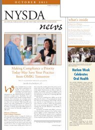
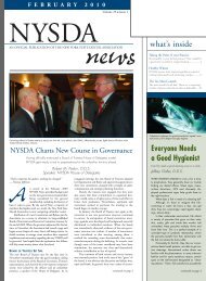
Figure 1. Orthopantographic exam shows presence of tooth.<br />
teeth and oral hygiene was poor. No edema or any soft tissue defect<br />
was present.<br />
In the extraoral examination, an edematous lesion, 1.5 x 2 cm<br />
in diameter, <strong>with</strong> a central fistular orifice located at the midline<br />
region of the chin, was observed. Bidigital palpation revealed that<br />
the area was endurated and the fistula tract was not occluded.<br />
The patient said the swelling on his chin was present for one<br />
week and had ruptured the day before.<br />
Conventional periapical radiographs showed no abnormality<br />
at the related part of the mandible. Orthopantographic examination<br />
showed the presence of a tooth in the soft tissue at the midline of<br />
the chin (Figure 1). The location and position of the tooth were<br />
evaluated by right cephalometric radiography (Figure 2). Also, the<br />
occlusal radiography showed the tooth at the midline of the chin<br />
(Figure 3).<br />
Treatment<br />
The area was cleaned <strong>with</strong> an antiseptic solution. An extraoral ring<br />
blockage was performed <strong>with</strong> an anesthetic solution containing 2 cc<br />
articain and epinephrine. An incision of 1.5 cm was made to the<br />
endurated area on the chin <strong>with</strong> a No.11 scalpel. Soft tissue around<br />
the affected area was dissected <strong>with</strong> a N0:2 cryohemostate. After<br />
that the tooth was removed from the dissected area by using hemostatic<br />
forceps. The dissected area was sutured subcutaneously using<br />
a 3/0, polyglycolic acid (Vicryl), rounded spiral, 20 mm suture<br />
material. The overlying skin was sutured <strong>with</strong> a 6/0 polyethilene<br />
propilen (Prolene) suture material. The patient was prescribed<br />
amoxicillin (1 gr) twice a day and Naproxene sodium (250 mg)<br />
three times daily after the operation.<br />
The patient was evaluated clinically at one-day intervals. On<br />
day seven, the skin sutures were removed, and the patient was<br />
referred to other clinics for further treatment.<br />
Discussion<br />
Post-traumatic teeth avulsions may result in serious complications if<br />
they are not correctly diagnosed and treated. An important complication<br />
of teeth avulsions is displacement of the avulsed tooth. There<br />
NYSDJ • APRIL 2005 27
Figure 2. Cephalometric radiography<br />
helps pinpoint tooth.<br />
Figure 3. Occlusal radiograph<br />
shows tooth at midline of chin.<br />
are several case reports in the literature describing impactions of<br />
the avulsed tooth into the larynx, nasopharynx, nasal cavity, maxillar<br />
sinus, frontal sinus, pyrifirm sinus and soft tissues of labia and<br />
cheek. 4-10 There are also a few cases described of the aspiration of the<br />
avulsed tooth. 11,12 In our literary search we have found no other case<br />
describing impaction into the soft tissue of the chin.<br />
An undiagnosed avulsed tooth embedded into the soft tissue<br />
may result in chronic, persistant infection, discharge and fibrosis.<br />
In the early period following trauma, the patient may not understand<br />
the severity and importance of the injury, or his low socioeconomic<br />
status may hinder urgent appearance at a dental clinic. In<br />
addition, inadequately performed intraoral or radiological examination<br />
may lead to delay in therapy and prognosis, as in our case.<br />
This case once more emphasizes the need for detailed clinical<br />
and radiographical examinations in patients who had maxillofacial<br />
traumas to make sufficient diagnosis before planning the treatment<br />
modality to prevent possible complications. ■<br />
REFERENCES<br />
1. Petersson EE, Andresson L, Sorensen S. Traumatic oral vs. non-oral injuries. Swed Dent<br />
J 1997;21:55-68.<br />
2. Forsberg CM, Tedestam G. Etiological and predisposing factors related to traumatic<br />
injuries to permanent teeth. Swed Dent J 1993;17:183-90.<br />
3. Andreasen JO, Andreasen FM. Classification, etiology and epidemiology of traumatic<br />
dental injuries. In: Andreasen JO, Andreasen FM, editors. Textbook and Color Atlas of<br />
Traumatic Injuries to the Teeth, 3rd Ed. Copenhagen:Munksgaard. 1994.<br />
4. Brudlo E, Sokalski J, Strozyk M. Tooth impaction into the nasal septum as a complication<br />
of facial injury. Czas Stomatol. 1986 Dec;39(12):826-9.<br />
5. Tung TC, Chen YR, Chen CT, Lin CJ. Full intrusion of a tooth after facial trauma. J Trauma<br />
1997 Aug;43(2):357-9.<br />
6. Thor AL. Delayed removal of a fully intruded primary incisor through the nasal cavity:<br />
a case report. Dent Traumatol 2002 Aug;18(4):227-30.<br />
7. Waugh R. Traumatic nasal impaction. Oral Surg Oral Med Oral Pathol 1970<br />
Dec;30(6):730-3.<br />
8. Clark JC, Jones JE. Tooth fragments embedded in soft tissue: a diagnostic consideration.<br />
Quintessence Int. 1997 Sept;18(9):653-4.<br />
9. Mody RN, Indurkar AD. Tooth in cheek. Oral Surg Oral Med Oral Pathol 1993<br />
Sept;76(3):388.<br />
10. McDonnell DG, Mc.Kiernan EX. Broken tooth fragments embedded in the tongue: a case<br />
report. Br J Oral Maxillofac Surg 1986 Dec;24(6): 464-6.<br />
11. Delap TG, Dowling PA, McGilligan T, Viljaya-Sekaran S. Bilateral pulmonary aspiration<br />
of intact teeth following maxillofacial trauma. Endod Dent Traumatol 1999 Aug<br />
15(4):190-2.<br />
12. Dhanrajani PJ, Swaify GA. Aspiration of a bridge and a tooth. J Craniomaxillofac Surg<br />
1992 Feb-Mar;20(2):91-2.<br />
28 NYSDJ • APRIL 2005
Figure 1. Charleston as it appeared about time Rodrigues family arrived in America.<br />
THE GREATEST ORAL SURGEON OF THE ANTEBELLUM SOUTH<br />
The Story of Benjamin A. Rodrigues<br />
Malvin E. Ring, D.D.S., M.L.S.<br />
Abstract<br />
The family of Benjamin A. Rodrigues fled Spain in the late<br />
18th century to escape persecution of Jews by the<br />
Inquisition. They settled in Charleston, South Carolina,<br />
where young Rodrigues was born. Showing great interest<br />
in medicine, he studied <strong>with</strong> one of the South’s leading<br />
physicians and then went on to study dentistry <strong>with</strong> an<br />
equally famous dentist. Feeling the need for a more thorough<br />
education, he enrolled in the Medical College of<br />
South Carolina, from which he was graduated in 1834 <strong>with</strong><br />
the M.D. degree. But his interests lay in dentistry, and<br />
upon his mentor’s emigrating to Europe, he took over the<br />
practice and became one of the leading dentists not only<br />
in South Carolina but in the entire South. He was active in<br />
his local and national dental societies and was a participant<br />
in their lively discussions. He soon earned a reputation<br />
as the leading oral surgeon of the southern states, and<br />
in 1850, the Baltimore College of <strong>Dental</strong> Surgery awarded<br />
him an honorary D.D.S.<br />
IN THE EARLY YEARS OF THE 19TH CENTURY there was a mass<br />
migration of Jews to the United <strong>State</strong>s from Spain, Portugal and<br />
other lands where church rulings and the Holy Inquisition<br />
oppressed and affected Jewish lives. Many of these immigrants,<br />
fleeing anti-Semitism and discrimination, ended up in the southern<br />
United <strong>State</strong>s,<strong>with</strong> Charleston,South Carolina,being a particularly<br />
favorite landing place.<br />
By 1800, there were more Jews living in South Carolina than<br />
anywhere else in the American colonies. 1 This was not by accident.<br />
The colony of South Carolina had entrusted the writing of its constitution<br />
to the renowned liberal philosopher, John Locke, who<br />
included in it guarantees of freedom of conscience to everybody<br />
including “Jews, heathens and dissenters.” 2 Among those settling in<br />
Charleston was the family Rodrigues, who arrived in this country<br />
sometime near the end of the 18th century (Figure 1). In 1815, a<br />
son was born to them, who was destined to become one of the<br />
greatest oral and maxillofacial surgeons of his time.<br />
His Personal Life<br />
Young Benjamin Rodrigues was educated in the public schools of<br />
Charleston and was graduated from Charleston High School. For<br />
a short time he attended Charleston College, but his interest lay<br />
in medicine. He found a mentor in Dr. Henry Frost, <strong>with</strong> whom he<br />
read medicine. In those days “reading medicine” <strong>with</strong> a practicing<br />
physician was also acceptable as medical training. Dr. Frost,<br />
who was a prominent and highly regarded physician, had<br />
30 NYSDJ • APRIL 2005
Figure 2. Dr. C. Starr Brewster, mentor of Dr.<br />
Rodrigues, at about time he left for Europe.<br />
Figure 3. Certificate, in Dr. Brewster’s hand, attesting to fact that Dr. Rodrigues had<br />
satisfactorily completed his dental studies.<br />
received his medical degree from the University of Pennsylvania<br />
in 1816. He strongly advocated the creation of a medical school in<br />
South Carolina; and it was ultimately established in 1824. Dr.<br />
Frost was named professor of materia medica, a position he held<br />
for 40 years. 3<br />
When young Rodrigues completed his studies <strong>with</strong> Dr. Frost,<br />
he started a preceptorship <strong>with</strong> one of Charleston’s leading dentists,<br />
Dr.C.Starr Brewster. 4 At that time there were no dental schools in<br />
existence anywhere (Figure 2). Upon completion of his apprenticeship<br />
he received from Dr. Brewster a certificate attesting that:<br />
“Mr. B. A. Rodrigues having pursued the study of Medicine and<br />
Surgery as a private pupil under the instruction of H. Frost, M.D.,<br />
Prof. of Materia Medica in the Medical College<br />
of SoCa, and having attended lectures<br />
in that institution has, for some time<br />
past, been a pupil in my office<br />
and acquired a knowledge of<br />
<strong>Dental</strong> Surgery which qualifies<br />
him to perform any<br />
operations therein. I<br />
therefore recommend<br />
him to my friends<br />
and the public as<br />
being fully entitled to<br />
their confidence.”<br />
It was signed<br />
by C. Starr Brewster,<br />
Surgeon Dentist, of<br />
Charleston, and dated<br />
March 1, 1832 (Figure<br />
3).<br />
What is amazing<br />
about this is that<br />
Rodrigues was only<br />
17 years old when he completed his studies <strong>with</strong> Dr. Brewster.<br />
Young Benjamin desired a formal degree, and soon after finishing<br />
his studies <strong>with</strong> Dr. Brewster, he matriculated at the medical<br />
school, which had been renamed The Medical College of South<br />
Carolina. On March 8, 1834, he received his M.D., <strong>with</strong> the distinction<br />
of being one of the new school’s first graduates (Figure 4).<br />
A short time after receiving his M.D. degree, Rodrigues<br />
married Cecelia Soloman, in February 1835, bought a house and<br />
settled down to married life. He and his wife became members<br />
of Beth Elohim synagogue, which had been founded in Charleston<br />
12 years earlier. This was the first<br />
synagogue in America of Reform<br />
Judaism, and is today one of the<br />
oldest continually functioning<br />
congregations in the country.<br />
The family became<br />
members, in spite of the<br />
fact that Rodrigues proclaimed<br />
himself a Deist<br />
and a follower of Tom<br />
Paine, who, he said,“drilled<br />
holes in the Bible<br />
through which any intellectual<br />
mind could see the<br />
absurdities” (Figure 5).<br />
The Rodrigues had a<br />
baby girl who died in<br />
infancy. Soon after, they<br />
adopted a baby girl. This<br />
child,upon reaching adulthood,<br />
married one of<br />
Benjamin’s first cousins,<br />
Figure 4. Medical College of South Carolina in Charleston at time Dr. Rodrigues was<br />
student there.<br />
Daniel Ottolengui, and<br />
they had three children,<br />
NYSDJ • APRIL 2005 31
one of whom, Rodrigues Ottolengui, became renowned as the editor,<br />
for more than half a century, of <strong>Dental</strong> Items of Interest, one of<br />
the most important dental journals in the world.<br />
A short time afterward, Dr. Brewster left America for Paris,<br />
where he became one of the most prominent American dentists; it<br />
was he who induced the notable Dr. Thomas W. Evans to leave<br />
America and come to Paris to practice. Before leaving for Europe,<br />
Dr. Brewster turned his Charleston practice over to Dr. Rodrigues,<br />
who soon became one of the foremost dentists, not only in South<br />
Carolina, but throughout the South. His contributions to mankind<br />
and the profession were recognized when the Baltimore College of<br />
<strong>Dental</strong> Surgery awarded him an honorary D.D.S. degree in 1850. His<br />
mentor, Dr. Brewster, had received a similar honor in 1842. 5<br />
organization in its last year. The convention was followed in 1859 by<br />
the formation of the American <strong>Dental</strong> Association at Niagara Falls,<br />
<strong>New</strong> <strong>York</strong>.<br />
When the South Carolina <strong>Dental</strong> Association was formally<br />
organized on April 5, 1870, Dr. Rodrigues was a charter member. He<br />
not only attended the sessions, but took a lively part in the discussions.<br />
The minutes of November 2, 1870, state that, “following the<br />
reading of an essay on ‘Alveola Abscess’ (sic), a lively discussion was<br />
held in which Dr. Rodrigues took part.” 9 The last meeting of the<br />
association that he attended was on April 11, 1871, shortly before<br />
his untimely death. Dr. Rodrigues had also been active in civic<br />
affairs and had served as a member of the Charleston Board of<br />
Health in 1849.<br />
Role in Organized Dentistry<br />
On August 18, 1840, a number of foresighted and progressive<br />
dentists came together at the American Hotel<br />
in <strong>New</strong> <strong>York</strong> City for the purpose of organizing the first<br />
national society of dentists in the world. A constitution<br />
and by-laws were adopted, and on the second<br />
day of the meeting, numerous dentists were declared<br />
charter members. These individuals were either in<br />
attendance at the meeting, or else had been invited by<br />
mail to become active members. Among this latter<br />
group was Dr. Rodrigues, one of the 62 active<br />
members of the newly formed American<br />
Society of <strong>Dental</strong> Surgeons. 6<br />
Dr. Rodrigues was aware of the importance<br />
of dentists being organized and valued<br />
the need for communication among members<br />
of the profession. In 1839, the organization launched the first<br />
dental journal in the world, the American Journal of <strong>Dental</strong> Science.<br />
Rodrigues was one of the first subscribers, and it was not long<br />
before the Journal carried several of his articles. 7, 8<br />
The American Society of <strong>Dental</strong> Surgeons went out of existence<br />
in 1856, primarily because of the battle over the propriety of<br />
using silver amalgam, the struggle known popularly as the<br />
“Amalgam War.” This first national society of dentists was followed<br />
by the establishment of the American <strong>Dental</strong> Convention on August<br />
2, 1855, in Philadelphia. Dr. Rodrigues was named secretary of this<br />
The Nature of His Practice<br />
Although his interests lay primarily in surgery, Dr.<br />
Rodrigues carried on a practice in all branches of<br />
dentistry.A surviving bill (Figure 6), sent to a patient<br />
in 1851, when he had been in practice for about 17<br />
years, indicates that he constructed advanced prosthetic<br />
dentures for which he was well compensated.<br />
The fee of $125 was handsome indeed, equivalent<br />
in purchasing power to almost $1,000 today. His<br />
description of the upper denture as having “suction<br />
gum[s]” probably meant that he had<br />
incorporated a “suction device” into the<br />
palate of the denture. This was a new<br />
innovation that was believed to give<br />
Figure 5. Dr. Benjamin A. Rodrigues<br />
at about age 24.<br />
added retention to an upper denture.<br />
The prevailing belief was that an upper<br />
denture was held in place by atmospheric pressure and that this<br />
could be augmented by creating a “vacuum chamber” in the mid-<br />
10, 11<br />
palatal area of the denture. In time, the value of this was disproved,<br />
but devices to create such additional “suction” were marketed to<br />
dentists until the early 20th century (Figure 7).<br />
We must speculate, however, as to the material from which the<br />
denture was made. The Goodyear <strong>Dental</strong> Rubber Company began<br />
marketing its new product, Vulcanite, to the dental profession in<br />
late 1851. Since the denture Rodrigues constructed was made<br />
months before that, it was likely that it was a swaged gold denture,<br />
Mr. Cain 1851<br />
To: B.A.Rodrigues, Dr.<br />
July 7 6 Extractions for Lady $6.00<br />
July 28 1 for Daughter $1.00<br />
Sept. 11 One upper suction gum art. teeth $125.00<br />
Sept. 19 One extraction for Daughter $1.00<br />
$ 133.00<br />
Rec’d payment Oct. 10, 1851 B.A.Rodrigues<br />
Figure 6. Bill for Dr. Rodrigues’s services in 1851.<br />
Figure 7. Package insert from device sold by Swiss company in<br />
1890s that allowed dentist to place metal-rubber ring in middle of<br />
denture’s palate, supposedly to create suction.<br />
32 NYSDJ • APRIL 2005
the type made by most ethical dentists. To produce it, an impression<br />
was taken and a cast poured in lead. Upon this cast a sheet of<br />
gold was placed, and hammered <strong>with</strong> a wooden mallet until the<br />
gold spread out and assumed the proper shape. (In order to achieve<br />
a “suction” cavity, additional lead would have been added to the<br />
palatal area of the cast.)<br />
Porcelain teeth had been introduced in 1809 by Guiseppangelo<br />
Fonzi, and by the 1850s, many improvements had been made in<br />
denture teeth. They generally had a platinum pin fused to the back<br />
of the tooth, and this would be soldered to the gold denture base<br />
(Figure 8).<br />
Since Dr. Rodrigues practiced in a large urban setting, a good<br />
portion of his practice was surgery, including many extractions.<br />
Rural areas, however, had access only to traveling itinerant dentists,<br />
and, thus, physicians in these areas did a fair number of extractions,<br />
as well as “bleedings.”The account book of a Dr. Andrew Hasell, who<br />
practiced at the same time in Georgetown County, South Carolina,<br />
shows that his fee for bleeding a white person was $2, while the<br />
charge for the same service for a slave was $1. He used the same fee<br />
scale for extractions. In his journal he described a visit to the<br />
Waverly Plantation near Georgetown, where he vaccinated a slave<br />
and also extracted a tooth and received a fee of $1 for each service. 12<br />
The problem of cleft palate, either congenital or acquired (as a<br />
result of the ravages of syphilis, for which a successful cure had not<br />
yet been found), taxed the ingenuity of dentists everywhere. One of<br />
the earliest of the modern researchers of this problem was Charles<br />
W. Stearns, who, although practicing in the United <strong>State</strong>s at the<br />
same time as Dr. Rodrigues, published four papers on his methods<br />
of creating obturators in the British journal, the Lancet. 13<br />
Rodrigues similarly crafted obturators for patients <strong>with</strong> large<br />
defects of the palate, although he said he thought it much more<br />
desirable, if possible, to correct them surgically. The minutes of the<br />
South Carolina <strong>Dental</strong> Association meeting, mentioned earlier,<br />
state that “Dr. Rodrigues then introduced a young lad for the purpose<br />
of demonstrating the operation of forming an artificial palate,<br />
and spoke at some length in explanation of various noteworthy<br />
operations that he had performed from time to time.”<br />
He was a skillful and dexterous surgeon from his earliest days<br />
of practice, <strong>with</strong> no problem seeming too difficult for him to tackle.<br />
One of his most remarkable cases was reported in the very first<br />
issue of the American Journal of <strong>Dental</strong> Science. 14 On August 18,<br />
1837, a young woman presented <strong>with</strong> excruciating pain in her face.<br />
She sought Dr. Rodrigues’s help after having all the teeth on the<br />
upper right side of her mouth extracted. Rodrigues assumed it was<br />
an abscess and inserted a trochar into the socket of the second<br />
molar, expecting a gush of pus, but instead met stiff resistance,<br />
which he assumed was an exostosis (in which diagnosis his medical<br />
colleagues concurred).<br />
He then “dissected away the gum from the canine tooth to the<br />
last molar, raised the flap, and <strong>with</strong> a trephine opened into the cavity.<br />
[The mass] was so general on the affected side as to reach from<br />
the second incisor anteriorly to the pterygoid process posteriorly….The<br />
bony tumor filled up and occupied [the antrum] and<br />
could be readily reached. The trephine was applied, the cavity<br />
Figure 8. Swaged<br />
gold denture of<br />
about 1840.<br />
Porcelain teeth,<br />
which had been<br />
introduced some<br />
20 years earlier,<br />
had platinum pins<br />
embedded, and<br />
these were<br />
soldered to<br />
denture base.<br />
enlarged, and the exostosis removed. It measured in circumference<br />
three inches, was light and cancellated on its surface, but dense and<br />
resisting in its more internal layers. There was little or no hemorrhage….After<br />
removing every spiculum of diseased bone and<br />
cleansing the cavity, the flap was replaced and to nature was<br />
entrusted the cure. Granulations opened up in full luxuriance and<br />
in the short period of four weeks, the woman was in the enjoyment<br />
of excellent health…To my recollection there has been no history<br />
recorded where the tumour was so large, where such extensive<br />
injury was inflicted on the adjacent parts, and where nature, after<br />
the cause of the malady had been removed, exerted her recuperative<br />
powers so benignly and so quickly.”<br />
This case is all the more remarkable in that the surgery was<br />
performed before anesthesia had been discovered, and even more<br />
amazing that it was carried out when the surgeon was a mere 22<br />
years old!<br />
Dr. Rodrigues performed many such difficult operations in the<br />
course of his professional life and was extolled throughout the<br />
southern United <strong>State</strong>s for his knowledge and skill. He practiced<br />
right up to his death from a stroke on October 19, 1871. He died at<br />
the age of 57, and is buried in Magnolia Cemetery in Charleston,<br />
South Carolina. 15 ■<br />
REFERENCES<br />
1. A Portion of the People: Three Hundred Years of Southern Jewish Life, Rosengarten,<br />
Theodore, Editor. Charleston: University of South Carolina Press, 2004.<br />
2. Learsi R. The Jews in America: A History. Cleveland: World Publishing Co., 1954. Page 33.<br />
3. Waring Jl. A History of Medicine in South Carolina. Columbia, SC: South Carolina<br />
Medical Association, 1967. Page 73.<br />
4. Asbell MB. 200 years of Jewish contribution to dentistry in America. Alpha Omegan Vol.<br />
69(2), September 1976. Page 31.<br />
5. Maryland <strong>State</strong> <strong>Dental</strong> Association. GM Anderson, Editor. Proceedings <strong>Dental</strong> Centenary<br />
Celebration, 1840-1940. Baltimore, 1940. Page1041.<br />
6. Ibid, page 1046.<br />
7. Rodrigues BA. Correction of malocclusion. Amer J <strong>Dental</strong> Science Vol.1, First Series,<br />
1839-41. Pages 175-6.<br />
8. Rodrigues BA. An essay on the mode of treating caries of the teeth. Amer J <strong>Dental</strong><br />
Science Vol. 1, First Series, 1839-41. Pages 282-89.<br />
9. Macaulay NW. History of the South Carolina <strong>Dental</strong> Association. Columbia, SC: South<br />
Carolina <strong>Dental</strong> Association. Page 26.<br />
10. Ames WB. Atmospheric pressure in the retention of entire dentures. Brit Dent J Vol. 6,<br />
1885:601-04.<br />
11. Koch CRE, Editor. History of <strong>Dental</strong> Surgery, Vol. 1. Chicago:National Art Publishing,<br />
1909. Pages 282-3.<br />
12. Account book of Dr. Andrew Hasell. South Caroliniana Library, Charleston, SC.<br />
13. Walter JD. Charles W. Stearns and Norman W. Kingsley and the Development of the<br />
Artificial Velum. Bulletin of the History of Dentist Vol. 29(2) Oct. 1981. Page 76.<br />
14. Rodrigues BA. Removal of a tumor from the antrum. Amer J Dent Science Vol. 1(1),<br />
1839. Pages 88-89.<br />
15. Obituary, Charleston Daily Courier Oct. 19, 1871.<br />
NYSDJ • APRIL 2005 33
PLATFORM SWITCHING AS A MEANS TO<br />
ACHIEVING IMPLANT ESTHETICS<br />
A CASE STUDY<br />
David M. Gardner, D.D.S.<br />
Abstract<br />
This article discusses the literature related to the dynamics<br />
that are in effect when an implant is placed in bone.<br />
Osseous and soft tissue changes can result subsequent to<br />
implant placement. These changes will determine the<br />
esthetic outcome. A technique will be presented to limit<br />
both osseous and soft tissue changes, thereby creating a<br />
predictable esthetic result.<br />
SINCE THE INTRODUCTION of osseointegrated implants for the<br />
restoration of missing or lost teeth, treatment options for the partially<br />
or fully edentulous patient have expanded exponentially.<br />
Removable prostheses are no longer the only available option to<br />
treat the edentulous state. In fact, this option is no longer the first<br />
to be considered. Furthermore, the destruction of the healthy dentition<br />
to provide tooth-borne fixed prostheses can be avoided <strong>with</strong><br />
predictability.<br />
Implant therapy offers increased longevity, improved function,<br />
bone preservation and quality of life. 1 Ten-year survival surveys of<br />
fixed prostheses on natural teeth reveal a survival rate of approximately<br />
75%. 2 Success rates for endosseous implants have been<br />
shown to be greater than 90%. 3,4<br />
As for all clinical modalities, implant restoration has many<br />
variables that must be carefully controlled. One particular aspect of<br />
implant therapy that can be most challenging is the placement and<br />
subsequent restoration in the esthetic zone.A comprehensive understanding<br />
of crestal bone changes around endosseous implants and<br />
subsequent soft tissue reaction to these osseous changes must be<br />
achieved to predictably realize our patients’ esthetic expectations.<br />
The author reviews pertinent literature on the subject of<br />
implants in the esthetic zone. Next, through a case study, he presents<br />
a concept to make esthetic outcomes more achievable.<br />
Key Determinant of Esthetics<br />
The dentogingival and peri-implant complex are similar in their<br />
cellular makeup; both consist of keratinized oral epithelium and<br />
non-keratinized junctional epithelium (Figure 1). 5 Histologically,<br />
their differences are seen in their ability to attach to their respective<br />
tooth/implant. While junctional epithelial attachment through glycoproteins<br />
is noted for the natural dentition, a pseudo attachment<br />
through hemidesmosones exists around endosseous implants. In<br />
addition, connective tissue fibers mechanically insert into root<br />
cementum. This phenomenon is not seen <strong>with</strong> implants; instead, a<br />
tight cuff is formed around the titanium implant. 6<br />
In their study of the periodontium, Garguilio, Wentz and<br />
Orban investigated an average histological dimension of the epithelial<br />
attachment (.97 mm) and connective tissue (1.07 mm) (Figure<br />
2). 7 Known as biologic width, this dimension is a key determinant<br />
of esthetics.<br />
The biologic width found around teeth is also characteristic of<br />
the peri-implant complex. These dimensions are similar to that of<br />
34 NYSDJ • APRIL 2005
Figure 1. Histological<br />
illustration of dentogingival<br />
complex.<br />
Figure 2. Illustrates components<br />
of biologic width.<br />
Figure 3. Illustrates stable biologic<br />
width around implants as compared<br />
to natural dentition.<br />
Figure 4. The Black Triangle.<br />
Figure 5. Measuring distance from contact<br />
point to IHB.<br />
Figure 6. Illustrates 1.5-2 mm of crestal bone loss<br />
from implant/abutment interface.<br />
Figure 7. Horizontal component<br />
of bone loss after<br />
abutment connection.<br />
teeth and are stable even after loading (Figure 3). 8 From these studies<br />
one can calculate the position of the gingival architecture in<br />
relation to the osseous crest. This position in fact follows the principles<br />
of biologic width.<br />
Much has been said on the topic of interdental papilla and its<br />
importance in esthetics. The interproximal height of bone has a distinct<br />
influence on interdental papilla by acting as a guidepost to soft<br />
tissue contours. Garber et al. demonstrated the existence of a predictable<br />
length as the achievable and maintainable papilla length in<br />
the maxillary anterior sextant, measured from the most coronal<br />
interproximal height of bone immediately adjacent to a tooth or<br />
implant fixture. The mean papilla length for an implant-tooth relationship<br />
is 6.5 mm and the implant-implant relationship is 4.5 mm. 9<br />
In further studies, the papilla length was determined to be 3.4 mm<br />
between adjacent implants. 10<br />
Dreaded Black Triangle<br />
The importance of soft tissue architecture cannot be overstated. In<br />
the interproximal region a deficiency will lead to the dreaded “black<br />
triangle” seen in patients <strong>with</strong> periodontal disease (Figure 4). Facial<br />
soft tissue deficiencies can create a tooth/implant that appears longer<br />
than desired and a gingival crest that is inconsistent <strong>with</strong> the remainder<br />
of the esthetic region. The predictable presence of interdental soft<br />
tissue is dependent upon the position of the interproximal bone<br />
height and its relation to the contact point. When the distance from<br />
the contact point to interproximal height of bone is greater than 5<br />
mm, avoiding the “black triangle” is difficult (Figure 5). 11<br />
Understanding how crestal bone responds to implant placement<br />
and subsequent abutment connection is critical in achieving<br />
an acceptable esthetic outcome. The position of crestal bone will<br />
influence the position of the soft tissue, which in turn dictates,<br />
along <strong>with</strong> other factors, the total esthetic outcome. Through<br />
research and clinical experience, we can conclude the following:<br />
● The rough/smooth border on one-piece non-submerged<br />
implants determines the first bone-to-implant contact.<br />
● In two-piece implants, the first bone-to-implant contact was<br />
located 1.5-2 mm apical to the microgap (Figure 6). 12 This represents<br />
the bone loss seen both clinically and radiographically<br />
around two-piece implants placed at or below the crest of bone.<br />
●<br />
Bony changes are present after the creation of a microgap at<br />
the implant abutment interface.<br />
Understanding this information is critical to achieving our<br />
patients’ esthetic expectations. Crestal bone loss circumferentially<br />
around an implant can lead to an increased distance from the contact<br />
point to the interproximal height of bone. Consequently, predictable<br />
papilla formation is more difficult. In addition, an increase<br />
in crestal bone loss will cause the facial soft tissue to recede as new<br />
biologic width is formed.<br />
Controlling Crestal Bone Loss<br />
The makeup of crestal bone loss around implants from the microgap<br />
to crestal bone has both horizontal and vertical components.<br />
Following abutment connection, crestal bone will move away from<br />
the microgap 1.3 mm to 1.4 mm in a horizontal direction (Figure 7). 13<br />
NYSDJ • APRIL 2005 35
Figure 8. Root<br />
resorption of #8.<br />
Figure 9. Preparing osteotomy for<br />
immediate placement.<br />
Figure 10. Custom healing abutment.<br />
Figure 11. Radiograph<br />
of custom healing<br />
abutment. Note apical<br />
position of implant<br />
platform.<br />
Figure 12. Provisional restoration. Figure 13. Ceramo-metal abutment. Figure 14. Final all-ceramic<br />
restoration.<br />
This can influence the interproximal height of bone if implants are<br />
not placed correctly adjacent to teeth (1.5 mm) or other implants<br />
(3 mm). 14 The loss of interproximal bone will increase the distance to<br />
the contact point, influencing esthetics.<br />
Clinicians, researchers and implant companies have dedicated<br />
time to finding ways to control crestal bone loss after abutment<br />
connection. Use of non-submerged implants to control or eliminate<br />
bone loss is a proven way to accomplish this. 15 A scalloped implant<br />
platform has been developed to follow osseous architecture and<br />
eliminate crestal bone loss. 16 Another method is altering the horizontal<br />
position of the microgap.<br />
As stated previously, crestal bone loss following abutment connection<br />
has both horizontal and vertical components. The horizontal<br />
component consists of the 1.3 mm to 1.4 mm of bone loss from the<br />
microgap to the crest of bone. If the horizontal component can be<br />
controlled or decreased, then crestal bone loss can also be decreased.<br />
Case Study<br />
A 37-year-old male <strong>with</strong> no significant medical history presented<br />
complaining of pain radiating from tooth #8. His upper anterior<br />
teeth had experienced trauma approximately 15 years earlier, and<br />
had been restored <strong>with</strong> porcelain-fused-to-metal crowns. All teeth<br />
were asymptomatic up until the present.<br />
Upon clinical examination, #8 presented <strong>with</strong> pertinent probing<br />
into the pulp chamber. A periapical radiograph was reviewed<br />
and revealed root resorption that left the tooth <strong>with</strong> a hopeless<br />
prognosis (Figure 8). Treatment options were presented; and a sin-<br />
gle tooth implant was selected as a means of restoration.<br />
The thorough understanding of osseous and soft tissue<br />
response to implant placement is required to predictably achieve an<br />
esthetic result. Failure to grasp these concepts can result in a catastrophic<br />
esthetic outcome.<br />
It was decided that the tooth would be extracted using a periotome,<br />
in order to perform this procedure in atraumatic fashion.<br />
In addition, an immediate implant was placed at the time of<br />
extraction (Figure 9). Immediate placement was selected to preserve<br />
the position and dimensions of the osseous structures. The<br />
anterior alveolar ridge can show up to 23% resorption <strong>with</strong>in the<br />
first six months following extraction; immediate placement is a<br />
way to prevent resorption of the alveolus. 17 Since little destruction<br />
to the crestal bone dimensions was observed, augmentation was<br />
not necessary. Instead, preservation was key to maintaining<br />
osseous and soft tissue contours.<br />
A 5.0 mm x 13 mm osseotite certain implant (3i, Palm Beach<br />
Gardens, Fla.) was placed <strong>with</strong> the platform level <strong>with</strong> the buccal crestal<br />
bone. Since crestal bone has a scalloped architecture, the implant was<br />
apical to the interproximal height of bone on the mesial and distal.<br />
Once primary stability was established, a temporary abutment was<br />
placed and composite resin formed around to maintain sulcular<br />
dimensions through osseointegration (Figures 10 & 11).<br />
Approximately four months were allowed for integration of the<br />
fixture, and a provisional was placed (Figure 12). A cementretained<br />
all-ceramic crown was used for the final restoration<br />
(Figures 13 & 14).<br />
36 NYSDJ • APRIL 2005
Figure 15. Implant abutment<br />
interface following platform<br />
switching. Note position<br />
of IHB.<br />
Figure 16. Final<br />
radiograph. Note<br />
IHB.<br />
According to the literature, several changes should occur after abutment<br />
connection. Resorption of bone approximately 1.5 mm from<br />
the implant abutment interface should occur circumferentially.<br />
This will result in 1.5 mm of bone loss on the buccal. Interproximately,<br />
however, osseous changes should result in even greater crestal<br />
bone loss. Resorption 1.5 mm apical to the microgap will result<br />
in even greater bone loss since the implant was placed apical to the<br />
interproximal height of bone.<br />
As a result of these osseous changes, the soft tissue architecture<br />
will be affected as biologic width is reestablished. This will be<br />
most prevalent in the interproximal areas, where bone loss will<br />
result in an increase in the distance between the height of bone and<br />
the contact. Using these absolute facts of the dynamics of implant<br />
therapy, could a variable be applied that would influence the<br />
changes that occur in bone and soft tissue when an implant is<br />
placed and restored<br />
The variable to be controlled is the dimension of the abutment<br />
to be connected to the implant platform. In particular, the width of<br />
the abutment to be used would control the bony changes around the<br />
implant.When an abutment is connected to a dental implant at the<br />
crestal level, bone loss around the fixture occurs. This bone loss has<br />
a horizontal component that results in 1.3 mm to 1.4 mm of resorption<br />
from the microgap to the bone in a horizontal direction. 18 If this<br />
horizontal component could be reduced or nullified, then bone<br />
preservation and acceptable soft tissue architecture would result.<br />
The concept of “platform switching” was introduced to restore<br />
this edentulous space. Instead of using an abutment that was 5<br />
mm in width, a standard abutment was used that measured 4.1<br />
mm (Figure 15). This results in a microgap that is 4.5 mm from the<br />
edge of the platform. Consequently, the horizontal component of<br />
bone loss from the microgap was reduced, and osseous dimensions<br />
were maintained (Figure 16). The resultant changes were<br />
minimal. The height of bone was maintained after abutment connection<br />
and one year after restoration. Buccal crestal bone was<br />
maintained in its original position following implant placement.<br />
Preservation of osseous dimensions led to maintenance of gingival<br />
architecture and pleasing esthetic results. Mesial and distal papillae<br />
filled the interproximal spaces because of the height of interproximal<br />
bone. The gingival crest is in the correct position as compared<br />
to the adjacent central incisor. Preoperative esthetic expectations<br />
were satisfied.<br />
Discussion<br />
“Platform switching” is a simple and effective way to control circumferential<br />
bone loss around dental implants. By altering the<br />
horizontal position of the microgap, the horizontal component of<br />
bone loss after abutment connection can be reduced. This technique,<br />
however, has its faults. Switching can only be used <strong>with</strong><br />
components that have similar designs, that is, the screw access<br />
hole must be uniform. In addition, sufficient space is needed to<br />
develop a proper emergence profile.<br />
Despite these facts,“platform switching” is a subject that needs<br />
more investigating. Can implants be placed closer than 3 mm from<br />
an adjacent implant while still maintaining interproximal height of<br />
bone Can implants be placed less than 1.5 mm from an adjacent<br />
tooth and still maintain interproximal bone Can implants be placed<br />
at or below the osseous crest and avoid bone loss to the first thread<br />
after abutment connection Can implant esthetics be improved<br />
through “platform switching” These questions and others need to<br />
be answered to determine the viability of this technique.<br />
Through positive results, future implant and abutment<br />
designs will be greatly influenced to preserve critical tissues<br />
around dental implants. ■<br />
The author thanks Alter Smiles <strong>Dental</strong> Studios (<strong>New</strong> Hyde Park, NY) for its commitment to<br />
excellence on this case.<br />
REFERENCES<br />
1. Misch CE. Contemporary Implant Dentistry. 2nd Edition. St. Louis, MO: Mosby; 1999:9-11.<br />
2. Walton JN, Gardner FM, Agar JR. A survey of crown and fixed partial denture failures:<br />
length of service and reason for replacement. J Prosthet Dent. 1986;56:416-421.<br />
3. Adell R. Clinical results of osseointegrated implants supporting fixed prostheses in<br />
edentulous jaws. J Prosthet Dent. 1983;50:251-254.<br />
4. Albrektsson T, Jansson T, Lekholm U. Osseointegrated dental implants. Dent Clin North<br />
Am. 1986;30:151-174.<br />
5. Linde J. The Anatomy of the Periodontium: Textbook of Periodontology. 1st Edition.<br />
Philadelphia: WB Saunders, Co. 1983:19-66.<br />
6. Branemark, PI. Osseointegration and its experimental background. J Prosthet Dent<br />
1983;50:399-410.<br />
7. Gargiulo AW, Wentz FM, Orban B. Dimensions and relations of the dentogingival junction<br />
in humans. J Periodontol 1961;32:262-7.<br />
8. Cochran, Hermann, Schenk, Higginbotom, Buser. Biologic width around titanium<br />
implants. A histomeric analysis of the implantogingival junction around unloaded and<br />
loaded non-submerged implants in the canine mandible. J Periodontol 1997;68:186-<br />
198.<br />
9. Salama H, Salama inMS, Garber D,Adar P. The interproximal height of bone: a guidepost<br />
to predictable aesthetic strategies and soft tissue contours in anterior tooth replacement.<br />
Pract Periodonticts Aesthet Dent. 1998 Nov-Dec:10(9):1131-41.<br />
10. Tarnow D, Elian N, Fletcher P, Froum S, Magner A, Cho SC, Salama M, Salama H, Garber<br />
DA. Vertical distance from the crest of bone to the height of the interproximal papilla<br />
between adjacent implants. J Periodontol 2003 Dec;74(12):1785-8.<br />
11. Tarnow DP, Magner AW, Fletcher P. The effect of the distance from the contact point to<br />
the crest of bone on the presence of absence of the interproximal dental papilla. J<br />
Periodontol 1992 Dec;63(12): 995-6.<br />
12. Herman, Cochran, Nummikoski, Buser. Crestal bone changes around titanium implants.<br />
A radiographic evaluation of unloaded non-submerged and submerged implants in the<br />
canine mandible. J Periodontol 1997:68;1117-1130.<br />
13. Tarnow DP, Cho SC, Wallace SS. The effect of inter-implant distance on the height of<br />
inter-implant bone crest. J Periodontol 2000;Apr:71(4): 546-9.<br />
14. Garber DA, Salama MS, Salama H. Immediate total tooth replacement. Compend Contin<br />
Educ Dent 2001 Mar;(3):210-6, 218.<br />
15. Hermann JS, Buser D, Schenk RK, Cochran DL. Crestal bone changes around titanium<br />
implants. A histometric evaluation of unloaded non-submerged and submerged<br />
implants in the canine mandible. J Periodontol 2000 Sep;71(9):1412-24.<br />
16. Wohrle PS. Nobel perfect esthetic scalloped implant: rationale for a new design. Clin<br />
Implant Dent Relat Res 2003:5 Suppl 1:64-73.<br />
17. Gelb DA. Immediate implant surgery: three-year retrospective evaluation of 50 consecutive<br />
cases. Int J Oral Maxillofac Implant 1993;8(4):388-99.<br />
18. Tarnow DP, Cho SC, Wallace SS. The effect of inter-implant distance on the height of<br />
inter-implant bone crest. J Periodontol 2000 Apr;71(4):546-9.<br />
NYSDJ • APRIL 2005 37