A Conversation - New York State Dental Association
A Conversation - New York State Dental Association
A Conversation - New York State Dental Association
- No tags were found...

Create successful ePaper yourself
Turn your PDF publications into a flip-book with our unique Google optimized e-Paper software.
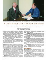
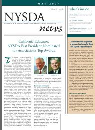
An amateur winemaker, Larry takes almost asmuch pleasure in designing labels for the bottlesas he does in filling them.Photo by Daniel BranchLarry and Judy Volland relax at home in Lockport.A <strong>Conversation</strong>WITH THE PRESIDENTNYSDA President Lawrence E. Volland reveals his plans forimproving the future of the profession and the <strong>Association</strong>.Kevin J. Hanley, D.D.S. Associate Editor, The NYSDJPhoto by Daniel BranchI SAT DOWN RECENTLY with NYSDA’s 2005 President, LawrenceE. Volland, for a discussion of important issues facing the dentalprofession and the <strong>Association</strong>. I was impressed with Dr. Volland’scandor and forthrightness, especially when addressing some of thethornier matters he’s likely to encounter in the coming year. Dr.Volland’s thoughts, perceptions and hopes for his term in office arecontained in the interview that appears below.Dr. Hanley: Hello, Larry. First, let me thank you for meetingwith me today. I appreciate your taking the time to talk with me andsharing your thoughts about the various issues facing our profession.Let me begin by asking, what do you believe is the mostimportant issue facing NYSDA and organized dentistry today?Dr.Volland: If you’re talking about organized dentistry from abroad perspective, it would be access to care. ADA PresidentRichard Haught has made access the theme for ADA action thisyear, and I doubt that there is a state in the nation that does nothave concerns in this critical area. <strong>New</strong> <strong>York</strong> is no exception. One ofmy first initiatives as NYSDA President will be to address the acuteaccess to care problems in the state’s rural areas.Facing NYSDA? I’d have to say relevance. Relevance to itsmembers. NYSDA has been very effective in the past in addressingmembers’ concerns. More importantly, NYSDA has done an incrediblejob of dealing with issues, often of potentially disastrousimpact to our members, well before they get on the radar screen ofthe average dentist. But we may not communicate that well.Consequently, the <strong>Association</strong> is often viewed in a light dimmed byignorance, and is seen as lacking relevance to the average dentistmember.Dr. Hanley: Access to care was an important issue discussed atthe 2004 Annual Session of the ADA House of Delegates. What arethe unique problems facing NYSDA and <strong>New</strong> <strong>York</strong> <strong>State</strong> with regardto access to care?Dr.Volland: We have urban access issues as well as rural ones,with each set of issues posing its own unique challenge. One wouldthink that in an urban environment, where there is a concentrationof dentists, that access to care would not be a problem. But, it is—from a socio-economic standpoint. In rural areas, access problemsare more geographic in nature, and, yet, socioeconomic barriersexist there, too.In <strong>New</strong> <strong>York</strong>, we went to great lengths to satisfy the urbanaccess problem when NYSDA negotiated a $576-million increase inthe Medicaid fee-for-service reimbursement. But many counties inrural areas of the state suffer from a dearth of dentists, and necessarydental treatment—not to mention preventive care—is notbeing delivered because dentists are not available.Dr. Hanley: Continuing with access to care, what can we do toimprove access to care in <strong>New</strong> <strong>York</strong> <strong>State</strong>?Dr. Volland: I don’t have all the answers to that, but, certainly,we need to continue to improve the delivery of dental care via theMedicaid system. Also, solving the problem in <strong>New</strong> <strong>York</strong> <strong>State</strong> may24 NYSDJ • JANUARY 2005
2005 NYSDA Presidentrequire an approach not contained in the “box”of possible solutionswe, as a profession, have been looking into up to this point. To thatend, the Board of Governors, at its meeting in November, authorizedme to set up a task force, if necessary, to study solutions. Initially,our Council on <strong>Dental</strong> Practice and the Executive Committee will beworking on it, but NYSDA may need to go outside the profession foranswers—and we should not be afraid to do that.If we don’t work to solve this problem, we run the risk of governmentofficials dictating a solution, not unlike what is happeningin Alaska, where the government is helping to train high schoolgraduates to provide dental services, including irreversible ones, tothe native population. It is a solution that was conceived to meet theneeds of native Alaskans who have been receiving little or no dentalcare because there are no dentists. Clearly, the profession is unhappywith this type of “fix” and is mustering its resources to fight it.But, quite frankly, it’s difficult to argue against non-dentists providingcare when the alternative is no care at all.Organized dentistry must work aggressively to provide dentalcare to those in need so that the Alaskan model does not become thedefault solution. I truly believe, given the choice, people would chooseto have their services provided by a qualified doctor of dentistry. It isour obligation to strive to make our services available. That’s why Iwill be placing the access issue on the front burner in 2005.Amalgam SeparatorsDr. Hanley: Shifting gears slightly, environmental mercury is a hotbutton issue today. Do you think we, as dentists, do enough toaddress this issue voluntarily in our offices?Dr. Volland: Yes, environmental mercury is an issue, but, aslinked to dentistry, it is a hot button one only for people enamoredwith junk science. Dentists, as individuals, have long been responsiblein their handling of mercury. And the profession has proactivelydealt with the management of mercury, whether elemental or asa component of dental amalgam, for years—particularly withregard to its disposal. In October of 2000, we published a specialissue of The <strong>New</strong> <strong>York</strong> <strong>State</strong> <strong>Dental</strong> Journal, in which we stressedthat obligation and reiterated best management practices withrespect to the disposal of dental amalgam.Dr. Hanley: What do you think about amalgam separators?Are they the answer to mercury pollution?Dr.Volland: You know what I think about separators? They sendthe wrong message. Sure, they can reduce mercury from, say, threeparts per million to two, but they are not cost-effective and serve noenvironmental purpose. Dentistry simply is not a measurable part ofthe mercury pollution issue. If every dental office in the state wereoutfitted with the most efficient separator known to science thereThe Vollands met when Judy applied for a job in Larry’s dentaloffice. The encounter is described as “love at first sight.”would be no measurable change in mercury pollution. Furthermore,we have invested an enormous amount of effort educating the publicthat dental amalgam is safe and that the mercury in amalgam is not“free.” To embrace separators, or even acquiesce grudgingly to theiruse, gives the impression that waste amalgam is not safe enough toexist in wastewater, let alone in the patient’s mouth.Want a change? Forget about amalgam separators and go afterthe fossil fuel burning plants in the Great Lakes Basin.Our members should know that the <strong>New</strong> <strong>York</strong> <strong>State</strong> Departmentof Environmental Conservation is currently developing regulationscovering the management of dental amalgam and that I willcontinue to lead NYSDA’s effort to ensure that such regulations donot mandate amalgam separators.At this point, the DEC is suggestingthat such a mandate could be implemented over time—perhapsa three-year period. And, while this graduated implementationwould be helpful, NYSDA is firm in its continuing oppositionto any mandate.The Role of Organized DentistryDr. Hanley: How important is organized dentistry to the daily routineof dentists in their offices?Dr. Volland: It’s extremely important, but, unfortunately, notalways perceived as such. Most dentists practicing in <strong>New</strong> <strong>York</strong>Photo by Daniel BranchNYSDJ • JANUARY 2005 25
2005 NYSDA President<strong>State</strong> would have difficulty relating membership in organized dentistryto their daily routine. But the key word here is “organized.”Strength in numbers is not a cliché. Dentists enjoy the quality oftheir practice and take satisfaction in what they do because they areorganized and have certain clout. Have you seen the current AARPad campaign? The tag line is,“If one person alone could do it, therewould be no need for AARP.” Substitute ADA or NYSDA for AARP,and you have a great membership campaign.Dr. Hanley: If the importance of organized dentistry is unclearto the average dentist, what can NYSDA do to help its members inthe daily activities in their offices? How can NYSDA help make itsmembers more efficient?Dr. Volland: NYSDA has no responsibility to make its membersmore efficient, but it can give them the tools needed in thisregard. Then it’s up to them. The <strong>Association</strong> already assists membersin their daily activities with downloadable form letters, a contractanalysis service, lending library, legal services panel, riskmanagement, help with third party payer disputes, to name just afew of the services available to members. NYSDA can alwaysimprove on its efforts, but your question implies that NYSDA is deficientin meeting the needs of membership, and it is not.Governing StructureDr. Hanley: A change in the governing structure of NYSDA wasproposed in 2000 and went before the membership for adoption.Although a majority of those voting favored this change in governance,it did not receive the required 2/3-majority vote needed foradoption. Should governance in NYSDA be reexamined?Dr. Volland: Yes, and it will be. The governance issue has beenkicking around for years. It’s my hope that 2005 will finally be theyear in which NYSDA changes its governing structure from the currentBoard of Governors to a House of Delegates.Dr. Hanley: What type of governing structure do you support?Dr. Volland: Our current governing structure is archaic to thepoint of being an anomaly. Why there has been resistance to changingto a more representative form of governance mystifies me. I believe thatthose who are opposed fear change. But this profession cannot affordnot to change. We must embrace it for the benefit of the membership.The fact remains that a bicameral form of governance has worked forthe ADA and nearly every other constituent society. Our state and federalgovernments use a variation of this very same governing approach.If NYSDA does not change the way it governs, it risks losing allcredibility. I strongly support the move to a House of Delegates andHelp Along the WayLARRY VOLLAND recalls his first day in dental school. “Iremember Dr. Richard Powell, long-time associate dean ofUB’s dental school, came in to address us and told a storyabout when, before he became a dentist, he worked as agravedigger. He was helping another gravedigger, an oldtimer,dig a grave. The old-timer wasworking on the corners of the grave,striving to make them perfectly perpendicular.Powell asked him why hewas spending so much time makingthe corners perfect if, in just an hour,they would be replacing all the dirtand covering up his perfect corners.After all, no one would ever know thecorners weren’t perfect. The old-timersimply told him, ‘I’ll know.’ Dr. Powell’spoint was, of course, that, in practice,26 NYSDJ • JANUARY 2005Larry and Judy take special pride in theirgrandchildren Tyler and Haleigh.no one would be standing over us,making sure everything was perfect.More importantly, his story instilled inme a ‘Do it right’ philosophy.”Lawrence E. Volland, D.D.S., newly installed president ofthe <strong>New</strong> <strong>York</strong> <strong>State</strong> <strong>Dental</strong> <strong>Association</strong>, lives by those words.Born and raised in the Western <strong>New</strong> <strong>York</strong> community ofKenmore, and now living in the Town of Lockport, not far fromthe city of Buffalo, he is the oldest of five children—two brothersand two sisters. Being the oldest has been “a lifetime ofmisery,” he says and laughs.Larry came about his career in dentistry rather serendipitously.Pursuing pre-med at St. John Fisher College in Rochester,his suitemate was Joseph Zambon, now a periodontistand associate dean for academic affairs atUB School of <strong>Dental</strong> Medicine. “Joe suggestedwe take the DAT being given inBuffalo as a warm-up for the MCATs and asa bit of insurance. Besides, what did wehave to lose; dentists were almost like realdoctors!” Both did very well and ended upat the dental school.“And, I haven’t had a moment’s regretabout choosing dentistry,” Dr. Volland said.Larry was exposed to organized dentistryvery early in his career. In 1973, theEighth District <strong>Dental</strong> Society wanted studentrepresentation on its executive council.Larry was junior class president, andwas recommended to the society by the administration of thedental school. “I think at the time, the Eighth District was theonly component society in the entire ADA to have a studentrepresentative on its governing body,” Larry said.Overwhelmed and hooked at the same time, he parleyedthis first position into a lifelong involvement in organized den-
Board of Directors, and I hope this “revolution” is accomplished thiscoming year. Our credibility and our continuing ability to be relevantto the membership are at stake.Dr. Hanley: How would such a change in governance benefitNYSDA’s members?Dr. Volland: In a nutshell, rank and file membership would bebetter represented. And, components would be more equitably representedas well.Currently, NYSDA has a Board of Governors with a total of 29representatives from its 13 components. The Executive Committeeis composed of the officers, the immediate past president, thechairman of the Committee on Finance, Budget & Audit, and twoadditional appointees of the president, for a total of nine people.With the proposed House of Delegates, there would be approximately101 delegates from the components, in addition to a Boardof Trustees that would be made up of one member from each of the13 components.EDPAC Protects the ProfessionDr. Hanley: You mentioned earlier that organized dentistry givesdentists a certain amount of clout. One area where such clout isimportant is the legislative arena. EDPAC is now funded through adues increase passed last year. Is EDPAC an effective voice forNYSDA members in Albany?Dr. Volland: Effective? Hell, yes! Without EDPAC, you and I,and all the dentists in <strong>New</strong> <strong>York</strong> <strong>State</strong> would be part of a professionso overly regulated and utterly disemboweled that it would be barelyrecognizable as a trade, let alone a profession.Our political action committee is the vehicle through whichwe, as dentists, help to elect politicians who listen to and actupon our concerns to the benefit of both us and our patients.EDPAC is the weapon we use to combat the likes of the <strong>New</strong> <strong>York</strong><strong>State</strong> Trial Lawyers <strong>Association</strong>, which is constantly trying todenigrate the profession, raise liability insurance rates andexploit our patients. Albany is a big league park, and EDPACallows us to play the game.Dr. Hanley: What are some of the recent lobbying victoriesNYSDA has attained for its members?Dr. Volland: Lobbying victories come in two forms: onerousproposals that are defeated, and beneficial ones that are passed.Recently, NYSDA achieved major licensure reform by requiringdental students to complete a clinically based general practice ortistry. After setting up his practice in Lockport, he assumedvarious positions in the Niagara County <strong>Dental</strong> Society, eventuallymoving through the chairs. That led to responsibilitiesand leadership positions at the Eighth District and, finally, atNYSDA and the ADA.There have been many mentors in his career, Larry said,all of them influential in one way or another. “Dr. GaryWieczkowski got me involved in research while I was a student,”he related. “UB was on the cutting edge of sponsoringstudent research and ‘Wiz’ taught me to think critically.” Hisfirst exposure to the ADA Annual Session came about as aresult of that involvement in research. He presented a tableclinic at the 1973 Annual Session, earning second place inthe clinical division of the student competition – the first timea student from UB had ever placed in the competition.Larry enjoys reminding one of his UB professors, HarveySprowl, a past president of NYSDA and current treasurer ofthe Eighth District, how he pushed him as a student (unreasonably,of course!). And he credits William Feagans, dean ofthe dental school at the time, with teaching him that there islife (and fun) outside of dentistry.Perhaps the strongest influence in Larry’s professionallife, after Dr. Powell, has been the man he calls his “godfather,”Joe Accardo of Niagara Falls. “Joe was an EighthDistrict leader when I was a student representative, and hehas nurtured me through the years—sometimes in a not-sosubtlefashion!” he said, adding that many other peoplealong the way have had a positive and meaningful impacton him.Larry met his wife, Judy, when setting up his dental office.“Judy’s next door neighbor was a patient in the practice I wasleaving. She told Judy about the dentist who was opening anew office in town, and suggested she apply for a job. Judydid, and it was love at first sight. We worked together for 20years, and I loved every minute of it.”Larry and Judy have four children, all grown and living ontheir own. They are: Judd, 35, of Lockport; Laura, 33, of Albany;Allison, 32, of Charlotte, NC; and Geoffrey, 29, of Lockport. Larryspeaks glowingly of his children and their accomplishments,admitting without hesitation that he is extremely proud of theirindividual successes in a variety of professions.But if you really want to see him beam, talk about hisgrandchildren. Larry and Judy are unabashed about howmuch they dote on their two grandchildren: 6-year-old HaleighLarry on outing with Tyler and Haleigh.NYSDJ • JANUARY 2005 27
Photo by Daniel Branchand 8-year-old Tyler. “They are the lights on the Christmastree,” Judy said with a twinkle in her eye. Larry has a specialrelationship with his grandson, Tyler. “I took Tyler on his firstfishing ‘expedition,’” he said. “He caught six fish that day to myone. Forget the fact that he enjoyed himself; he’s more proudof the fact that he ‘beat Papa.’”Tyler helps Larry cork the wine he makes; they collectinsects together; and Larry has already walked him throughdissecting a frog. “No grandson of mine is going to grow upwithout biology!” he said.When he isn’t involved in his practice or with his duties asan officer of NYSDA, Larry loves to garden. “There’s somethingtherapeutic about getting your hands dirty,” he said.And there is no project he isn’t willing to try at least once,whether building a pond for his 30-plus Koi, creating a flagstonepatio or laying a half-acre of sod. Another hobby iswinemaking, something he started several years ago with theencouragement of a dental classmate. As many amateurwinemakers do, Larry designs his own labels and has wontwo awards for his creations from WineMaker magazine. “Ithink I enjoy creating the labels as much as making the wine!”he said. He built his own wine cellar and also enjoys woodworking,particularly turning the wood.“He’s sort of compulsive when it comes to organization,”Judy chimed in. “He has all the plants in the garden photographedand organized on a computer program by locationand scientific name. In our basement, there are 92 30-galloncontainers, all labeled with their contents. When we built ourhouse, Larry built a scale model of the design he wanted. Thebuilders couldn’t believe it when he walked in with this modelof our house, complete with a removable second story showingroom layout. They told us if Larry ever got tired of dentistryto call them and they would put him to work!”“It all goes back to my first day of dental school and Dr.Powell’s story,” Larry said with conviction. “I do my best,because if I don’t, I’ll know.” It is a trait that will serve him wellas president of NYSDA.Kevin J. Hanley28 NYSDJ • JANUARY 2005Larry, who is notedfor his precision andorganizational skills,finds woodworking tobe an appropriateoutlet for his talents.specialty residency of one year’s duration as a requisite for licensure.In addition, we have:● Obtained an exemption for dentists from <strong>New</strong> <strong>York</strong> <strong>State</strong>’sSharps Safety Act.● Passed legislation protecting residents from losing their limitedpermits.● Forced managed care plans to use objective and fair criteria foradmitting dentists to a plan.● Required managed care plans to have a formal due processsystem.● Passed legislation requiring insurers to use the standardclaim form.● Tightened the laws against the unauthorized practice of dentistry.● Passed dental malpractice reform legislation.● Negotiated an additional $576 million for the Medicaid feefor-servicedental program.● Enacted a prohibition against insurers requiring radiographicverification of treatment.● Overseen legislation regulating dental anesthesia and conscioussedation.Moreover, we have defeated every attempt by the <strong>New</strong> <strong>York</strong><strong>State</strong> Trial Lawyers <strong>Association</strong> to eliminate the statute of limitationsand create new categories of non-economic damage awards;defeated all initiatives to allow denturism in the state; defeated legislationallowing the independent practice of dental hygiene; and,Yard work gives Larry a chance to establish a therapeuticconnection with the outdoors.Photo by Daniel Branch
2005 NYSDA Presidentstalled the requirement for amalgam separators. And that is just toname a few of our legislative victories.We achieve these victories because we have a strong andskilled lobbyist and the political action funds to back up our lobbyingactivities.Dr. Hanley: How can individual members of NYSDA be aneffective voice in EDPAC’s efforts?Dr.Volland: By virtue of the fact that most members contribute$75 to EDPAC, they’re effective. Dollars talk. Members can be moreeffective by being aware of legislative agendas, by becoming active in“grassroots” lobbying, and by being effective communicators toEDPAC regarding local office holders/candidates. Local “stars”become state office holders, and early support from EDPAC oftenmakes them friends for life. One note about grassroots or individualefforts: Members are urged to check first with the <strong>Association</strong> tomake sure they are articulating the proper position on an issue.NYSDA’s ImageDr. Hanley: Even with all of NYSDA’s accomplishments, we hear complaintsfrom some that NYSDA doesn’t do enough for its members.Are such complaints valid, and how can NYSDA counter them?Dr. Volland: No, such a characterization is not valid. You’realways going to have whiners and complainers—it’s the nature ofan association. I’m not sure it is entirely this <strong>Association</strong>’s responsibilityto change the perception of those members, particularlywhen it is usually discovered that these members are not readingthe newsletters and journals NYSDA puts out, or visiting the<strong>Association</strong> Web site. My experience is that these complainers havea chronic “glass is half-empty” view of all things in their life, bothprofessional and private.NYSDA will continue, under this leadership, to act and advocatein a manner that is in the best interest of its members and thepatients they serve. I can live with whatever “image” ensues.Dr. Hanley: What do you hope to accomplish as NYSDAPresident?Dr. Volland: My goal for the next year is to provide effectiveleadership for the <strong>Association</strong>. Accomplishments are the product ofthe efforts of staff, volunteers on councils and committees, effectivelobbying and, sometimes, good fortune. It’s sexy to imagine, whenyou get to this position, that you can be some kind of driving force,but the truth is, you’re pretty much lashed to the wheel of the boat,and a whole bunch of other “stuff ” is determining your direction.I’ll be proud of what is accomplished by NYSDA this comingyear, but everyone should know that it is a collaborative effort andnot one man’s doing.Dr. Hanley: Thank you very much, Larry, for sharing yourthoughts on these issues. I am sure our readers will be very interestedin your answers. Good luck with your year as president.Dr. Volland: Thank you, Kevin. I look forward to serving ourmembers to the best of my abilities. You have my word on that. ■NYSDJ • JANUARY 2005 29
An Unusual Caseof an Embedded Foreign Body in the FaceHarry Dym, D.D.S.; Orett Ogle, D.D.S.AbstractAn unusual case is discussed in which a child presentedwith a significant facial deformity that was thought to bea soft tissue tumor. After surgical exploration it wasfound to be a fibrous encapsulated mass containing aforeign body.PEDIATRIC TRAUMATIC INJURIES to the face and oral cavity are,of course, common everyday occurrences. These traumatic injuriesare often caused by a variety of flying missiles, such as glass, pebbles,darts or BB pellets.When allowed to stay in place, they often goon to cause cosmetic defects. It is also not unusual for retained foreignbodies in the mouth or face to develop into an acute inflammatoryresponse with abscess formation and cavitation, which willrequire I&D, exploration and removal of the foreign bodies.However, more often than not, these embedded foreign artificialinvaders can lie dormant in the soft tissues, with the bodywalling off the objects and no resulting cosmetic or functionaldefects occurring.Case DiscussionA 5-year-old girl from South America presented to the outpatientambulatory facility with a right cheek facial swelling. The child’smother said it was present over one year and fluctuated in size. Themother gave no history of any facial trauma, and said the child sufferedfrom no medical conditions and was on no medications. Onexam the child was afebrile, pleasant in appearance and said she hadno pain from the facial swelling.The mass was nodular, approximately 3x2 centimeters in size,with a doughy consistency that was not painful to palpation. Therewas normal salivary flow from both right and left parotid ducts,and the patient opened fully with no trismus or pain. The massappeared to be lateral to the buccinator muscle.The patient was sent for ultrasound imaging (Figure 1) and aCT scan (Figure 2), which revealed a nodular mass with possiblecalcifications.Our clinical differential diagnosis included the following:● Calcified lymph node● Sebacious cyst●●Healed reactive fistula from previous infectionPossible insect biteThe patient was scheduled for an excisional biopsy in theoperating room under general anesthesia. A small one-centimeterincision was made in the right cheek through skin and subcutaneoustissues. A small hemostat was used to develop a skin flap,and the mass was encountered immediately. No pus was found, butthere was serous exudatc seen, along with a spongy encapsulatedmass. The mass was removed completely and found to consist of a30 NYSDJ • JANUARY 2005
Figure 1: Ultrasound, which was positive for superfacial lesion.Figure 2: CT scan showing nodular mass with calcifications, right cheek.This case is most unusual in that what appearedto be a facial mass was in fact an encapsulatedforeign body.piece of rolled plastic, consistent with a plastic drinking straw(Figure 3).The patient did very well following surgery with no resultantfacial defects, scars or trimus developing (Figure 4).CommentsThis case is most unusual in that what appeared to be a facialmass was in fact an encapsulated foreign body. This was notpart of our differential diagnosis because the child’s motherdenied any history of facial trauma or any accident or lacerationsto the face. Additionally, there was no scar seen on the faceor inside of the mouth, which would have suggested an embeddedforeign body.We hypothesized that the patient was playing with a straw inher oral cavity and was hit simultaneously in the mouth, resultingin a piece of the straw shearing off and being forced into the rightcheek intraorally. Over time this firm plastic mass worked its waylaterally and initiated a chronic reactive inflammatory responsethat resulted in fibrous encapsulation and resultant cosmetic facialdefect and nodular mass.Diagnostic imaging was not very helpful in clearly identifyingthis patient’s mass as a foreign body. Surgical intervention forexploration proved to be the treatment of choice. ■The authors would like to thank Dr. Rebecca Brevard and Dr. John Yu for supplying the picturesthat accompany this article.Figure 3: 1.5 centimeter plastic straw removed from patient’s cheek.Figure 4: Immediate postsurgical appearance showing locationof cheek incision.NYSDJ • JANUARY 2005 31
Low Carb EndodonticsJames K. Bahcall, D.M.D., M.S.AbstractThe field of endodontics has seen great technologicaladvances and changes in thought processes over the lastdecade. Unfortunately, with these advances and changeshas come an overload of information (too many carbs!).This article will help to reduce your intake of carbohydrateinformation and provide you with what you need to knowabout conventional endodontic treatment.WITH APPROXIMATELY 80% of the general dentists in this countryproviding endodontic treatment, it is important to try to sortthrough the abundance of endodontic procedure information andidentify what is pertinent to everyday practice. Much in the waythat Americans have become obsessed with lowering their dietarycarbohydrate intake, I have attempted to remove the unwanted“sugar” hype of today’s clinical endodontic practice and to presentthe “protein” of what you need to know when performing conventionalendodontic treatment on your patients.DiagnosisProviding the best high tech endodontic treatment on the wrongtooth does not do you or your patient any good. This is why goodendodontic treatment begins with proper diagnosis. It is paramountthat etiology be identified prior to any endodontic treatment. Thisinvolves listening to the patient’s perception of the problem (subjective),followed by a dentist’s clinical testing (objective) to reproducethe patient’s subjective pain symptoms prior to any treatment.Also, it is important to review a patient’s medical and dental historyas part of the diagnostic process.There is often confusion among practitioners as to the correlationof objective clinical tests to endodontic diagnosis. There arefive objective clinical tests that need to be incorporated into thediagnostic evaluation. They are:● Cold, 1 electric pulp tester (ept) 2 and/or heat tests for pulp vitality● Percussion testing to determine the status of the pdl● Palpation testing to evaluate the gingival tissue and corticaland medullary bone for infection or inflammation● Periodontal examination that includes probings and tooth●mobilityRadiographic examination that is current with multiple angleperiapical and/or bitewing films when indicated.Basic, inflammatory pulp diagnosis is reversible or irreversiblepulpitis. Reversible pulpitis is pain from an inflamed pulp that canbe treated without the removal of the pulp tissue. It should be notedthat this is not a disease but a symptom. Classic clinical symptomsare sharp, quick pain that subsides as soon as stimulus is removed.Physiologically, it is the A-Delta fibers that are firing, not the C-fibers of the pulp. A-Delta fibers are the myelinated, low threshold,sharp/pricking pain nerve fibers that reside principally in the pulpdentinjunction. They are stimulated by cold and ept and cannotsurvive in a hypoxic environment. Reversible pulpitis does notinvolve unprovoked (spontaneous) response.Irreversible pulpitis is an inflamed pulp that cannot be treatedexcept by removal of the pulp tissue. Classic clinical symptoms arelingering of cold/hot stimulus greater than five seconds and/orpatient reporting of spontaneous tooth pain. Physiologically, it canbe the A-Delta fibers and/or the C-fibers firing neural impulses. C-fibers are the unmyelinated, high threshold, aching pain nerveThe author has no financial, economic or professional interest in anyof the instruments or materials cited in this article.32 NYSDJ • JANUARY 2005
each. Sodium hypochlorite should be used during canal instrumentation.The literature supports the use of sodium hypochloritefrom 1% to 5.25% (full-strength bleach). 6 The common clinicalusage is 2.5% sodium hypochlorite. The simple formula to reachthis concentration is taking 50% bleach and mixing it with 50% water.RC-Prep® (Premier <strong>Dental</strong> Products, Norristown, PA) (Figure 4) shouldbe used in conjunction with sodium hypochlorite during instrumentation.RC-Prep is made up of 15% EDTA (ethylenediaminetetra-acetic acids) and 10% urea peroxide. The EDTA is a chelatingagent (removes calcium ions) that helps remove the smear layer(dentin debris and pulp tissue left on the canal walls from instrumentation)in combination with sodium hypochlorite. 7 Theremoval of the smear layer allows the antibacterial irritants andintracanal medicaments into the dentinal tubules. The combinationof RC-Prep and sodium hypochlorite acts as a lubricant and effervescentto aid in the flotation of dentinal particles from the rootcanal. 8 The urea peroxide produces hydroxyl radicals that can causecell death.It is a common misconception among clinicians that RC-Prepshould be used in instrumentation of necrotic root canal systemsonly. It is important to use RC-Prep with vital cases too. It preventscollagen plugs from occurring during instrumentation in vitalcases. A collagen plug is the result of an instrument packing downthe pulp into the root canal. This collagen blockage can be just ashard to negotiate through as a dentinal chip blockage.Recent literature has suggested the use of chlorhexidine as anendodontic irrigant in non-vital (necrotic) teeth. Chlorhexidine hasa broad-spectrum antimicrobial action and is relatively absent oftoxicity. Its major disadvantage is that it does not dissolve pulp tissue.A study by Kuruvilla and Kamath 9 demonstrated that use of acombination of 2.5% sodium hypochlorite and 0.2% chlorhexidinegluconate significantly reduced microbial flora as compared to theuse of 2.5% sodium hypochlorite alone.ObturationThe purpose of obturation is to eliminate all avenues of leakagefrom the oral cavity or the periradicular tissue into the root canalsystem. Also, obturation should help seal in any irritants (that is,necrotic tissue) or pulp tissue that could not be fully removed duringcanal cleaning and shaping procedures, so that these pulpalremnants do not leak out of the root canal system and cause breakdownof the periradicular tissues. 10A study by Brothman 11 demonstrated that there is no statisticaldifference in filling efficacy when warm vertical and lateralcompaction of gutta-percha was compared in cross sections. But,radiographic examination of these specimens revealed that warmvertical compaction techniques had nearly doubled the number oflateral and accessory canals.Also, the warm vertical technique produceda denser endodontic filling than the lateral gutta-perchacompaction technique.As we think about tapered canal preparations in three dimensions,the same must hold true for obturation. The reason whywarm gutta-percha has gained much acceptance over the last 10years is that it adapts better to the three dimensions of the taperedroot canal systems that we are preparing today.There are many types of thermoplastic gutta-percha deliverysystems on the market. The use of Thermafil® (Tulsa, Tulsa, OK)(Figure 5) seems to be the thermoplastic gutta-percha of choice formany general dentists, yet only a handful of the estimated 4,500endodontists in the U.S. use this obturation system. It is importantto understand that any product has advantages and disadvantages.The advantage of Thermafil is that it shortens considerably thetime needed to obturate a canal when compared to lateralcompaction. The disadvantages are:1. There is no margin of error with placement;2. It has greater propensity to overfill; and3. It can be difficult to retreat.The obturation technique used by a majority of endodontistsis warm vertical compaction. A 0.6 or 0.4 taper gutta-percha cone(Figure 6) is calibrated to the master apical files (largest file placedto working length) size and taper. Kerr EWT® sealer (SybronEndodontics, Glendora, CA) is recommended and should be mixed tobreak approximately one inch off the mixing slap (Figure 7). KerrEWT sealer is zinc-oxide eugenol cement. When it sets, it formszinc eugenate, a prostaglandin inhibitor. Prostaglandins are knownpain mediators of the body’s inflammatory response. 12 This is thereason why, although overfills will occur with warm vertical guttaperchacompaction, the patient does not have an increase in postoperativeflare-ups. The System B® (Sybron Endodontics, Glendora, CA)Figure 6: Tapered gutta-percha cones.Figure 7: Kerr EWT® sealer.NYSDJ • JANUARY 2005 35
(Figure 8) is used as a heat source; and the Obtura®(Obtura Corp.,Fenton, MO) (Figure 9) is used to backfill the canal with warm guttaperchaafter the master gutta-percha cone is seared with the SystemB. Luks pluggers (Figure 10) are used to down pack the master coneafter searing and the warm gutta-percha backfill from the Obtura.The Luks plugger tip should be wiped with alcohol prior to placementin the canal, to prevent it from sticking to the gutta-perchaand sealer.There is a new trend toward use of resin-based obturationmaterials (Figure 11) that is infiltrating the endodontic marketplace.Unfortunately, there is minimal clinical research to supportthese new products. I would not recommend switching materialsbecause of the new product hype and eliminating gutta-percha andFigure 8: System B®heat source.Figure 9: Obtura® forwarm gutta-perchabackfill.Figure 10: Lukspluggers.Figure 11: Resin-basedobturation material.zinc oxide-eugenol sealer from your obturation armamentarium.In time, if scientific and clinical studies prove resin-based obturationmaterials to be significantly better than gutta-percha, thenchanging obturation materials would be prudent.Endodontic-Restorative RelationshipEndodontics and restorative treatment are among the most importantinterdisciplinary relationships in dentistry. The best endodonticallyprepared and obturated tooth will ultimately be doomed tofailure if a proper final restoration is not completed in a timelymanner. Today, it is better understood that coronal leakage is amain cause for endodontic failure. 13 It is for this reason that a dentistshould consider at the time of obturatation to restore theendodontically treated tooth by post and core, core alone or to placea permanent restoration while the rubber dam is still on. This willhelp prevent saliva leakage into the canal(s) and ensure propersealing of the endodontic fill. The built-up tooth can then have apermanent crown fabricated at a subsequent appointment.SummaryThe amount of information on conventional endodontic treatmentmade available over the last 10 years has caused most dentists tobecome indecisive about which technique or material they shouldincorporate into their everyday practice. By making proper diagnosisand eliminating the “high carbohydrate” hype of manufacturerson the “latest and greatest” instruments and obturation material,yet focusing on which proven techniques and materials can bemore efficient and consistent in providing conventional endodontictreatment and injunction with good restorative dentistry, clinicianswill ultimately provide a better long-term prognosis from the conventionalendodontic treatment they perform on their patients. ■REFERENCES1. Rickoff B, Trowbridge H, et al. Effects of thermal vitality tests on human dental pulp. JEndodon 1988;14:482-485.2. Lado E, Richmond A, Marks R. Reliability and validity of a digital pulp tester as a test formeasuring sensory perception. J Endodon 1988;14:352-356.3. Okeson J. Orofacial Pain. Carol Stream, IL: Quintessence Publishing Co., Inc. 1996:89.4. Malamed S. Handbook of Local Anesthesia, 4th Ed. St. Louis, MO: CV Mosby. 1997:14-17.5. Bahcall J, Barss J. Understanding and evaluating the endodontic file. General Dentistry2000;48:691.6. Baumgartner J, Cuenin P. Efficacy of several concentrations of sodium hypochlorite forroot canal irrigation. J Endodon 1992;18:605-611.7. Baumgartner J, Mader C. A scanning electron microscope evaluation of four root canalirrigation regimens. J Endodon 1987;13:147-157.8. Heling I, Irani E, et al. In vitro antimicrobial effect of RC-prep within dentinal tubules. JEndodon 1999;25:782-785.9. Kuruvilla J, Kamath M. Antimicrobial activity of 2.5% sodium hypochlorite and 0.2%chlorhexidine gluconate separately and combined, as endodontic irrigants. J Endodon1998;24:472-476.10. Gutmann J, Witherspoon DE. Obturation of the Cleaned and Shaped Root Canal System.In: Pathways of the Pulp, 8th Ed. Cohen S, Burns RC, eds. St. Louis MO: CV Mosby2002:295.11. Brothman P. A comparative study of the vertical and the lateral condensation of guttapercha.J Endodon 1981;7: 27-30.12. Ruddle C. Three-dimensional Obturation: The Rationale and Application of WarmGutta-percha with Vertical Condensation. In: Pathways of the Pulp, 6th Ed. Cohen S,Burns RC, eds. St. Louis, MO: CV Mosby 1994:245.13. Goodacre C, Spolnick K. The prosthodontic management of endodontically treatedteeth: a literature review. Part II: Maintaining the apical seal. J Prosthodontics.1995;4(1):51-3.36 NYSDJ • JANUARY 2005
WE’RE JUST ABOUT AVERAGEPutting <strong>New</strong> <strong>York</strong> <strong>State</strong> Residents’ Healthand Social Conditions In PerspectiveH. Barry Waldman, D.D.S., M.P.H., Ph.D.ABSTRACTA series of reports on health andsocial standards place <strong>New</strong> <strong>York</strong> <strong>State</strong>residents at a mid position in comparisonto national statistics and datafor other states. The question is raised,should we be satisfied with being“just average”?NEW YORK STATE RANKED 25th (in 2001)among all states in terms of 10 indicators ofchild well-being. The state was about averagefor two-thirds of a series of 15 indicatorsof adult well-being.(1,2)YoungstersSince the mid-1990s (through 2001), in <strong>New</strong><strong>York</strong> <strong>State</strong> and nationally, fewer babies aredying, children are less likely to be living inpoverty and fewer youngsters are droppingout of school. Nationally, except for increasesin the percent of low birth-weight babies(associated to some degree with the increasein multiple births) and the percent of singleparentfamilies with children, there has beenimprovement in a series of health and socialchild well-being indicators reported by theAnnie E. Casey Foundation. 1,2 Thirty-fivestates and the District of Columbiaimproved on at least 6 out of 10 indicators.Despite these improvements, nearly one insix young adults, 3.8 million residentsbetween 18 and 24 years, were not enrolledin school, had no job and held no degreebeyond a high school diploma.Since the mid-1990s, in <strong>New</strong> <strong>York</strong> <strong>State</strong>,there were improvements in 8 of the 10 indicators,with no change in the percent of lowbirth-weight babies and the percent of teenhigh school dropouts. By 2001, compared tonational averages, a somewhat smaller percentof <strong>New</strong> <strong>York</strong> children had no healthinsurance, infant and child mortality rateswere lower, and teen birth rates were lower.In other indicators, <strong>New</strong> <strong>York</strong> rates wereequal to, or slightly higher than, the nationalaverage (Table 1).The sum total of these factors is that<strong>New</strong> <strong>York</strong> <strong>State</strong> is the exact middle of all staterankings: 25th. We’re just average!AdultsThe “2003 <strong>State</strong> Health Profiles” reportissued by the Centers for Disease Controland Prevention (CDC) provides a detailedcomparison of a series of health andsocial demographic characteristics. (1)Once again, <strong>New</strong> <strong>York</strong> <strong>State</strong> residents don’tdiffer that much from national averages,except for:● Higher rates of AIDS (the Districtof Columbia has the nation’s highestrate—almost four-times the <strong>New</strong> <strong>York</strong>rate), heart disease-related deaths (theDistrict of Columbia has the nation’shighest rate) and the proportion of thepopulation that lives in areas thatexceed ozone standards (CT, DC, DE,●MA, NJ, RI have the highest rates).Lower rates of stroke deaths, motorvehicle accidents and, yes, somewhatlower rates of homicide (Table 1).(23 states and the District of Columbiahave higher homicide rates than <strong>New</strong><strong>York</strong>. [1])Once again, we’re just about average!The CDC report provides a cumulativelisting of the years of potential life lostbefore age 75 years by leading causes ofdeath (cancer, heart disease, unintentionalinjuries, suicide and homicide). <strong>New</strong> <strong>York</strong><strong>State</strong> rates are lower than the national averagein each of these categories. The state is38 NYSDJ • JANUARY 2005
The challenges faced by thestate include a high infectiousdisease rate.also lower than the national average forpotential years lost by all races, but higherfor the Hispanic population.(1)Total NY Population RankingThe United Health Foundation’s report forstate health composite rankings for 2003places <strong>New</strong> <strong>York</strong> <strong>State</strong> at a slightly lower thanaverage position as compared to the otherstates: We’re 31st. 3,4 The challenges faced bythe state include high infectious disease rate,low access to adequate prenatal care (e.g.,only 55% of pregnant black women receiveadequate prenatal care, compared to 71% ofwhite women), high rate of death from heartdisease and low high school graduation rate.CommentsYes, we are doing better, as is the rest of thenation. But I’m not satisfied with being“just about average.” Are you? ■REFERENCES1. Centers for Disease Control and Prevention. 2003 <strong>State</strong>Health Profiles. Atlanta, GA: Department of Health andHuman Services, 2003.2. Annie E. Casey Foundation. Kids Count 2004 Data BookOnline. Web site: www.aecf.org. Accessed June 4, 2004.3. America’s Health: <strong>State</strong> Health Rankings. Minnetonka,MN: United Health Foundation, 2003. Available atwww.unitedhealthfoundation.org1 The child well-being indicators include: percent of lowbirth-weightbabies, infant morality rates, child deathrates, rate of teen deaths by accident, homicide and suicide,teen birth rates, percent of teens who are highschool dropouts, percent of teens not attending schooland not working, percent of children in families whereno parent has full-time year-round employment, percentof children in poverty, percent of families with childrenheaded by a single parent.2 The foundation is a private charitable organization dedicatedto helping build better futures for disadvantagedchildren in the United <strong>State</strong>s. (2)3 Composite measurement data include: prevalence ofsmoking, motor vehicle deaths, violent crimes, risk forheart disease, high school graduation, children inpoverty, adequacy of prenatal care, lack of health insurance,support for public health care, occupational fatalities,limited activity days, cancer deaths, infectious disease,total mortality, infant mortality and prematuredeath. (3)4 The United Health Foundation, the American PublicHealth <strong>Association</strong> and the Partnership for Preventionsupported the report.TABLE 1NY <strong>State</strong> and US Residents by Health and Social Demographics: 2001, 2002 (1,2)NEW YORKUNITED STATESChildren (primarily)Access to health care 88% 87%Children without health insurance 10% 12%Prenatal care (first trimester)/ live births 80% 83%Low birthweight < 2,500 8% 8%Infant mortality/1,000 live births 5.8 6.8Child death rate/100,000 pop. 1-14 yrs 18 22Teen birth rate/100,000 females 15-17 yrs 18 25Regular physical activity – children 66% 65%Vaccination coverage among children 77% 74%Single-parent families 31% 28%Children in poverty 19% 16%Extreme child poverty 13 yrs 41.3 17.5Heart disease related deaths/100,000 pop. 243 196Stroke deaths/100,000 pop. 40 61Motor vehicle deaths/100,000 pop. 8.2 15.2Homicides/100,000 pop. 5.5 6.1Note: Percents have been roundedNYSDJ • JANUARY 2005 39
The Digital Revolution, Images and X-raysLawrence F. Emmott, D.D.S.AbstractThe digital revolution is upon us. Are you ready to takeadvantage of it? Do you even know what it means? If youbegin with the basics, the rest falls into place. In thisoverview of the digital revolution and its impact on dentistry,we start from the beginning of an amazing, and magical,quest to capture the best images.IT IS FASHIONABLE to talk of the “digital revolution” as if itsmeaning was obvious to everyone. In fact, it is obvious in someways. But if you don’t understand the basic concepts, it can also besomething of a mystery. As Arthur C. Clark famously said,“Any sufficientlyadvanced technology is indistinguishable from magic.”Explaining the magic starts with basic definitions. Onceunderstood, they can be applied to some of the digital technologiesnow being used in dentistry.You might think you already know thisbasic stuff, and maybe you do. But the fact is, you don’t know whatyou don’t know. Understanding the basics will make these technologieseasier to comprehend. And, it will help you be aware ofwhat might be coming in the future.Digital and Analog?In the most literal sense, to digitize something means to turn it intodigits or numbers. In a more practical sense, it means turning somethinginto the electronic language a computer can understand.Digital information can be many things, including words,numbers, photos, sounds, movies or X-rays. Any object can be digitizedby breaking it down into discrete bits of information. Forinstance, photographs in newspapers consist of an array of dotsthat are either black or white (a classic digital format). From a distance,the viewer does not see the dots, but only lines and shading,which appear to be continuous. Although digital representationsare approximations of analog events, they are extremely usefulbecause they are easy to control electronically.Once an item is digitized, there are three significant things youcan do with it. You can store, transmit and manipulate, or enhancethe information electronically. That’s it. That’s the digital revolution.At first glance it may not seem too important, but in action it literallychanges everything.The opposite of digital is analog. A good example of ananalog device is a wristwatch with hands that move continuouslyaround the face. A wristwatch is capable of indicatingevery possible time of day. In contrast, a digital clock is capableof representing a limited number of times (every tenth of a second,for example).Humans experience an analog world.Vision is an analog experience,because we see infinitely smooth gradations of shapes andcolors. However, most analog events can be simulated digitally.40 NYSDJ • JANUARY 2005
In practical terms, a digital X-ray may look like magic, but it isjust a digital simulation of the same kind of image we have beenviewing on film for more than 100 years.Image AcquisitionWe mentally distinguish between two image types—radiographsand photographs. However, in many ways there really is no differenceas far as the computer is concerned. Just as a 35mm color slideand an X-ray transparency are different versions of the same photographictechnology, a digital color image and a digital X-ray aredifferent versions of the same digital technology. The primary differenceis in how the image is acquired. Yet once it is digitized, thecomputer software needed to work with either an X-ray or visiblelight image is really the same.There are three common ways to create or acquire a digitalimage in the dental office. They are: video capture, digital cameraand radiography sensor.Video CaptureVideo capture converts an analog video image to a computerizeddigital image. There are many ways to do this, but most dentaloffices will use a video capture card installed in the treatment roomcomputer. Any existing intraoral camera can be used with a videocapture card. It is not the camera that makes it a digital system; it iswhat you plug it into. In old analog systems, you plugged the camerainto the printer. In the new digital systems, you plug the camerainto a card in the back of the computer.Once the video image is captured, the computer electronicscan be used to freeze, store, enhance or print the image. In otherwords, the computer takes the place of the video printer or mavographthat was at the heart of old analog video camera systems.Using a video capture card makes the intraoral camera muchmore effective and saves thousands of dollars over hard-wired video(TV) systems.An analog video mavograph costs at least $1,500, plusthe cost of wiring docking stations and TVs. A video capture cardcosts less than $200 and plugs into an existing computer.A new and possibly better approach is a direct USB-connectedcamera. These cameras eliminate the need for an expensivedocking station, light source, power source, fiber optics and capturecard. They plug directly into the USB port of any computer.The USB port provides the power and captures the images. Thecameras have tiny LED lights built in, which are powered by theUSB connection. Images are frozen with a button on the camera.The cameras are light, extremely portable and inexpensive. Somesell for less than $2,000. The only problem has been limited focusand image quality.NYSDJ • JANUARY 2005 41
Intraoral cameras are great for looking at back teeth, but don’tdo a good job of taking a smile image. For that you need to considera digital camera.Digital CameraThe second way to create a digital image is with a digital camera.These cameras use traditional photographic cameras and lensesbut capture the image with a chip, usually a CCD (charge coupleddevice) or CMOS (complimentary metal oxide semiconductor),which converts the light image to a digital format. The digital imageis then stored on a removable transfer media, which takes the placeof traditional photographic film. The most common media areCompact Flash or SmartMedia. A newer, even smaller media, is SD(Secure Digital), which is smaller than a business card, can storehundreds of images, and can be used over and over again.Remember all those bits or dots needed to turn the analogphoto into a digital photo? In digital photography, each one of thosedots is called a pixel. Pixel is short for picture element. The morepixels an image contains, the greater the image detail or resolution.A million pixels equal one mega pixel. For example, if your camerahas a maximum image size of 3,072 pixels by 2,048 pixels, it has aresolution of 6.3 mega pixels (multiply 3,072 x 2,048, and you get6.3 million).As a general rule, any mega pixel camera will do well in thedental office. Many newer cameras are capable of 7 to 10 megapixel resolutions. However, this high resolution is not needed andactually slows down the process.Small, low-resolution images are best for screen display.Computer monitors display between 1/2 a mega pixel (800 x 600)and two mega pixels (1,200 x 1,600).An image larger than the monitorcan display is wasteful when viewing on-screen. The very largeimage occupies excess storage space and takes a long time toprocess. Often, images as small as 640 x 512 (about 1/3 mega pixel)are adequate for screen display.The only time a higher resolution image may be useful onscreen is if the user wants to zoom in from a full arch to a singletooth.Large, higher resolution images are better for cosmetic imaging.A large image gives the user maximum detail and color informationto start with. As a result, simulations appear more naturaland appealing.Choose the image size that’s most appropriate for its intendeduse. Use small images for on-screen display, E-mail attachments orsmall prints. Use large images for high zoom magnification, cosmeticsimulations or large prints. If you are not sure how yourimage will be used, take it in a larger size and then reduce its size inan image-editing program.To effectively use digital photography in the dental office, thedentist will need the proper camera as well as lens adaptors, aremote ring flash or diffuser, memory media, a media reader, batteries,possibly a battery charger, and, most importantly, instructions.Finding the right camera and putting all the accessoriestogether can be an expensive, time-consuming process. There areseveral dental packages that include a good camera and all theaccessories.The third way to acquire a digital image is with digital radiographysensors.A sensor converts X-rays to light and then uses chipssimilar to a digital camera to create a digital image.Radiography SensorsThere are two distinct types of sensors: corded sensors, which captureand digitize the image directly; and non-corded sensors, alsocalled wireless, which capture the image indirectly.Traditionally, so-called wireless systems have used a phosphor-coatedplate (PSP) and a scanner to acquire a digital radiographicimage. Rather than wireless, a better name for thesewould be indirect. Corded systems have used a solid-state sensorwith a silicon chip and a direct connection to the computer. A fewsystems connect to the computer not with a cord but via a radio signal.These are truly wireless, but they aren’t what have traditionallybeen called wireless. A better name for the chip-based sensors,whether they use a cord or not, would be direct.The corded sensors look like chubby black film packs with acord attached. The wireless PSP sensors look like the typical whitefilm packs we are used to.The corded sensors have a computer chip receptor embeddedin them. There are two types of chips currently in use—CCD orCMOS. The latest from Kodak is Super CMOS. What’s the difference?Functionally, there are many differences; however, in actualclinical use, there does not appear to be a significant advantage toone chip over the other.The wireless sensors are made from a plastic plate coated witha phosphor material sensitive to X-rays. These plates are called PSP(photostimulable phosphor plate). The PSP sensor is exposed to X-rays, which creates a latent image stored on the plate. This image isthen digitized with a special scanner. The plates must be cleared bylight exposure for several minutes before they can be used again.In most cases, a direct sensor provides greater functionality forless cost. Both systems will provide a low radiation digital image.The difference is in ease of use and speed of acquisition. Using adirect system there are three steps to acquire an image, and it will beavailable for viewing and diagnosis in less than five seconds. Usingan indirect PSP system there are as many as 10 steps, and the imagetakes minutes to become available for viewing and diagnosis.Once an image is acquired, whatever method is used, imagemanagement software is needed to store and enhance the image.Image StorageThe higher the image resolution, the more electronic memory isneeded to store the image. At one time this was a concern. The fearwas that dental offices would run out of electronic hard drive storageand need to store images on alternative media like CDs. Don’tworry. This is not going to happen. The growth of memory storagecapacity has far outpaced our ability to fill it up.42 NYSDJ • JANUARY 2005
Digital images are stored in one of several image formats.The format is indicated by the extension, which is the three lettersfollowing the dot after a file name. Common image formatsinclude .jpg, .tif, .gif or .bmp. As a general rule, in dentistry, photographsshould be stored as a .jpg, and X-rays as a .tif. As long asthe image is stored in a common format, there is no concern.However, if the software insists on using a proprietary formatunique to that program, the user may be limited in how theimages may be used or transmitted.Image EnhancementThe ability to store and transmit digital images electronically isnice, but the ability to alter or enhance the image is what reallymakes digital image management a powerful tool. It is similar tothe difference between typing and word processing.In its simplest form, image enhancement can be used to makean average image great. For example, if the image is too dark or thecolor is off, it is a simple matter with image management softwareto alter the picture to an ideal state. The user can also zoom into aspecific area of concern and crop out any unwanted elements.With digital X-rays, the ability to enhance the image greatlyincreases our diagnostic efficiency. For years there has been a discussionin dentistry as to whether digital X-rays are as good as film.The answer is, they aren’t as good; they are better! A digital X-rayhas more data than our eyes can see, and using software to analyzethat data makes us better diagnosticians.For example, a study reported in the July 2002 Journal of theAmerican <strong>Dental</strong> <strong>Association</strong> found that using caries detectionsoftware to examine digital radiographs enabled dentists to find20% more caries penetrating into dentin than they were able to findwithout it. In addition, they did not mistreat any healthy teeth.Digital technology isn’t really magic, but it sure looks like it.The digital revolution is just beginning.We will be seeing a lot morein the future. The future is coming, and it will be amazing! ■Dr. Emmott is in Phoenix, AZ, and can be reached by calling (607) 279-1641. Or, visit his Website, www.drlarryemmott.com.NYSDJ • JANUARY 2005 43
PAROTID ENLARGEMENTin Patient with HIVLouis Mandel, D.D.S.; John Vakkas, D.D.S.AbstractPatients with bilateral parotid swellings are seen in thedental office. The swellings may be initiated by HIV.Diagnosis demands a thorough clinical examination combinedwith imaging. Usually, multiple parotid cysts arepresent. However, the authors wish to call attention tothose HIV patients whose swellings are caused by a lymphocyticinfiltration. A case report is used to illustratesigns and symptoms.MULTIPLE ORGAN INVOLVEMENT and symptomatology are therule in patients with HIV.As can be expected, the oral cavity and itssurrounding structures often are targeted by the virus. Head- andneck-related symptoms are seen in 41% of HIV-positive patients attheir initial visit, 1 with parotid salivary gland enlargement presentin 5% to 10% of all HIV patients. 2,3 The parotid involvement is usuallybilateral and is associated with cervical lymphadenopathy.The bilateral parotid swelling is most frequently a reflection ofthe presence of multiple lymphoepithelial cysts within the glandproper. Occasionally, it reflects an aggressive intraglandular lymphoproliferation.Both processes, the development of cysts andlymphoidal infiltration, are responses to active viral replicationwithin the parotid. 4 They probably represent different morphologicmanifestations of an exuberant reactive lymphoid hyperplasia. 5Itescu 4 et al. have noted that patients with bilateral parotidswelling and cervical lymphadenopathy also have an elevated andpersistent circulating CD8 lymphocytosis and a diffuse visceralCD8 lymphocytic infiltration, which frequently cause an interstitialpneumonitis. This symptom complex has been defined as the diffuseinfiltrative CD8 lymphocytosis syndrome (DILS), 4 and it isconsidered a subset of HIV patients. DILS patients do not adhere tothe standard CD4/CD8 cell depletion scenario. Surprisingly, theyhave a significant CD8 elevation. Additionally, they seem to have amore favorable prognosis, which appears to be a result of a geneticallydetermined immune response to HIV. 4With the virus infecting increasing numbers of individuals,many positive patients will inevitably be seen in the dental office. Itis the purpose of this paper to alert dental professionals to the needto closely scrutinize the parotid glands of their patients to ascertainthe possible existence of parotid swelling. Such swellings mandateimaging procedures to determine their nature. In an HIV-positivepatient, cysts are usually present, but as stated, the parotid enlargementcan also be caused by an intense CD8 lymphocytic infiltration.6,7 This florid lymphoid response may originate from hyperplasticactivity of intraparotid lymph nodes, from a proliferation ofnormally present intraglandular lymphocytes and/or from anextraglandular infiltration into salivary gland tissue. 8 Although the44 NYSDJ • JANUARY 2005
With the virus infecting increasing numbersof individuals, many positive patients willinevitably be seen in the dental office.salivary gland lymphoproliferation mostly consists of CD8 lymphocytes,20% to 25% of these T cells are CD4 in lineage, and some areeven B cells. 9Case ReportA 45-year-old HIV-positive female was referred to the SalivaryGland Center (SGC) of Columbia University School of <strong>Dental</strong> andOral Surgery. In November 2003, she had sought medical care andwas treated with antibiotics for a pulmonary infection.At that time,she was found to be HIV-positive. Upon subsidence of her pulmonaryproblem, she returned (December 2003) for a consultationregarding bilaterally swollen parotid glands. The infectious diseaseclinic ordered a CT scan, anticipating the presence of parotid lymphoepithelialcysts. No such cysts were seen. The CT scan reportstated that both glands were enlarged with foci of increased densitypresent (Figure 1). In addition, multiple enlarged lymph nodeswere seen in the submandibular and cervical areas. Infiltrativechanges in the lung were also reported.The patient was tentatively diagnosed with the DILS variant ofHIV after blood studies indicated a CD4 cell count of 445/MM³(normal 390-1770/MM³) and an elevated CD8 cell count of1630/MM³ (normal 240-1200/MM³) . Because the CT scan did notvisualize the expected pathognomonic cysts, further investigationof the parotid swellings was requested, and the patient was referredto the SGC.When seen in the SGC (February 2004), the patient, as yetuntreated for HIV, was in apparent good health. The patient voicedno complaints associated with her respiratory system. She was concernedabout the cosmetic effect of the bilateral parotid swellings,which she said had been present for one year. Questioning indicatedthat the swellings caused little discomfort, and they did not fluctuatein size during meals.Extraorally, parotid swellings were obvious with the rightparotid larger than the left (Figure 2). Palpation revealed that bothparotids were firmer than normal and essentially painless, with abilateral lymphadenopathy present in both the submandibular triangleand the cervical area.Intraorally, there were no signs of mucosal pathology. Themucosa was normally moist, with a free salivary flow evident fromeach parotid duct orifice.A labial salivary gland biopsy was performed as an aid in rulingout a systemic pathologic process (Sjögren’s syndrome, sarcoid)other than DILS. The histologic examination revealed the presenceof a mixed chronic inflammatory infiltrate with a marked predominanceof lymphocytes (Figure 3). A sialadenitis similar to that seenFigure 1. CT scan, axial view. Enlarged parotid (P) glands. Mostmarked on right side.Figure 2. Clinical view of bilateral parotid swellings (arrows).Figure 3. Microscopic view of labial gland. Diffuse lymphocytic infiltration(L) replacing glandular mucous acini (A) (Hematoxylin-eosinstain x250).NYSDJ • JANUARY 2005 45
in Sjögren’s syndrome (SS) was noted. It was initially interpreted asa Chisholm grade 3.5, positive for SS. 10 Subsequent immunocytochemicalfindings indicated that the lymphocytic infiltrate wasdominated by CD8 cells, thus confirming a diagnosis of DILS. It nowcould be assumed from the serologic, imaging and histologic evidencethat the parotid swellings were caused by a profound CD8 cellinfiltration. With this definitive diagnosis, the patient was referredback to her physician for antiviral therapy and care.DiscussionClinically bilateral parotid swellings are seen in DILS. Even whenthe problem is clinically unilateral, imaging usually reveals bilateralinvolvement. The submandibular salivary gland is only occasionallyinvolved. Our patient did not have the classic parotid cystsassociated with HIV, and this created some doubt regarding a DILSdiagnosis. Palpation did indicate firmness of both parotids, whichprobably was a reflection of the massive cellular lymphoidal infiltrate.In turn, the glandular infiltration mirrors and confirms thefinding of the elevated circulating CD8 cell count.Clinically, serology readily differentiates HIV from SS, but theparotid histologic pattern initially can be confused with SS.Differentiation rests in the fact that CD4 cells predominate in SS,while CD8 cells are the dominant feature in the salivary glands ofthe DILS variant of HIV. Serum counts in the classic HIV-positiveuntreated patient usually show a decrease in the CD4 cell countwith a moderate lowering of the CD8 cells. Because our patient hadDILS, a normal level of CD4 cells and an elevation of the CD8 cellswere demonstrated. Furthermore, it can be assumed that the pneumonitisseen on the CT scan is representative of the visceral CD8infiltration associated with DILS.DILS most commonly affects the salivary glands and lungsand, less frequently, the liver, kidney and gastrointestinal tract. 4 Thesalivary glands and lungs contain abundant macrophage lineagecells. It is believed that the preferential CD8 cell infiltration intothese locations reflects an immunologic response to the virus withinthe tissue macrophages. 11 The minor salivary glands also have anextensive network of periacinar macrophages that have beenshown to be infected with HIV in DILS patients. 11 The increasedpresence of CD8 lymphocytes is likely to have originated in theexpanded population of circulating CD8 lymphocytes respondingto HIV-related antigens within salivary gland macrophages. 11In any systemic disease that implicates salivary gland tissue,the pathologic process can be expected to include both major andminor salivary glands. In this case, the labial gland histologyserved to mirror the existing parotid pathology. The accessibilityand availability of labial salivary gland tissue make it a readilyavailable diagnostic tool that can be used not only in the diagnosisof DILS, but also for the diagnosis of diverse conditions such as SSand sarcoid.In the past, management of these parotid enlargements wasaccomplished by surgical gland removal. 12 Today, a successfulapproach can be achieved with highly active antiretroviral therapy(HAART), a cocktail of viral inhibitors. Therapy with HAART resultsin cessation of viral replication, a consequent decrease in viral loadand a return to more normal CD4/CD8 cell counts. It also has succeededin eradicating parotid swellings and has become the therapeuticmeasure of choice. 7,13,14 Unfortunately, treatment is lifelongbecause the immune system cannot totally clear residual virus, andviral rebound can be expected with interruption of therapy.The natural history of the parotid lymphocytic hyperplasiaseen in DILS is not fully known. Observation of these patients ismandatory because of the predisposition of HIV patients to developmalignant lymphomas. 9,15 Fine-needle aspiration biopsy canserve as an effective diagnostic monitoring procedure. 16 Becausethere were no clinical, radiologic or cytologic indications of malignancyin our patient, observation with periodic fine-needle aspirationbiopsy was recommended 5,17 in conjunction with HAART. ■REFERENCES1. Marcusen D, Sooy C. Otolaryngologic and head and neck manifestations of acquiredimmune defiency syndrome (AIDS). Laryngoscope 1988;95:401-5.2. Schiodt M, Greenspan D, Daniels TE, Nelson J, Legott PJ, Warra DW, et al. Parotid glandinvolvement with xerostomia-associated labial sialadenitis in HIV-infected patients. JAutoimmun 1989;2:415-25.3. Soberman N, Leonidas JC, Berdon W, Bonagura V, Haller JO, Posner M, et al. Parotidenlargement in children seropositive for human immunodeficiency virus. AJR Am JRoentgenol 1991;157:553-6.4. Itescu S, Brancato LJ, Buxbaum J, Gregersen PK, Rizk CC, Croxson TS, et al.A diffuse infiltrativeCD8 lymphocytosis syndrome in human immunodeficiency virus (HIV) infection:a host immune response associated with HLA-DR5. Ann Int Med 1990;112:3-10.5. Casiano RR, Cooper JD, Gould E, Ruiz P, Uttamchandani R. Value of needle biopsy indirecting management of parotid lesions in HIV-positive patients. Head Neck 1991;13:411-4.6. Mandel L, Kim D, Uy C. Parotid gland swelling in HIV diffuse infiltrative CD8 lymphocytosissyndrome. Oral Surg Oral Med Oral Pathol Oral Radiol Endod 1998;85:565-8.7. Mandel L, Surattanont F. Regression of HIV parotid swellings after antiviral therapy:case reports with computed tomographic scan evidence. Oral Surg Oral Med Oral PatholOral Radiol Endod 2002;94:454-9.8. Schiodt M. HIV-associated salivary gland disease: a review. Oral Surg Oral Med OralPathol 1992;73:164-7.9. Itescu S, Winchester R. Diffuse infiltrative lymphocytosis syndrome: a disorder occurringin human immunodeficiency virus-1 infection that may present as a sicca syndrome.Rheum Dis Clin North Am 1992;18:683-97.10. Chisholm DM, Mason DX. Labial salivary gland biopsy in Sjögren’s disease. J Clin Pathol1968;21:656-60.11. Itescu S, Dalton J, Zhang H, Winchester R. Tissue infiltration in a CD8 lymphocytosissyndrome associated HIV-1 infection has the phenotypic appearance of an antigenicallydriven response. J Clin Invest 1993;91:2216-25.12. Finfer MD, Schinella RA, Rothstein SG, Persky MS. Cystic parotid lesions in patients atrisk for the acquired immunodeficiency syndrome. Arch Otolaryngol Head Neck Surg1988; 114:1290-4.13. Craven DE, Duncan RA, Stram JR, O’Hara CJ, Steger KA, Jhamb K, et al. Response of lymphoepithelialparotid cysts to antiretroviral treatment of HIV-infected adults. Ann InternMed 1998;128:455-9.14. Uccini S, D’Offizi G,Angelici A, Prozzo A, Riva E,Antonelli G, et al. Cystic lymphoepitheliallesions of the parotid gland in HIV-1 infection. AIDS Patient Care STDS 2000;14:143-7.15. Moulignier A, Authier FJ, Baudremont M, Pialoux G, Belec L, Polioka M, et al. Peripheralneuropathy in human immunodeficiency virus-infected patients with diffuse infiltrativelymphocytosis syndrome. Ann Neurol 1997;41:438-45.16. Elliott JN, Oertel YC. Lymphoepithelial cysts of the salivary glands. Am J Clin Pathol1990;93:39-43.17. Huang RD, Pearlman S, Friedman VM, Loree T. Benign cystic vs. solid lesions of theparotid gland in HIV patients. Head Neck 1991;13:522-7.46 NYSDJ • JANUARY 2005
Dento-Skeletal Implicationsof Klippel-Feil Syndrome[ A Case Report ]Violet Barbosa, M.D.S., D.D.S.; Anthony L. Maganzini, D.D.S., M.S.D.; Lewis G. Nieberg, D.D.S., M.S.D., M.S.AbstractKlippel-Feil syndrome is a disorder characterized by failureof normal segmentation of any two of the seven cervicalvertebrae. It presents with a high frequency of cleft lipand/or palate and occasional oligodontia in both the primaryand permanent dentition, craniofacial asymmetry,maxillary constriction and velopharyngeal insufficiency.KLIPPEL-FEIL SYNDROME is also known as congenital cervicalsynostosis or cervical vertebral fusion. It was first described byKlippel and Feil 1,2,3 in 1912 as a classic triad comprising a short neck,low posterior hairline and painless limitation of head movement. Itis a disorder characterized by failure of normal segmentation of anytwo of the seven cervical vertebrae. 4The correct incidence of this syndrome is unknown; 5 and themost consistent findings are limited cervical motion. 4The craniofacial findings include an appearance of the headlocated on the thorax, 3 cleft vertebrae and fusion of the atlas withthe occipital bone.Additional anomalies are hemifacial microsomiaand prominent nasal bridge. 6According to Helmi, 15% of the KFS cases reported had palatalclefts. 7 Oral findings include oligodontia in both the primary andpermanent dentition, micrognathia and bruxism. 8 Abnormal bonymass in the ramus and zygomatic region have been reported. 3Sudden onset of velopharyngeal insufficiency and hoarseness followinga minor neck torsion injury has been reported in one case. 9This was due to compression of the brain stem by the odontoidprocess through basilar invagination.Other abnormalities reported include neurological and cardiovascularanomalies, ear deformities, hearing loss, tortiocollis,scoliosis, Sprengel’s deformity (malformation of the scapula),renal and urogenital anomalies, respiratory problems, speech andocular defects.Case ReportPast Medical HistoryNo abnormalities were noticed in the Caucasian male baby at birth.When discharged from the hospital ND was referred to pediatricorthopedics because of his “tight hands and neck.” The diagnosis ofKFS was made during his first year. Developmental milestonesinvolving standing and walking were delayed, and his speechwas nasal.Past diagnostic investigations included an MRI examination ofthe brain and spine at 10 months of age, which revealed a normalbrain, normal cervical spine and spinal cord. The skull base wasasymmetric with the possibility of an asymmetric atlanto-axialcoalition or other congenital cervico-cranial junction abnormality.The right floor of the posterior fossa and the petrous bone48 NYSDJ • JANUARY 2005
appeared to be higher than the left. A repeat MRI at age 5 yearsrevealed stable asymmetry of the skull base and petrous bones,normal alignment of the cervical spine and underdeveloped mastoidsinuses. There appeared to be fusion of C2-3 vertebrae, consistentwith Klippel-Feil Syndrome.X-rays of the spine at age 4 and 9 displayed fusion of C2-C3.The remainder of his vertebral bodies and posterior elementsthroughout the entire spine were unremarkable. Hands and wristradiographs at age 4 revealed the first metacarpals to have a somewhatshort, broad configuration.Bilateral hearing loss was 40% in both ears, and was restrictedto low tones.The patient, now 11 years 10 months, presented for orthodontictreatment. His height and intelligence were normal. He revealeda short neck, restricted head movements and a low neckline, allthese comprising the classic triad for Klippel Feil syndrome. He hadright side torticollis. Extraorally, he presented an absence ofeyebrows, asymmetry in the orbital area with inadequate infraorbitaland malar support on the right side, a deficient midface, bilateralptosis and downward obliquity of the palpebral fissures (Figure 1).The pinna were normal. The external auditory canals weretortuous and vertically oriented. His neck was short and webbed(Figure 2).Intraorally, he presented in the late mixed dentition withnumerous congenitally missing permanent teeth; over-retained,severely worn-out primary teeth; minimal overjet; a deep bite;large, conical maxillary central incisors; and a crossbite of upperpermanent lateral incisors (Figure 3). Occlusion was Angle Class IIDiv I, with moderate spacing in both arches, a cant of the occlusalplane and a deep palatal vault. No hypertrophied tonsillar tissuewas noted. His mouth opening was restricted to 25mm. TMJ examinationrevealed bilateral clicking. He had difficulty chewing andvelopharyngeal insufficiency.Examination of the extremities revealed decreased extensionat both elbows. His left leg appeared longer than the right, thus,forcing a pelvic tilt when the patient walked.Figure 1. Asymmetry of facein orbital area and lack ofinfra-orbital support onpatient’s right, suggestingmild hemifacial microsomia.Figure 2. Short neck, low posteriorhairline.Figure 3. Upper midline deviated to left, cant of occlusal plane,deep bite, large conical upper centrals and crossbite of laterals.Figure 4. Panorex: Impacted #6, 11, 22 and 27; Missing #1, 2, 4,13, 15, 16, 17, 18, 20, 24, 25, 31, 32; Over-retained #A, C, H, J, K,M, N, O, P, R and T.Diagnostic ToolsAt the current appointment, radiographs included a panoramic X-ray (Figure 4), which revealed oligodontia with missing maxillarysecond premolars and second and third molars bilaterally. Theupper cuspids were impacted. Mandibular second and third molarswere bilaterally absent, as were both central incisors and a lower leftsecond premolar. Additionally, over-retained primary canines werenoted in each quadrant, as well as three over-retained mandibularprimary incisors. Both the mandibular canines and the left secondbicuspid were impacted.Occlusal radiograph of the maxillary arch showed the presenceof a submucosal cleft (Figure 5).The lateral cephalogram (Figure 6) revealed an abnormality inthe cranial base in the region of the occiput, C1, C2 and C3. The posteriorcranial base seemed to be somewhat vertically inclined, lead-Figure 5. Upper occlusalradiograph showing presenceof submucosal cleft.Figure 6. Lateral cephalogram.Note abnormality in posteriorcranial base and C1-3, low facialgrowth with decreased verticaldevelopment.NYSDJ • JANUARY 2005 49
ing to a decreased saddle angle. Basion was clearly demarcated andwas superiorly positioned. The lateral cephalometric analysis(Table 1) revealed a normal relationship between the skeletal basesof the maxilla and the mandible in the anterior-posterior plane. Inthe vertical plane, the patient displayed a hypodivergent growthpattern. He had a decreased midface, mildly proclined maxillaryand mildly retroclined mandibular anteriors.The PA cephalogram (Figure 7) revealed asymmetry of thecranial base in the occiput region, with the right side being higher.The right side of the facial skeleton measured smaller than the left,confirming the presence of a mild hemifacial microsomia. Thepresence of a submucosal palatal cleft was evident.DiscussionIt has not been determined whether the craniofacial and oral findings,namely, hemifacial microsomia, oligodontia, velopharyngealinsufficiency and trismus, are of a casual association, or if they aretruly related by any malformation mechanism with KFS. Clefts ofTABLE 1Cephalometric AnalysisSteiner AnalysisRef NormPatientSNA 82° 88°SNB 80° 88°ANB 2° 0°U1 to NA 22°/4mm 27°/8mmL1 to NB 25°/4mm 19°/1mmInterincisal Angle 130° 135°Occl to SN 14° 4°GoGn to SN 32° 21°Quadrilateral AnalysisRef NormPatientMax. Length Same as mand. length 51mmMax. Posterior Leg Same as mand. posterior leg 106mmMax. Total Length Same as total mand. length 157mmMand. Length Same as max. length 50mmMand. Posterior Leg Same as max. posterior leg 109mmMand. Total Length Same as total max. length 159mmAnt. LFH 56% 69mm/60%Post LFH 2/3xAnt LFH 48mmAvg. LFH58.5mmAnt UFH 44% 45mm/40%Facial Angle 165° to 178° 176°GoGn-PP 23° 25°Bjork AnalysisRef NormPatientSaddle Angle 123° 112°Articular Angle 143° 142°Gonial Angle 131° 125°Sum of the Angles 396° 379°50 NYSDJ • JANUARY 2005
ConclusionKlippel Feil syndrome presents a wide spectrum of signs andsymptoms, namely, asymmetry of the face, temporomandibulardysfunction, restrictive mouth opening, palatal clefts, includingsubmucosal types and oligodontia, which are of interest to thedental surgeon. ■Figure 7. Posterior anterior cephalogram. Note asymmetry inmaxilla, mandible and occipital base, suggesting mild hemifacialmicrosomia. Also note presence of cleft palate (submucous cleft)and short neck.the palate have often been found in KFS cases, but an interestingfact is that the malformation of the cervical vertebrae (in theabsence of other stigma of KFS) occurs in higher frequency inpatients with cleft palate with or without cleft lip than in the generalpopulation. 6 It has been suggested that there might be a functionaldevelopmental relationship between cleft palate and thecervical vertebrae. 10,11 Individuals with this syndrome withoutcleft palate tend to have high arched palates. A submucosal cleftwas revealed on the occlusal radiograph and on the PA cephalogram,although clinically, it was difficult to see any evidence forthe same.Spinal deformities are of interest to the dental surgeon, asinability to extend the neck often leads to difficulties duringdental treatment. 11 However, the cervical deformity is not considereda contraindication to treatment. Treatment for Klippel-Feil syndrome is symptomatic; and the prognosis for mostindividuals is good if the disorder is detected early and treatedappropriately. Early detection helps to render necessary care,but detection is often complicated by the fact that in youngpatients, the presence of cervical fusion cannot be readily determinedbecause ossification has not yet progressed to indicatelack of segmentation. 9Orthodontic treatment for ND has been staged with thePhase 1 plan designed to extract all primary canines, thus creatingspace to encourage eruption of the permanent canines. Thelower right primary lateral incisor was extracted to create spacefor the succedeneous lateral, which was in linguoversion. A maxillaryHawley appliance with coffin springs and anterior bite platewas delivered, and the patient was asked to wear it for at leasteight hours a day. Upon activation, the coffin springs will flare theincisors, thus obtaining a normal overjet. The anterior bite platewill encourage eruption of the posterior teeth, thus opening thebite and stopping the incisal wear. Comprehensive orthodontictreatment will follow.REFERENCES1. Klippel M, Feil A. Un cas d’absence des vertebres cervicales, Nouv Iconogr Salpet,1912,25,223-250.2. Feil A. L’absence et la diminution Des vertebres cervicales. Thesis, Libraire Litteraire etMedicale, Paris.1919.3. Gorlin RJ,Cohen Jr MM, Hennekam RCM. Syndromes of the Head and Neck (FourthEdition). Oxford University Press. 2001.4. Curcione PJ, Mackenzie W. Klippel-Feil Syndrome. Clinical Case Presentation,Orthopaedic Department, Wilmington, Delaware. The Alfred I. Dupont Institute.September 1995.5. Gray SW, Romaine CB. Skandalaskis, Congenital fusion of the cervical vertebrae. SurgGynecol Obstet. 1964;118 (2):373-385.6. Ozdiler E, Akcam MO, Sayin MO. Craniofacial characteristics of Klippel-Feil syndromein an eight-year-old female. J Clin Pediatr Dent 2000;24(3):249-54.7. Helmi C, Pruzansky S. Craniofacial and extracranial malformations in the Klippel-Feilsyndrome. Cleft Palate J 1980;17(1):65-88.8. Papagrigorakis MJ, Synodinos PN, daliouris CP, Metaxotou C. De Novo inv(2)(p12q34)associated with Klippel-Feil anomaly and hypodontia. Eur J Pediatr 2003;162(9):594-7.9. Derkay CS, Grundfast KM, McCullough DC. Sudden onset of velopharyngeal insufficiencyin Klippel-Feil syndrome. Ear Nose Throat J 1990;8(8):548-52.10. Ross RB, Lindsay WK. The cervical vertebrae as a factor in the etiology of cleft palate.Cleft Palate J 1965;2(36):273-281.11. Cooper JC. The Klippel-Feil syndrome: a rare cause of cervico-facial deformity. Brit DentJ. 1976;140(8):264-8.NYSDJ • JANUARY 2005 51
Robert Peskin, Nassau County, chair, Council on <strong>Dental</strong> Education& Licensure, addresses Board of Governors at its Annual Meetingin November in <strong>New</strong> <strong>York</strong> City.Elliott Moskowitz, editor, The NYSDJ, follows proceedings.NYSDA Board Holds Annual MeetingResolutions receiving approval include measures aimed at clarifying role of auxiliaries.Photos by Joel Friedman, DDSTHE NYSDA BOARD OF GOVERNORS at its Annual Meeting inNovember in <strong>New</strong> <strong>York</strong> City tackled a broad range of issues, but thegovernors were most emphatic in their support of measures aimedat clarifying the role of dental auxiliaries. Four of the 42 measuresacted upon by the Board concerned responsibilities carried out bydental assistants or dental hygienists. They included the following,all of which received the blessing of the Board:● That NYSDA support legislation and/or regulations to eliminatethe “laundry list” of duties for certified dental assistantsdrawn up by the <strong>New</strong> <strong>York</strong> <strong>State</strong> Department of Education infavor of an exclusionary statement prohibiting assistants fromdiagnosing and performing irreversible dental procedures.● That NYSDA use its considerable influence to expand postdoctoraldental training programs to include certification coursesfor dental hygienists wishing to administer local anesthesiaand nitrous oxide analgesia.● That the <strong>Association</strong>’s councils on <strong>Dental</strong> Practice and <strong>Dental</strong>Education and Licensure investigate how other states handlesupervision of dental hygiene, including associated benefits●or problems.That NYSDA encourage the <strong>New</strong> <strong>York</strong> <strong>State</strong> <strong>Dental</strong> Foundationand any other appropriate agency to develop an alternativepathway for dental assisting certification. The Board notedthat it hoped the foundation would work “expeditiously” onthis task.In addition to being shorter than previous sessions—twodays instead of the traditional three—the 2004 Annual Meetingoffered several new features. Among these were two continuingeducation sessions, devoted to amalgam wastewater managementand micro-dentistry for the pediatric patient; a luncheon sponsoredby the <strong>New</strong> <strong>York</strong> <strong>State</strong> <strong>Dental</strong> Foundation that featured a presentationon cultural diversity and dentistry; and presentation ofNYSDA’s highest honors, the Jarvie-Burkhart and DistinguishedService awards.Resolutions Meet With Board ApprovalThe Board of Governors, which met for a full day on Saturday, considered42 resolutions, almost all of which received the board’sapproval. Among these were two that could have a significantimpact on dental education and the practice of dentistry if carriedthrough to a final resolution. In the first instance, NYSDA wasdirected to work toward thwarting the establishment of a nationalclinical exam that uses live patients, and in the second, NYSDA wasinstructed to introduce legislation to amend the scope of dentalpractice to allow qualified dentists to perform procedures in theoral and maxillofacial region that are not limited to conditions ofthe mouth or oral cavity.What follows is a synopsis of other, major items passed bythe Board.● That NYSDA adopt the <strong>New</strong> <strong>York</strong> <strong>State</strong> <strong>Dental</strong> Foundation draftemergency plan (see NYSDJ June/July 2004) as its official policystatement.● That NYSDA amend <strong>New</strong> <strong>York</strong> <strong>State</strong> law to require that dentistsuse thyroid collars when taking dental X-rays except when thenature of the patient or type of X-ray being taken dictatesagainst use of the collar.12 NYSDJ • JANUARY 2005
<strong>Conversation</strong>al break involves, from left, Andrew Vorassi,Seventh District; Richard Andolina, Seventh District;President Brian Kennedy.Executive Director Roy Lasky offers his opinion on resolution beingconsidered by Board.●●●●●●●That the NYSDA election for ADA Trustee take place at theAnnual Board of Governors Meeting one year prior to expirationof the sitting Trustee’s term.That NYSDA evaluate its current endorsement of Web MD withthe objective of pursuing endorsement of an electronic claimsclearinghouse that is capable of state-of-the-art dental transactionsand has the ability to develop solutions to the currentadministrative and managed-care obstacles faced by dentistswho treat Medicaid patients.That NYSDA propose legislation to require that third-partypayers honor patient authorizations to make payment directlyto their dentists.That NYSDA seek legislation and/or explore funding mechanismsto expand current loan forgiveness programs toinclude dental graduates who become members of faculty inpredoctoral and postdoctoral dental education programs in<strong>New</strong> <strong>York</strong> <strong>State</strong>.That NYSDA initiate legislation that would allow deployed militarydentists serving on active duty to have their MCE requirementwaived.That NYSDA nominate and aggressively support Lucy Hobbsfor inclusion in the Women’s Hall of Fame in Seneca Falls.That $15,000 be donated to dentists in Florida who sufferedfinancial setbacks as a result of the hurricanes that struck theSouth this past spring.The Board of Governors will convene next June 9-12 in Buffalo.For further information about the meeting or any of the measuresaddressed at the Annual Meeting, call NYSDA. ■Lidia Epel, left, governor from Nassau County and chair, ADACouncil on Membership, with Sheila Thorne, president, TheMulticultural Healthcare Marketing Group, guest speaker at<strong>Dental</strong> Foundation luncheon.Secretary-Treasurer John Asaro.NYSDJ • JANUARY 2005 13