Chlamydia trachomatis IgG
Chlamydia trachomatis IgG
Chlamydia trachomatis IgG
You also want an ePaper? Increase the reach of your titles
YUMPU automatically turns print PDFs into web optimized ePapers that Google loves.
Code:<br />
<strong>Chlamydia</strong> <strong>trachomatis</strong> <strong>IgG</strong><br />
50410002<br />
0459<br />
Format: 3 x 12 tests<br />
For In vitro Diagnostic Use only<br />
Intended Use<br />
The ImmunoComb ® <strong>Chlamydia</strong> <strong>trachomatis</strong> <strong>IgG</strong> Kit is a rapid test<br />
intended for the semi-quantitative determination of <strong>IgG</strong> antibodies to<br />
<strong>Chlamydia</strong> <strong>trachomatis</strong> in human serum or plasma. Thirty-six tests<br />
may be performed with one kit.<br />
Introduction<br />
<strong>Chlamydia</strong>e are nonmotile, gram-negative bacteria with an obligate<br />
intracellular life cycle in eukaryotic cells. The genus <strong>Chlamydia</strong> consists<br />
of four species, C. <strong>trachomatis</strong>, C. psittaci, C. pneumoniae, and<br />
C. pecorum, all of which cause a broad spectrum of well-known and<br />
characterized human and animal diseases. All four share a common<br />
genus-specific lipopolysaccharide (LPS) antigen, in addition to<br />
species-specific outer membrane protein antigens.<br />
<strong>Chlamydia</strong> <strong>trachomatis</strong> was known as a causative agent of trachoma.<br />
However, genital infections due to C. <strong>trachomatis</strong> are the most<br />
common sexually transmitted diseases (STD) in many countries. The<br />
most frequent types of C. <strong>trachomatis</strong>-induced STD are urogenital<br />
infections, in particular non-gonococcal urethritis (NGU) and<br />
epididymitis in men, and pelvic inflammatory disease (PID) in women.<br />
Although usually asymptomatic, undiagnosed infection in women may<br />
lead to acute salpingitis, with a high risk of ectopic pregnancy or tubal<br />
infertility. Neonatal conjunctivitis and pneumonia, probably acquired<br />
during passage through an infected birth canal, have also been<br />
reported.<br />
The traditional approach to laboratory diagnosis for C. <strong>trachomatis</strong><br />
infections is isolation in cell culture. However, culture requires stringent<br />
collection and transport conditions as well as technical expertise and<br />
expensive equipments. Direct antigen detection methods, such as<br />
enzyme immunoassays (EIA) and direct fluorescence assays (DFA)<br />
are still suffering from lack of specimen adequacy, which affect test<br />
performance, mainly sensitivity. Nucleic acid-based hybridization and<br />
amplification tests offer high levels of specificity and sensitivity.<br />
However, with the exception of urine analysis, there is still a sampling<br />
bias due to specimen collection. Moreover, molecular methods are<br />
costly and require a high level of skill to perform and analyze properly.<br />
Serological detection of antibodies to chlamydiae constitutes a more<br />
convenient and yet highly sensitive approach to the diagnosis of<br />
chlamydial infections. It facilitates diagnosis also in cases of problematic<br />
physical access and are been used as complimentary tests to antigen<br />
detection. In most tests, however, inter-species cross-reactivity impedes<br />
clinically significant interpretation of the results. The<br />
micro-immunofluorescence (MIF) test, which is considered as reference<br />
technique and enables discrimination between the species, requires a<br />
high level of skill to perform and to interpret properly.<br />
Depending upon the titer level and its kinetics, presence of<br />
anti-C. <strong>trachomatis</strong> <strong>IgG</strong> may be indicative for a past chlamydial<br />
infection or for the active status in acute, chronic and recurrent<br />
chlamydial infections.<br />
The ImmunoComb ® <strong>Chlamydia</strong> <strong>trachomatis</strong> <strong>IgG</strong> kit employs broadly<br />
cross-reacting L2 serotype genus-specific antigens for identification<br />
and quantification of anti-C. <strong>trachomatis</strong> <strong>IgG</strong> antibodies.<br />
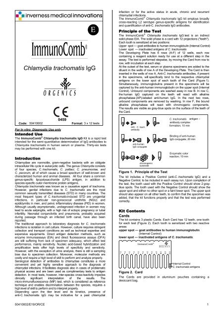
Principle of the Test<br />
The ImmunoComb ®<br />
<strong>Chlamydia</strong> <strong>trachomatis</strong> <strong>IgG</strong> test is an indirect<br />
solid-phase EIA. The solid phase is a card with 12 projections ("teeth").<br />
Each tooth is sensitized at two positions:<br />
Upper spot — goat antibodies to human immunoglobulin (Internal Control)<br />
Lower spot — inactivated antigens of C. <strong>trachomatis</strong><br />
The Developing Plate has 6 rows (A-F) of 12 wells, each row<br />
containing a reagent solution ready for use at a different step in the<br />
assay. The test is performed stepwise, by moving the Card from row to<br />
row, with incubation at each step.<br />
At the outset of the test, serum or plasma specimens are added to the<br />
diluent in the wells of row A of the Developing Plate. The Card is then<br />
inserted in the wells of row A. Anti-C. <strong>trachomatis</strong> antibodies, if present<br />
in the specimens, will specifically bind to the respective chlamydial<br />
antigens on the lower spot of each tooth of the Card (Figure 1).<br />
Simultaneously, immunoglobulins present in the specimens will be<br />
captured by the anti-human immunoglobulin on the upper spot (Internal<br />
Control). Unbound components are washed away in row B. In row C,<br />
the human <strong>IgG</strong> captured on the teeth will react with alkaline<br />
phosphatase (AP)-labeled anti-human <strong>IgG</strong>. In the next two rows,<br />
unbound components are removed by washing. In row F, the bound<br />
alkaline phosphatase will react with chromogenic components.<br />
The results are visible as gray-blue spots on the surface of the teeth of<br />
the Card.<br />
AP<br />
anti-human<br />
<strong>IgG</strong> conjugate<br />
AP<br />
anti-<br />
C. trach.<br />
antibody<br />
chromogenic<br />
substrate<br />
AP<br />
C. <strong>trachomatis</strong> antigen –<br />
antibody complex<br />
formation; 10 min<br />
Binding of anti-human<br />
<strong>IgG</strong> conjugate; 20 min<br />
Enzymatic color<br />
reaction; 10 min<br />
Figure 1. Principle of the Test<br />
The kit includes a Positive Control (anti-C. <strong>trachomatis</strong> <strong>IgG</strong>) and a<br />
Negative Control, to be included in each assay run. Upon completion of<br />
the test, the tooth used with the Positive Control should show 2 grayblue<br />
spots. The tooth used with the Negative Control should show the<br />
upper spot and either no other spot or a faint lower spot. The upper spot<br />
should also appear on all other teeth, to confirm that the specimen was<br />
added, that the kit functions properly and that the test was performed<br />
correctly.<br />
Kit Contents<br />
Cards<br />
The kit contains 3 plastic Cards. Each Card has 12 teeth, one tooth<br />
for each test (Figure 2). Each tooth is sensitized with two reactive<br />
areas:<br />
upper spot — goat antibodies to human immunoglobulin<br />
(Internal Control)<br />
lower spot — inactivated antigens of C. <strong>trachomatis</strong>.<br />
ImmunoComb ®<br />
10 10 10 10 10 10 10 10 10 10 10 10<br />
Internal Control<br />
C. <strong>trachomatis</strong> antigens<br />
Figure 2. Card<br />
The Cards are provided in aluminum pouches containing a<br />
desiccant bag.<br />
50410002/E19/OR/CE 1
Developing Plates<br />
The kit contains 3 Developing Plates covered with aluminum foil.<br />
Each Developing Plate (Figure 3) contains all reagents needed for the<br />
test. The Developing Plate consists of 6 rows (A–F) of 12 wells each.<br />
The contents of each row are as follows:<br />
Row A specimen diluent<br />
Row B washing solution<br />
Row C alkaline phosphatase - labeled goat anti-human<br />
<strong>IgG</strong> antibodies<br />
Row D washing solution<br />
Row E washing solution<br />
Row F chromogenic substrate solution containing<br />
5-bromo-4-chloro-3-indolyl phosphate (BCIP) and<br />
nitro blue tetrazolium (NBT)<br />
Figure 3. Developing Plate<br />
Positive Control — 1 vial (red-colored cap) of 0.25 ml<br />
heat-inactivated human plasma, diluted to an ImmunoComb® titer<br />
of 1:32 for anti-C. <strong>trachomatis</strong> <strong>IgG</strong>.<br />
Negative Control — 1 vial (green-colored cap) of 0.2 ml diluted,<br />
heat-inactivated human plasma, negative for antibodies to chlamydiae.<br />
Perforator — for perforation of the aluminum foil, covering the<br />
wells of the Developing Plate.<br />
CombScale — for reading test results.<br />
Safety and Precautions<br />
• Human source materials used in the preparation of the kit were<br />
tested and found to be non-reactive for hepatitis B surface<br />
antigen, and for antibodies to HIV and to hepatitis C virus. Since<br />
no test method can give complete assurance of the absence of<br />
viral contamination, all reference solutions and all human<br />
specimens should be handled as potentially infectious.<br />
• Wear surgical gloves and laboratory clothing. Follow accepted<br />
laboratory procedures for working with human serum or plasma.<br />
• Do not pipette by mouth.<br />
• Dispose of all specimens, used Cards * , Developing Plates,<br />
and other materials used with the kit as biohazardous waste.<br />
• Do not mix reagents from different lots.<br />
• Do not use kit after the expiry date.<br />
Storage and Stability of the Kit<br />
• The kit is shipped at 2 - 8 o C. During transport the kit can be<br />
kept at ≤ 30 o C for short time periods not exceeding a total of 48<br />
hours. The internal controls indicate that the kit has not been<br />
damaged during transport.<br />
• Store the kit in its original box at 2 - 8 o C.<br />
• Do not freeze the kit.<br />
• Following the first opening of the Kit the components have to be<br />
stored at 2 - 8 o C.<br />
• Performance of the Kit after the first opening is stable up to the<br />
expiry date of the Kit, when stored at 2 - 8 o C.<br />
• After first use, the card and plate cannot be used for more than<br />
three times.<br />
Handling of Specimens<br />
• You may test either serum or plasma.<br />
• Specimens may be stored for 7 days at 2°–8°C before testing.<br />
To store for more than 7 days, freeze specimens at –20°C or<br />
colder.<br />
• All frozen specimens must be centrifuged at 10,000 g for 5 min<br />
at room temperature. Carefully remove the test sample from the<br />
supernatant. If a lipid layer is formed on the surface of the<br />
liquid, ensure that the sample is taken from the clear liquid<br />
below that layer. Avoid repeated freezing and thawing.<br />
• Anti-coagulants such as Heparin, EDTA and Sodium Citrate<br />
were found to have no effect on test results.<br />
* Unless stored for documentation<br />
Test Procedure<br />
Equipment Needed<br />
• Precision pipettes with disposable tips for dispensing 10 μl<br />
• Scissors<br />
• Laboratory timer or watch<br />
Preparing the Test<br />
Bring all components, developing plates, cards, reagents and<br />
specimens to room temperature and perform the test at room<br />
temperature (22°-26°C).<br />
Preparing the Developing Plate<br />
1. Incubate the Developing Plate in an incubator at 37°C for<br />
30 minutes; or leave at room temperature (22°-26°C) for<br />
3 hours. Bring all reagents (cards, samples, sample diluent) at<br />
room temperature.<br />
2. Cover the work table with absorbent tissue to be discarded as<br />
biohazardous waste at the end of the test.<br />
3. Mix the reagents by shaking the Developing Plate.<br />
Note: Do not remove the foil cover of the Developing Plate.<br />
Break the foil cover by using the perforator, or the disposable tip of<br />
the pipette, only when instructed to do so by the Test Instructions.<br />
Preparing the Card<br />
Caution: To ensure proper functioning of the test, do not touch the<br />
teeth of the Card.<br />
1. Tear the aluminum pouch of the Card at the notched edge.<br />
Remove the Card.<br />
2. You may use the entire Card and Developing Plate or only a<br />
part. To use part of a Card:<br />
a. Determine how many teeth you need for testing the<br />
specimens and controls. You need one tooth for each test.<br />
Each tooth displays the code number "10" of the kit,<br />
to enable identification of detached teeth.<br />
b. Bend and break the Card vertically or cut with scissors<br />
(see Figure 4) to detach the required number of teeth<br />
(No. of tests including 2 controls).<br />
c. Return the unused portion of the Card to the aluminum<br />
pouch (with desiccant bag). Close pouch tightly, e.g.,<br />
with a paper clip, to maintain dryness. Store the Card in<br />
the original kit box at 2°–8°C for later use.<br />
ImmunoComb ®<br />
10 10 10 10 10 10 10 10 10 10 10 10<br />
Figure 4. Breaking the Card<br />
Test Instructions<br />
Antigen–Antibody Reaction (Row A of the Developing Plate)<br />
1. Pipette 10 μl of a specimen. Perforate the foil cover of one well<br />
of row A of the Developing Plate with the pipette tip or<br />
perforator and dispense the specimen at the bottom of the well.<br />
Mix by repeatedly refilling and ejecting the solution. Discard<br />
pipette tip.<br />
2. Repeat step 1 for the other specimens and the two controls.<br />
Use a new well in row A and change pipette tips for each<br />
specimen or control.<br />
3. a. Insert the Card (printed side facing you) into the wells<br />
of row A containing specimens and controls.<br />
Mix: Withdraw and insert the Card in the wells several times.<br />
b. Leave the Card in row A for 10 minutes. Set the timer.<br />
Near the end of 10 minutes, perforate the foil of row B using<br />
the perforator. Do not open more wells than needed.<br />
c. At the end of 10 minutes, take the Card out of row A. Absorb<br />
adhering liquid from the pointed tips of the teeth on clean<br />
absorbent paper. Do not touch the front surface of the teeth.<br />
First Wash (Row B)<br />
4. Insert the Card into the wells of row B. Agitate:<br />
Vigorously withdraw and insert the Card in the wells for at least 10<br />
seconds to achieve proper washing. Repeat agitation several times<br />
during the course of 2 minutes; meanwhile perforate the foil of row<br />
C. After 2 minutes, withdraw the Card and absorb adhering liquid<br />
as in step 3c.<br />
50410002/E19/OR/CE 2
Binding of Conjugate (Row C)<br />
5. Insert the Card into the wells of row C. Mix as in step 3a. Set timer<br />
for 20 minutes. Perforate the foil of row D. After 20 minutes,<br />
withdraw the Card and absorb adhering liquid.<br />
Second Wash (Row D)<br />
6. Insert the Card into the wells of row D. Repeatedly agitate during<br />
2 minutes, as in step 4. Meanwhile perforate the foil of row E. After<br />
2 minutes, withdraw the Card and absorb adhering liquid.<br />
Third Wash (Row E)<br />
7. Insert the Card into the wells of row E. Repeatedly agitate during<br />
2 minutes. Meanwhile perforate the foil of row F. After 2 minutes,<br />
withdraw the Card and absorb adhering liquid.<br />
Color Reaction (Row F)<br />
8. Insert the Card into the wells of row F. Mix. Set the timer for<br />
10 minutes. After 10 minutes, withdraw the Card.<br />
Stop Reaction (Row E)<br />
9. Insert the Card again into row E. After 1 minute, withdraw the Card<br />
and allow it to dry in the air.<br />
Storing Unused Part of Kit<br />
Developing Plate<br />
If you have not used all the wells of the Developing Plate, you may<br />
store it for future use:<br />
• Seal used wells with wide adhesive tape so that nothing can spill<br />
out of the wells, even if the Developing Plate is tipped over.<br />
Other Kit Materials<br />
• Return remaining Developing Plate(s), Card(s), perforator,<br />
controls, and instructions to the original kit box. Store at 2º– 8ºC.<br />
Test Results<br />
Validation<br />
In order to confirm the proper functioning of the test and to<br />
demonstrate that the results are valid, each one of the following<br />
four conditions must be fulfilled (see Figure 5):<br />
1. The Positive Control must produce two spots on the Card<br />
tooth.<br />
2. The signal of the lower spot of the Positive Control should be<br />
approximately equal to the second color frame starting from the<br />
left, if assessed using a CombScale.<br />
3. The Negative Control must produce an upper spot (Internal<br />
Control). The lower spot will either not appear or appear faintly,<br />
without affecting the interpretation of the results.<br />
4. Each specimen tested must produce an upper spot (Internal<br />
Control). This will also confirm that the specimen was added.<br />
If any of the four conditions are not fulfilled, the results are invalid,<br />
and the specimens and controls should be retested.<br />
Positive<br />
Controle<br />
Negative<br />
Controle<br />
Figure 5. Test Validation<br />
Invalid<br />
Result<br />
Reading and Interpretation of the Results<br />
Semiquantitative Interpretation by Visual Reading<br />
The level of species-specific antichlamydial <strong>IgG</strong> in each specimen may<br />
be assessed by comparing the color intensity of the lower spot on each<br />
tooth, with the color scale on the CombScale provided with the kit.<br />
This is performed as follows (Figure 6):<br />
1. Calibrate the CombScale for assessment of the<br />
anti-C. <strong>trachomatis</strong> <strong>IgG</strong> titers. Place the lower spot on the<br />
Positive Control tooth underneath the most similar color<br />
intensity of the color scale. Adjust the ruler so that "1/32; C+"<br />
appears in the window above the selected color intensity.<br />
2. Read results without changing the calibrated position of the ruler.<br />
Match the color intensity of each lower spot with the most similar<br />
intensity on the color scale. Record the value displayed in the window<br />
above that intensity, as the approximate titer of <strong>IgG</strong> antibodies to<br />
C. <strong>trachomatis</strong> for the corresponding specimen.<br />
Calibrate the<br />
CombScale<br />
Orgenics CombScale<br />
CHLAMYDIA TRACHOMATIS <strong>IgG</strong><br />
1/ 1/ 1/ 1/ 1/<br />
16 32 64 128 256<br />
C+<br />
Figure 6. CombScale<br />
Interpretation of Results<br />
Table 1. Interpretation of Results<br />
ImmunoComb® Titer<br />
Interpretation<br />
< 1:16 Negative<br />
1:16 Weakly positive<br />
≥ 1:32<br />
Positive<br />
Either ≥ 1:64 or a 4-fold<br />
increase in titer of specimens<br />
collected at a 2 - 3 weeks<br />
interval*<br />
Strongly positive, indicating<br />
active chlamydial infection<br />
*After 2 - 3 weeks collect new serum specimen and test both first and second specimens<br />
simultaneously.<br />
Note:<br />
• Simultaneous testing for C. <strong>trachomatis</strong> IgA antibodies, which are<br />
more prominent in active infections, is highly recommended.<br />
Documentation of Results<br />
As the color developed on the Card is stable, you may store the<br />
Cards for documentation.<br />
Limitations<br />
As with other tests intended for in vitro diagnostic use, the results of<br />
this test should be evaluated in relation to all symptoms, clinical<br />
history and other laboratory findings for the patient.<br />
In addition, some degree of cross-reaction with <strong>Chlamydia</strong><br />
pneumoniae may occur.<br />
Performance Characteristics*<br />
Sensitivity and Specificity of the ImmunoComb ® <strong>Chlamydia</strong><br />
<strong>trachomatis</strong> <strong>IgG</strong> have been evaluated in several laboratories.<br />
A total of 748 samples were analysed and compared with MIF as<br />
reference.<br />
The results are summarized in Table 2:<br />
Table 2. Test results of Clinical Evaluations<br />
ImmunoComb ® <strong>Chlamydia</strong> <strong>trachomatis</strong> <strong>IgG</strong><br />
Reference Assays Positive Negative<br />
Positive 318 29<br />
Negative 60 341<br />
Sensitivity – 91.6 %<br />
Specificity – 85 % (based on a study of 401 blood donors)<br />
Repeatability<br />
Ten cards were chosen at random from various parts of a<br />
production lot. One serum was assayed 12 times on these<br />
10 cards. In all cards the same <strong>IgG</strong> titer was observed for<br />
<strong>Chlamydia</strong> <strong>trachomatis</strong>.<br />
Reproducibility<br />
Three samples were assayed on cards taken from three different<br />
production lots. The specimens were assayed in duplicates. In all<br />
cards the same <strong>IgG</strong> titer was observed for <strong>Chlamydia</strong> <strong>trachomatis</strong>.<br />
Cross Reaction<br />
Cross-reactivity with positive samples for other pathologies<br />
(Mycoplasma Pneumoniae, HBs Ag, HIV, Rubella, EBV,<br />
Rheumatoid Factor, Auto Immune Diseases, HCV, Legionella and<br />
Coxiella burneti) was found to be insignificant.<br />
Very slight interferences with samples positive for CMV IgM,<br />
Toxoplasmosis <strong>IgG</strong>, <strong>Chlamydia</strong> Pneumoniae and <strong>Chlamydia</strong><br />
Psitacci cannot be excluded.<br />
* Detailed data available upon request<br />
50410002/E19/OR/CE 3
Interference<br />
Interference with hemolytic (hemoglobin up to 10 mg/ml), lipemic<br />
(cholesterol up to 281.6 mg/dl; triglycerides 381 mg/dl) and high<br />
bilirubin (up to 20 mg/dl) samples was found to be insignificant.<br />
Bibliography<br />
Barnes RC 1989. Laboratory diagnosis of human chlamydial<br />
infections. Clin Microbiol Rev 2:119-136.<br />
Bjercke S, Purvis K. 1993. Characteristics of women under fertility<br />
investigation with IgA/ <strong>IgG</strong> seropositivity for <strong>Chlamydia</strong> <strong>trachomatis</strong>.<br />
Eur J Obstet Gynecol Reprod Biol 51:157-161.<br />
Chutivongse S, Kozuh-Novak C, Annus J, Ward M, Cates Jr W,<br />
Rowe PJ, Farley TMM. WHO task force on the prevention and<br />
management of infertility. 1995 Tubal infertility: Serological<br />
relationship to past chlamydial and gonococcal infection.<br />
Sex Transm Dis 22:71-77.<br />
Clad A., Flecken U, Petersen EE. 1994. <strong>Chlamydia</strong>l serology in<br />
genital infections: InmunoComb versus Ipazyme.<br />
Infection 22:165-173.<br />
Csángo PA, Sarov B, Schiotz, H, Sarov I. 1988. Comparison<br />
between cell culture and serology for detecting <strong>Chlamydia</strong><br />
<strong>trachomatis</strong> in women seeking abortion. J Clin Pathol 41:89-92.<br />
Moss T, Darougar S, Woodland R, Nathan M, Dines RJ,<br />
Cathrine V. 1993. Antibodies to <strong>Chlamydia</strong> species in patients<br />
attending a genitourinary clinic and the impact of antibodies to C.<br />
pneumoniae and C. psittaci on the sensitivity and the specificity of<br />
C. <strong>trachomatis</strong> serology tests. Sex Transm Dis 20:61-65.<br />
Odland JØ, Ånestad G, Rasmussen S, Lungren, Dalaker K.<br />
1993. Ectopic pregnancy and chlamydial serology. Int J. Gynaecol<br />
Obstet 43:271-275.<br />
Samra Z, Sofer Y. 1992. <strong>IgG</strong> antichlamydia antibodies as a<br />
diagnostic tool for monitoring of active chlamydial infection. Eur J<br />
Epidemiol 8:882-884.<br />
Sarov I, Kleinman D, Holoman D, Potashnik G, Insler V, Cevenini<br />
R, Sarov B. 1986. Specific <strong>IgG</strong> and IgA antibodies to <strong>Chlamydia</strong><br />
<strong>trachomatis</strong> in infertile women. Int J Fertil 31: 193-197.<br />
Schachter J. 1991. <strong>Chlamydia</strong>e In: Balows A, Hausler WJ,<br />
Herrmann KL, Isenberg HD, Shadomy HJ, eds. Manual of Clinical<br />
Microbiology, Fifth edition. American Society for Microbiology,<br />
Washington, DC. pp. 1045-1058.<br />
Sellors JW, Mahony JB, Chernesky MA, Rath DJ. 1988. Tubal<br />
factor infertility: an association with prior chlamydial infection and<br />
asymptomatic salpingitis. Fertil Steril 49:451-457.<br />
Sweet RL, Schachter J, Landers DV. 1983. <strong>Chlamydia</strong>l infections<br />
in obstetrics and gynecology. Clin Obstet Gynec 26:143-164.<br />
Theunissen JJH, Minderhout-Basssie W, Wagenvoort JHT,<br />
Stolz E, Michel MF, Huikeshoven FJM. 1994. <strong>Chlamydia</strong><br />
<strong>trachomatis</strong>-specific antibodies in patients with pelvic inflammatory<br />
disease: comparison with isolation in tissue culture or detection<br />
with polymerase chain reaction. Genitourin Med 70:304-307.<br />
Symbols Legend<br />
IVD<br />
Σ<br />
n<br />
ImmunoComb® Card<br />
Developing Plate<br />
Positive Control<br />
Negative Control<br />
Perforator<br />
CombScale <br />
Consult Instructions for Use<br />
Caution,consult accompanying<br />
documents<br />
In Vitro Diagnostic Medical<br />
Device<br />
Temperature limitation<br />
Contains sufficient for n tests<br />
Manufacturer<br />
Authorized Representative in the<br />
European Community<br />
Catalogue number<br />
Batch code<br />
Use by<br />
Serial number<br />
Version: 50410002/E19/OR/CE<br />
(03/2009)<br />
50410002/E19/OR/CE 4
Summary of Main Test Procedures<br />
Preincubate the Developing Plate:<br />
3 hrs. at room temperature or 20 min.<br />
at 37°C<br />
1 2 3 4<br />
Draw specimens and<br />
controls<br />
Add specimens and<br />
controls to row A. Mix<br />
Remove Card from pouch<br />
Insert Card and mix in row A.<br />
Incubate<br />
5 6 7 8<br />
Open row B<br />
Absorb adhering liquid<br />
from teeth<br />
Insert Card and agitate in<br />
row B. Incubate<br />
After mixing/agitating &<br />
incubating in rows<br />
C, D and E…<br />
9 10<br />
Color reaction in row F Results<br />
Summary of the Test Procedure<br />
The abbreviated instructions below are for experienced users of the ImmunoComb ®<br />
<strong>Chlamydia</strong> <strong>trachomatis</strong> <strong>IgG</strong> Kit.<br />
(For detailed instructions please refer to complete text inside)<br />
1. Bring all reagents and specimens to room temperature and perform the test at room<br />
temperature.<br />
2. Dispense 10 μl of each specimen and the two controls into the wells of row A of the<br />
Developing Plate and mix.<br />
3. Insert Card in row A and continue as described in Table. 1.<br />
Table 1. Summary of test procedure<br />
Step Row Proceed as follows<br />
Antigen-antibody reaction A Mix; incubate 10 minutes; absorb.<br />
Wash B Agitate; incubate 2 minutes; absorb.<br />
Binding of conjugate C Mix; incubate 20 minutes; absorb.<br />
Wash D Agitate; incubate 2 minutes; absorb.<br />
Wash E Agitate; incubate 2 minutes; absorb.<br />
Color reaction F Mix; incubate 10 minutes.<br />
Stop reaction E Incubate 1 minute; dry in air.<br />
A<br />
B<br />
Bending and breaking the<br />
Card<br />
50410002/E19/OR/CE 5


