
You also want an ePaper? Increase the reach of your titles
YUMPU automatically turns print PDFs into web optimized ePapers that Google loves.
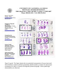
<strong>Protocol</strong> for immunohistochemical staining of Paraffin sections to unmask<br />
epitopes (AR) with amplification, using catalyzed signal amplification<br />
A. Tissue Controls:<br />
1. Positive control: Cells or tissue expected to be positive. Positive controls may also be the<br />
cells that are know to express the epitope, that are grown in tissue culture up to about 100<br />
million, taken and made into a pellet, fixed, paraffin embedded and sectioned.<br />
2. Negative control: Cells or tissue expected to be negative.<br />
3. Blocking peptide (see below)<br />
B. Reagent controls:<br />
1. 1% BSA/PBS reagent negative control for secondary reagents<br />
2. Rabbit IgG Dako Cat.# 1505 Negative control reagent at 5 ug/ml<br />
3. Rabbit anti human vWf Dako Cat# 0936 (pre-diluted). Positive for blood vessels in tissue.<br />
4. Mouse anti Vimentin Dako Catalog# N1521 prediluted, ready to use, as a technical positive<br />
control when using mouse antibodies as test<br />
5. Mouse IgG at 5 ug/ml<br />
6. Further controls included staining of cells expressing higher levels of epitope in question, and<br />
using the immunizing peptide (1 mg/ml) at a 1:50 dilution to compete out the reactivity of the<br />
antibody, resulting in a negative stain.<br />
C. Deparaffinization:<br />
----Label slides with date and reagent to be applied and take to the fume hood where the<br />
deparaffinization will occur<br />
-----BMRUAD= briefly move rack up and down. Move slide holder up and down so that the slides get<br />
“washed” before leaving the slide rack in the dish for the specified amount of time.<br />
1. Dip into Xylene (in green tub in the fume hood) ---BMRUAD and then leave for 10 minutes, and<br />
repeat two more times<br />
2. Dip into 100% alcohol, BMRUAD and then leave for 5 minutes, and repeat one more time<br />
3. Dip into 95% alcohol, BMRUAD and then leave for 5 minutes, and repeat one more time<br />
4. Dip into 70% alcohol, BMRUAD and then leave for 5 minutes, and repeat one more time<br />
5. Dip into TBST buffer, BMRUAD and then leave for 5 minutes, and repeat two more times<br />
TBST= Tris buffered saline with 0.05% Tween 20<br />
May also use PBS pH 7.1 but TBST helps to disperse the reagent evenly over entire section<br />
D. Antigen Retrieval AR (should use the steamer because the microwave method is patented)<br />
1. fill green tub with AR buffer of choice<br />
2. fill slide holder with “dummy” slides and intersperse the test slides among the dummy slides<br />
so that the slide holder is full of slides—this allows even heating<br />
3. cover and place in larger container with water that goes halfway up the tub, and heat for 5<br />
minutes on high.<br />
4. check levels and repeat a second time, for 5 more minutes<br />
5. remove slide container from microwave, (Careful—IT WILL BE HOT) remove lid and let the<br />
container sit on bench top for 20 minutes, to cool<br />
6. Dip into TBST buffer, BMRUAD and then leave for 5 minutes, Repeat two more times<br />
E. Block ENDOGENOUS PEROXIDASES . Immerse slides in slide holder in 0.3% H 2 O 2 in PBS for<br />
30 minutes at room temperature, followed with 3 buffer washes