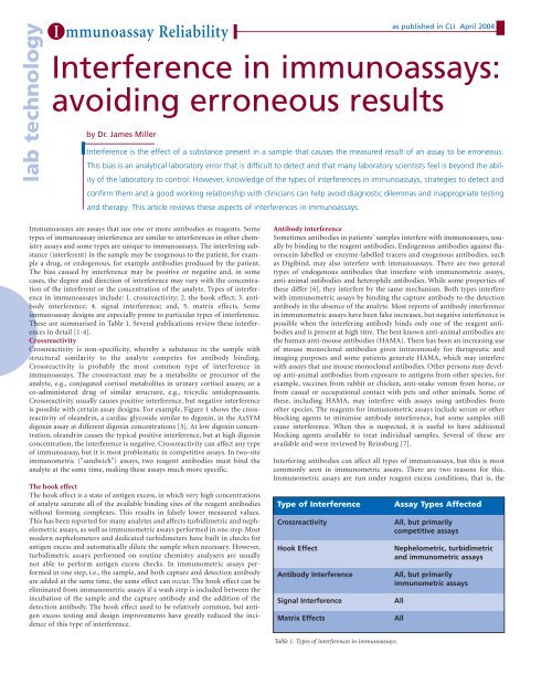
Interference in immunoassays: avoiding erroneous results
Interference in immunoassays: avoiding erroneous results
Interference in immunoassays: avoiding erroneous results

Create successful ePaper yourself
Turn your PDF publications into a flip-book with our unique Google optimized e-Paper software.
lab technology<br />
I mmunoassay Reliability<br />
<strong>Interference</strong> <strong>in</strong> <strong>immunoassays</strong>:<br />
avoid<strong>in</strong>g <strong>erroneous</strong> <strong>results</strong><br />
by Dr. James Miller<br />
as published <strong>in</strong> CLI April 2004<br />
<strong>Interference</strong> is the effect of a substance present <strong>in</strong> a sample that causes the measured result of an assay to be <strong>erroneous</strong>.<br />
This bias is an analytical laboratory error that is difficult to detect and that many laboratory scientists feel is beyond the ability<br />
of the laboratory to control. However, knowledge of the types of <strong>in</strong>terferences <strong>in</strong> <strong>immunoassays</strong>, strategies to detect and<br />
confirm them and a good work<strong>in</strong>g relationship with cl<strong>in</strong>icians can help avoid diagnostic dilemmas and <strong>in</strong>appropriate test<strong>in</strong>g<br />
and therapy. This article reviews these aspects of <strong>in</strong>terferences <strong>in</strong> <strong>immunoassays</strong>.<br />
Immunoassays are assays that use one or more antibodies as reagents. Some<br />
types of immunoassay <strong>in</strong>terference are similar to <strong>in</strong>terferences <strong>in</strong> other chemistry<br />
assays and some types are unique to <strong>immunoassays</strong>. The <strong>in</strong>terfer<strong>in</strong>g substance<br />
(<strong>in</strong>terferent) <strong>in</strong> the sample may be exogenous to the patient, for example<br />
a drug, or endogenous, for example antibodies produced by the patient.<br />
The bias caused by <strong>in</strong>terference may be positive or negative and, <strong>in</strong> some<br />
cases, the degree and direction of <strong>in</strong>terference may vary with the concentration<br />
of the <strong>in</strong>terferent or the concentration of the analyte. Types of <strong>in</strong>terference<br />
<strong>in</strong> <strong>immunoassays</strong> <strong>in</strong>clude: 1. crossreactivity; 2. the hook effect; 3. antibody<br />
<strong>in</strong>terference; 4. signal <strong>in</strong>terference; and, 5. matrix effects. Some<br />
immunoassay designs are especially prone to particular types of <strong>in</strong>terference.<br />
These are summarised <strong>in</strong> Table 1. Several publications review these <strong>in</strong>terferences<br />
<strong>in</strong> detail [1-4].<br />
Crossreactivity<br />
Crossreactivity is non-specificity, whereby a substance <strong>in</strong> the sample with<br />
structural similarity to the analyte competes for antibody b<strong>in</strong>d<strong>in</strong>g.<br />
Crossreactivity is probably the most common type of <strong>in</strong>terference <strong>in</strong><br />
<strong>immunoassays</strong>. The crossreactant may be a metabolite or precursor of the<br />
analyte, e.g., conjugated cortisol metabolites <strong>in</strong> ur<strong>in</strong>ary cortisol assays; or a<br />
co-adm<strong>in</strong>istered drug of similar structure, e.g., tricyclic antidepressants.<br />
Crossreactivity usually causes positive <strong>in</strong>terference, but negative <strong>in</strong>terference<br />
is possible with certa<strong>in</strong> assay designs. For example, Figure 1 shows the crossreactivity<br />
of oleandr<strong>in</strong>, a cardiac glycoside similar to digox<strong>in</strong>, <strong>in</strong> the AxSYM<br />
digox<strong>in</strong> assay at different digox<strong>in</strong> concentrations [5]. At low digox<strong>in</strong> concentration,<br />
oleandr<strong>in</strong> causes the typical positive <strong>in</strong>terference, but at high digox<strong>in</strong><br />
concentration, the <strong>in</strong>terference is negative. Crossreactivity can affect any type<br />
of immunoassay, but it is most problematic <strong>in</strong> competitive assays. In two-site<br />
immunometric ("sandwich") assays, two reagent antibodies must b<strong>in</strong>d the<br />
analyte at the same time, mak<strong>in</strong>g these assays much more specific.<br />
The hook effect<br />
The hook effect is a state of antigen excess, <strong>in</strong> which very high concentrations<br />
of analyte saturate all of the available b<strong>in</strong>d<strong>in</strong>g sites of the reagent antibodies<br />
without form<strong>in</strong>g complexes. This <strong>results</strong> <strong>in</strong> falsely lower measured values.<br />
This has been reported for many analytes and affects turbidimetric and nephelometric<br />
assays, as well as immunometric assays performed <strong>in</strong> one step. Most<br />
modern nephelometers and dedicated turbidimeters have built <strong>in</strong> checks for<br />
antigen excess and automatically dilute the sample when necessary. However,<br />
turbidimetric assays performed on rout<strong>in</strong>e chemistry analysers are usually<br />
not able to perform antigen excess checks. In immunometric assays performed<br />
<strong>in</strong> one step, i.e., the sample, and both capture and detection antibody<br />
are added at the same time, the same effect can occur. The hook effect can be<br />
elim<strong>in</strong>ated from immunometric assays if a wash step is <strong>in</strong>cluded between the<br />
<strong>in</strong>cubation of the sample and the capture antibody and the addition of the<br />
detection antibody. The hook effect used to be relatively common, but antigen<br />
excess test<strong>in</strong>g and design improvements have greatly reduced the <strong>in</strong>cidence<br />
of this type of <strong>in</strong>terference.<br />
Antibody <strong>in</strong>terference<br />
Sometimes antibodies <strong>in</strong> patients' samples <strong>in</strong>terfere with <strong>immunoassays</strong>, usually<br />
by b<strong>in</strong>d<strong>in</strong>g to the reagent antibodies. Endogenous antibodies aga<strong>in</strong>st fluoresce<strong>in</strong>-labelled<br />
or enzyme-labelled tracers and exogenous antibodies, such<br />
as Digib<strong>in</strong>d, may also <strong>in</strong>terfere with <strong>immunoassays</strong>. There are two general<br />
types of endogenous antibodies that <strong>in</strong>terfere with immunometric assays,<br />
anti-animal antibodies and heterophile antibodies. While some properties of<br />
these differ [6], they <strong>in</strong>terfere by the same mechanism. Both types <strong>in</strong>terfere<br />
with immunometric assays by b<strong>in</strong>d<strong>in</strong>g the capture antibody to the detection<br />
antibody <strong>in</strong> the absence of the analyte. Most reports of antibody <strong>in</strong>terference<br />
<strong>in</strong> immunometric assays have been false <strong>in</strong>creases, but negative <strong>in</strong>terference is<br />
possible when the <strong>in</strong>terfer<strong>in</strong>g antibody b<strong>in</strong>ds only one of the reagent antibodies<br />
and is present at high titre. The best known anti-animal antibodies are<br />
the human anti-mouse antibodies (HAMA). There has been an <strong>in</strong>creas<strong>in</strong>g use<br />
of mouse monoclonal antibodies given <strong>in</strong>travenously for therapeutic and<br />
imag<strong>in</strong>g purposes and some patients generate HAMA, which may <strong>in</strong>terfere<br />
with assays that use mouse monoclonal antibodies. Other persons may develop<br />
anti-animal antibodies from exposure to antigens from other species, for<br />
example, vacc<strong>in</strong>es from rabbit or chicken, anti-snake venom from horse, or<br />
from casual or occupational contact with pets and other animals. Some of<br />
these, <strong>in</strong>clud<strong>in</strong>g HAMA, may <strong>in</strong>terfere with assays us<strong>in</strong>g antibodies from<br />
other species. The reagents for immunometric assays <strong>in</strong>clude serum or other<br />
block<strong>in</strong>g agents to m<strong>in</strong>imise antibody <strong>in</strong>terference, but some samples still<br />
cause <strong>in</strong>terference. When this is suspected, it is useful to have additional<br />
block<strong>in</strong>g agents available to treat <strong>in</strong>dividual samples. Several of these are<br />
available and were reviewed by Re<strong>in</strong>sburg [7].<br />
Interfer<strong>in</strong>g antibodies can affect all types of <strong>immunoassays</strong>, but this is most<br />
commonly seen <strong>in</strong> immunometric assays. There are two reasons for this.<br />
Immunometric assays are run under reagent excess conditions, that is, the<br />
Type of <strong>Interference</strong><br />
Crossreactivity<br />
Hook Effect<br />
Antibody <strong>Interference</strong><br />
Signal <strong>Interference</strong><br />
Matrix Effects<br />
Assay Types Affected<br />
All, but primarily<br />
competitive assays<br />
Nephelometric, turbidimetric<br />
and immunometric assays<br />
All, but primarily<br />
immunometric assays<br />
All<br />
All<br />
Table 1. Types of <strong>in</strong>terferences <strong>in</strong> <strong>immunoassays</strong>.
I mmunoassay Reliability<br />
as published <strong>in</strong> CLI April 2004<br />
concentration of both reagent antibodies is much greater than<br />
the usual concentration of analyte. This drives the reaction to<br />
completion, mak<strong>in</strong>g this type of assay very rapid and analytically<br />
sensitive. However, any antibodies <strong>in</strong> the sample that<br />
have even weak aff<strong>in</strong>ity for both reagent antibodies may cause<br />
measurable complexes to form <strong>in</strong> the absence of analyte. In<br />
addition, a large number of these assays are performed.<br />
Almost all analytes large enough to b<strong>in</strong>d two antibodies<br />
simultaneously are assayed by immunometric assays and a few<br />
of these assays, e.g., chorionic gonadotrop<strong>in</strong>, thyrotrop<strong>in</strong>, and<br />
cardiac markers, are performed on large numbers of <strong>in</strong>dividuals.<br />
The reported frequency of heterophile and anti-animal<br />
antibodies varies greatly depend<strong>in</strong>g on the method of detection.<br />
Heterophile antibodies are present <strong>in</strong> more than 10% of<br />
patients [8] and 41% of patients who received <strong>in</strong>jections of<br />
mur<strong>in</strong>e monoclonal antibodies developed HAMA [9].<br />
However, with adequate block<strong>in</strong>g agent <strong>in</strong> the assay the frequency<br />
of cl<strong>in</strong>ically significant <strong>in</strong>terference may be less than<br />
0.05% [6, 10].<br />
Apparent Digox<strong>in</strong> (nmol/L)<br />
Other types of <strong>in</strong>terference<br />
The two other types of <strong>in</strong>terference <strong>in</strong> <strong>immunoassays</strong> are signal<br />
<strong>in</strong>terference and matrix effects. Some samples may conta<strong>in</strong><br />
compounds that artifactually <strong>in</strong>crease or decrease the magnitude<br />
of the detection mechanism, e.g., fluorescence. This is<br />
called signal <strong>in</strong>terference. Because there are many types of<br />
<strong>in</strong>dicator mechanisms used <strong>in</strong> <strong>immunoassays</strong>, there are many<br />
subtypes of signal <strong>in</strong>terference. The matrix of a sample is the<br />
environment of the analyte and <strong>in</strong>cludes properties like pH,<br />
ionic strength, and the prote<strong>in</strong> and lipid concentrations of the<br />
sample. Matrix effects are caused by variations <strong>in</strong> the reactivity<br />
of the analyte due to variations <strong>in</strong> its environment <strong>in</strong> the sample.<br />
Immunoassays are often quite sensitive to the matrix due to effects on antigen<br />
antibody b<strong>in</strong>d<strong>in</strong>g, efficiency of separation of bound and unbound fractions,<br />
and the extent of nonspecific b<strong>in</strong>d<strong>in</strong>g. Further details about signal<br />
<strong>in</strong>terference and matrix effects can be found <strong>in</strong> references 1-4.<br />
Detect<strong>in</strong>g and check<strong>in</strong>g <strong>in</strong>terference<br />
Detect<strong>in</strong>g <strong>in</strong>accurate <strong>results</strong> due to <strong>in</strong>terference is difficult because the true<br />
result is not known. However, there are several mechanisms that might be<br />
<strong>in</strong>cluded <strong>in</strong> an <strong>in</strong>terference screen<strong>in</strong>g program. Results from patients with<br />
cl<strong>in</strong>ical conditions commonly associated with more frequent occurrences of<br />
<strong>in</strong>terference, such as liver disease, renal failure, or pregnancy, may be scrut<strong>in</strong>ised<br />
for possible <strong>in</strong>terference. If certa<strong>in</strong> drugs are know to <strong>in</strong>terfere with a<br />
particular assay, collection of samples from patients tak<strong>in</strong>g those drugs might<br />
be timed to co<strong>in</strong>cide with trough concentrations of the drugs or delayed until<br />
the drugs are discont<strong>in</strong>ued. Samples should be <strong>in</strong>spected visually for haemolysis,<br />
icterus, and lipaemia, or better, evaluated objectively us<strong>in</strong>g serum<br />
<strong>in</strong>dices. Any <strong>results</strong> with <strong>in</strong>strument error codes should be <strong>in</strong>vestigated for<br />
possible <strong>in</strong>terference. In addition, unexpected changes over time <strong>in</strong> a patient's<br />
<strong>results</strong> (delta checks), <strong>in</strong>consistencies among physiologically related <strong>results</strong><br />
(e.g., thyrox<strong>in</strong>e and thyroid stimulat<strong>in</strong>g hormone), and exceptionally extreme<br />
<strong>results</strong> may be caused by <strong>in</strong>terferences. F<strong>in</strong>ally, <strong>in</strong>quiries from cl<strong>in</strong>icians<br />
regard<strong>in</strong>g test <strong>results</strong> that do not fit the patient's cl<strong>in</strong>ical picture may provide<br />
clues to possible <strong>in</strong>terferences.<br />
When suspect samples are identified by these mechanisms, three checks are<br />
especially helpful for decid<strong>in</strong>g whether the <strong>results</strong> are accurate: 1. test for l<strong>in</strong>ear<br />
dilution; 2. assay the sample by another method; and, 3. treat the sample<br />
to remove, destroy, or <strong>in</strong>hibit potentially <strong>in</strong>terfer<strong>in</strong>g substances. Interferents<br />
frequently dilute non-l<strong>in</strong>early and respond differently among assays, but<br />
these characteristics are not always present. In addition, one can treat the<br />
sample to remove, destroy, or <strong>in</strong>hibit the <strong>in</strong>terfer<strong>in</strong>g substance. Possible<br />
treatments <strong>in</strong>clude extraction, chromatography, ultrafiltration and addition<br />
of a heterophile block<strong>in</strong>g agent. Addition of a heterophile block<strong>in</strong>g preparation<br />
is quite easy and quick, but the other techniques may be time consum<strong>in</strong>g<br />
and their effect on the analyte needs to be known or checked with control<br />
samples.<br />
4<br />
3<br />
2<br />
1<br />
DIG 3.83 nM<br />
DIG 2.56 nM<br />
DIG 0.64 nM<br />
DIG 0.0 nM<br />
0<br />
0 30 60 90 120 150<br />
Added oleandr<strong>in</strong> (umol/L)<br />
Figure 1. Negative crossreactive <strong>in</strong>terference caused by the presence of oleandr<strong>in</strong> <strong>in</strong> the MEIA (AxSYM) digox<strong>in</strong><br />
immunoassay. Various mixtures of oleandr<strong>in</strong> and digox<strong>in</strong> were prepared and analysed. Notice that lower<br />
than expected values of digox<strong>in</strong> were measured at digox<strong>in</strong> concentrations of 2.56 and 3.83 nM. At lower digox<strong>in</strong><br />
concentrations, oleandr<strong>in</strong> exhibited typical positive <strong>in</strong>terference <strong>in</strong> this assay. Reproduced from [5] with permission.<br />
Consequences of <strong>in</strong>terference<br />
Falsely abnormal immunoassay <strong>results</strong>, whether falsely <strong>in</strong>creased or falsely<br />
decreased, suggest the presence of disease and may lead to the order<strong>in</strong>g of<br />
additional diagnostic tests and unnecessary therapy, <strong>in</strong>clud<strong>in</strong>g surgery. The<br />
most strik<strong>in</strong>g and most publicised examples have been persistently false positive<br />
chorionic gonadotrop<strong>in</strong> <strong>results</strong> due to <strong>in</strong>terfer<strong>in</strong>g antibodies suggest<strong>in</strong>g<br />
post-gestational choriocarc<strong>in</strong>oma or trophoblast disease and lead<strong>in</strong>g to<br />
unnecessary chemotherapy and surgery [11]. On the other hand, falsely normal<br />
<strong>results</strong> may contribute to an unrecognised diagnosis and failure to treat.<br />
For example, Steimer, et al. [12)]reported on a case of digox<strong>in</strong> toxicity that<br />
was masked by negative crossreactive <strong>in</strong>terference by canrenoate adm<strong>in</strong>istered<br />
simultaneously. This occurred by the same mechanism as that shown <strong>in</strong><br />
Figure 1.<br />
Manufacturers cont<strong>in</strong>ually try to improve the specificity of their assays and<br />
laboratories have begun to be more vigilant <strong>in</strong> screen<strong>in</strong>g for spurious <strong>results</strong>.<br />
These efforts have reduced the risk of diagnostic errors due to <strong>in</strong>terferences.<br />
However, this type of laboratory error cannot be abolished. Because of the<br />
<strong>in</strong>creas<strong>in</strong>g number of analytes measured by immunoassay and the <strong>in</strong>creas<strong>in</strong>g<br />
demand for rapid turn around times and analytically sensitive assays, <strong>erroneous</strong><br />
<strong>results</strong> due to <strong>in</strong>terferences will cont<strong>in</strong>ue to be a problem.<br />
Consequently, it is only through better communication between cl<strong>in</strong>icians<br />
and laboratory scientists that diagnostic misadventures and patient catastrophes<br />
are go<strong>in</strong>g to be avoided. Laboratory scientists and manufacturers need to<br />
educate cl<strong>in</strong>icians about the limitations of assay performance and the potential<br />
for <strong>in</strong>accurate <strong>results</strong>. Cl<strong>in</strong>icians should notify the laboratory when order<strong>in</strong>g<br />
tests on patients known or at risk for hav<strong>in</strong>g <strong>in</strong>terfer<strong>in</strong>g antibodies or<br />
other potential causes of <strong>in</strong>terference. Cl<strong>in</strong>icians should also consult with a<br />
laboratory scientist whenever test <strong>results</strong> do not fit the cl<strong>in</strong>ical picture. In<br />
such cases, further test<strong>in</strong>g can be arranged to <strong>in</strong>vestigate the accuracy of the<br />
<strong>results</strong>. F<strong>in</strong>ally, laboratorians should confer with the cl<strong>in</strong>ician whenever the<br />
possibility of <strong>in</strong>terference is identified by the screen<strong>in</strong>g mechanisms described<br />
above. Hopefully, through this improved collaborative dialogue between laboratory<br />
scientists and cl<strong>in</strong>icians, diagnostic dilemmas and <strong>in</strong>appropriate test<strong>in</strong>g<br />
and therapy can be avoided.
I mmunoassay Reliability<br />
as published <strong>in</strong> CLI April 2004<br />
References<br />
1. Weber TH, Kapyaho KI, Tanner P. Endogenous <strong>in</strong>terference <strong>in</strong> <strong>immunoassays</strong> <strong>in</strong><br />
cl<strong>in</strong>ical chemistry. A review. Scand J Cl<strong>in</strong> Lab Invest Suppl 1990; 201: 77-82.<br />
2. Miller JJ, Lev<strong>in</strong>son SS. <strong>Interference</strong>s <strong>in</strong> Immunoassays. In: Diamandis EP,<br />
Christopoulos TK, eds. Immunoassay. San Diego: Academic Press, 1996: 165-90.<br />
3. Selby C. <strong>Interference</strong> <strong>in</strong> immunoassay. Ann Cl<strong>in</strong> Biochem 1999; 36: 704-21.<br />
4. Miller JJ, Lev<strong>in</strong>son SS, El<strong>in</strong> RJ. <strong>Interference</strong>s <strong>in</strong> Laboratory Tests. In: Ward-Cook<br />
KM, et al., eds. Cl<strong>in</strong>ical Diagnostic Technology: The Total Test<strong>in</strong>g Process, Volume 2<br />
2004; The Analytical Phase. Wash<strong>in</strong>gton, D. C.: AACC Press, <strong>in</strong> press.<br />
5. Jortani SA, Miller JJ, Helm RA, Johnson NA, Valdes R, Jr. Suppression of immunoassay<br />
<strong>results</strong> by crossreactivity. J Cl<strong>in</strong> Ligand Assay 1997; 20(2): 177-9.<br />
6. Lev<strong>in</strong>son SS, Miller JJ. Towards a better understand<strong>in</strong>g of heterophile (and the like)<br />
antibody <strong>in</strong>terference with modern <strong>immunoassays</strong>. Cl<strong>in</strong> Chim Acta 2002; 325(1-2):<br />
1-15.<br />
7. Re<strong>in</strong>sberg J. Different efficacy of various block<strong>in</strong>g reagents to elim<strong>in</strong>ate <strong>in</strong>terferences<br />
by human antimouse antibodies with a two-site immunoassay. Cl<strong>in</strong> Biochem 1996; 29:<br />
145-8.<br />
8. Klee GG. Human anti-mouse antibodies. Arch Pathol Lab Med 2000; 124: 921-3.<br />
9. Dillman RO, Shawler DL, McCallister V, Halpern SE. Muman anti-mouse antibody<br />
response <strong>in</strong> cancer patients follow<strong>in</strong>g s<strong>in</strong>gle low-dose <strong>in</strong>jections of radiolabeled<br />
mur<strong>in</strong>e monoclonal antibodies. Cancer Biother. 1994; 9: 17-28.<br />
10. Ward G, McK<strong>in</strong>non L, Badrick T, Hickman PE. Heterophilic antibodies rema<strong>in</strong> a<br />
problem for the immunoassay laboratory. Am J Cl<strong>in</strong> Pathol 1997; 108: 417-21.<br />
11. Cole LA, R<strong>in</strong>ne KM, Shahabi S, Omrani A. False-positive hCG assay <strong>results</strong> lead<strong>in</strong>g<br />
to unnecessary surgery and chemotherapy and needless occurrences of diabetes and<br />
coma. Cl<strong>in</strong> Chem 1999; 45: 313-4.<br />
12. Steimer W, Mueller C, Eber B, Emmanuilidis K. Intoxication due<br />
to negative canrenone <strong>in</strong>terference <strong>in</strong> digox<strong>in</strong> drug monitor<strong>in</strong>g.<br />
Lancet 1999; 354: 1176-7.<br />
The author<br />
James J. Miller, Ph.D.<br />
Associate Professor of Pathology & Laboratory Medic<strong>in</strong>e<br />
Associate Director of Cl<strong>in</strong>ical Chemistry & Toxicology<br />
University of Louisville Hospital Laboratory<br />
University of Louisville School of Medic<strong>in</strong>e<br />
Louisville, KY 40292 ,USA<br />
Email: jmiller@louisville.edu