Serum free light chain ratio predicts outcome in MGUS
Serum free light chain ratio predicts outcome in MGUS
Serum free light chain ratio predicts outcome in MGUS
You also want an ePaper? Increase the reach of your titles
YUMPU automatically turns print PDFs into web optimized ePapers that Google loves.
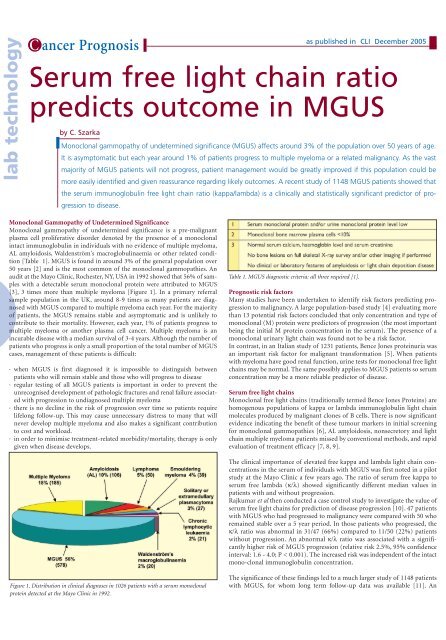
lab technologyC ancer Prognosis<strong>Serum</strong> <strong>free</strong> <strong>light</strong> <strong>cha<strong>in</strong></strong> <strong>ratio</strong><strong>predicts</strong> <strong>outcome</strong> <strong>in</strong> <strong>MGUS</strong>by C. Szarkaas published <strong>in</strong> CLI December 2005Monoclonal gammopathy of undeterm<strong>in</strong>ed significance (<strong>MGUS</strong>) affects around 3% of the population over 50 years of age.It is asymptomatic but each year around 1% of patients progress to multiple myeloma or a related malignancy. As the vastmajority of <strong>MGUS</strong> patients will not progress, patient management would be greatly improved if this population could bemore easily identified and given reassurance regard<strong>in</strong>g likely <strong>outcome</strong>s. A recent study of 1148 <strong>MGUS</strong> patients showed thatthe serum immunoglobul<strong>in</strong> <strong>free</strong> <strong>light</strong> <strong>cha<strong>in</strong></strong> <strong>ratio</strong> (kappa/lambda) is a cl<strong>in</strong>ically and statistically significant predictor of progressionto disease.Monoclonal Gammopathy of Undeterm<strong>in</strong>ed SignificanceMonoclonal gammopathy of undeterm<strong>in</strong>ed significance is a pre-malignantplasma cell proliferative disorder denoted by the presence of a monoclonal<strong>in</strong>tact immunoglobul<strong>in</strong> <strong>in</strong> <strong>in</strong>dividuals with no evidence of multiple myeloma,AL amyloidosis, Waldenström's macroglobul<strong>in</strong>aemia or other related condition[Table 1]. <strong>MGUS</strong> is found <strong>in</strong> around 3% of the general population over50 years [2] and is the most common of the monoclonal gammopathies. Anaudit at the Mayo Cl<strong>in</strong>ic, Rochester, NY, USA <strong>in</strong> 1992 showed that 56% of sampleswith a detectable serum monoclonal prote<strong>in</strong> were attributed to <strong>MGUS</strong>[3], 3 times more than multiple myeloma [Figure 1]. In a primary referralsample population <strong>in</strong> the UK, around 8-9 times as many patients are diagnosedwith <strong>MGUS</strong> compared to multiple myeloma each year. For the majorityof patients, the <strong>MGUS</strong> rema<strong>in</strong>s stable and asymptomatic and is unlikely tocontribute to their mortality. However, each year, 1% of patients progress tomultiple myeloma or another plasma cell cancer. Multiple myeloma is an<strong>in</strong>curable disease with a median survival of 3-4 years. Although the number ofpatients who progress is only a small proportion of the total number of <strong>MGUS</strong>cases, management of these patients is difficult:· when <strong>MGUS</strong> is first diagnosed it is impossible to dist<strong>in</strong>guish betweenpatients who will rema<strong>in</strong> stable and those who will progress to disease· regular test<strong>in</strong>g of all <strong>MGUS</strong> patients is important <strong>in</strong> order to prevent theunrecognised development of pathologic fractures and renal failure associatedwith progression to undiagnosed multiple myeloma· there is no decl<strong>in</strong>e <strong>in</strong> the risk of progression over time so patients requirelifelong follow-up. This may cause unnecessary distress to many that willnever develop multiple myeloma and also makes a significant contributionto cost and workload.· <strong>in</strong> order to m<strong>in</strong>imise treatment-related morbidity/mortality, therapy is onlygiven when disease develops.Table 1. <strong>MGUS</strong> diagnostic criteria: all three required [1].Prognostic risk factorsMany studies have been undertaken to identify risk factors predict<strong>in</strong>g progressionto malignancy. A large population-based study [4] evaluat<strong>in</strong>g morethan 13 potential risk factors concluded that only concent<strong>ratio</strong>n and type ofmonoclonal (M) prote<strong>in</strong> were predictors of progression (the most importantbe<strong>in</strong>g the <strong>in</strong>itial M prote<strong>in</strong> concent<strong>ratio</strong>n <strong>in</strong> the serum). The presence of amonoclonal ur<strong>in</strong>ary <strong>light</strong> <strong>cha<strong>in</strong></strong> was found not to be a risk factor.In contrast, <strong>in</strong> an Italian study of 1231 patients, Bence Jones prote<strong>in</strong>uria wasan important risk factor for malignant transformation [5]. When patientswith myeloma have good renal function, ur<strong>in</strong>e tests for monoclonal <strong>free</strong> <strong>light</strong><strong>cha<strong>in</strong></strong>s may be normal. The same possibly applies to <strong>MGUS</strong> patients so serumconcent<strong>ratio</strong>n may be a more reliable predictor of disease.<strong>Serum</strong> <strong>free</strong> <strong>light</strong> <strong>cha<strong>in</strong></strong>sMonoclonal <strong>free</strong> <strong>light</strong> <strong>cha<strong>in</strong></strong>s (traditionally termed Bence Jones Prote<strong>in</strong>s) arehomogenous populations of kappa or lambda immunoglobul<strong>in</strong> <strong>light</strong> <strong>cha<strong>in</strong></strong>molecules produced by malignant clones of B cells. There is now significantevidence <strong>in</strong>dicat<strong>in</strong>g the benefit of these tumour markers <strong>in</strong> <strong>in</strong>itial screen<strong>in</strong>gfor monoclonal gammopathies [6], AL amyloidosis, nonsecretory and <strong>light</strong><strong>cha<strong>in</strong></strong> multiple myeloma patients missed by conventional methods, and rapidevaluation of treatment efficacy [7, 8, 9].The cl<strong>in</strong>ical importance of elevated <strong>free</strong> kappa and lambda <strong>light</strong> <strong>cha<strong>in</strong></strong> concent<strong>ratio</strong>ns<strong>in</strong> the serum of <strong>in</strong>dividuals with <strong>MGUS</strong> was first noted <strong>in</strong> a pilotstudy at the Mayo Cl<strong>in</strong>ic a few years ago. The <strong>ratio</strong> of serum <strong>free</strong> kappa toserum <strong>free</strong> lambda (κ/λ) showed significantly different median values <strong>in</strong>patients with and without progression.Rajkumar et al then conducted a case control study to <strong>in</strong>vestigate the value ofserum <strong>free</strong> <strong>light</strong> <strong>cha<strong>in</strong></strong>s for prediction of disease progression [10]. 47 patientswith <strong>MGUS</strong> who had progressed to malignancy were compared with 50 whorema<strong>in</strong>ed stable over a 5 year period. In those patients who progressed, theκ/λ <strong>ratio</strong> was abnormal <strong>in</strong> 31/47 (66%) compared to 11/50 (22%) patientswithout progression. An abnormal κ/λ <strong>ratio</strong> was associated with a significantlyhigher risk of <strong>MGUS</strong> progression (relative risk 2.5%, 95% confidence<strong>in</strong>terval: 1.6 - 4.0; P < 0.001). The <strong>in</strong>creased risk was <strong>in</strong>dependent of the <strong>in</strong>tactmono-clonal immunoglobul<strong>in</strong> concent<strong>ratio</strong>n.Figure 1. Distribution <strong>in</strong> cl<strong>in</strong>ical diagnoses <strong>in</strong> 1026 patients with a serum monoclonalprote<strong>in</strong> detected at the Mayo Cl<strong>in</strong>ic <strong>in</strong> 1992.The significance of these f<strong>in</strong>d<strong>in</strong>gs led to a much larger study of 1148 patientswith <strong>MGUS</strong>, for whom long term follow-up data was available [11]. An
C ancer Prognosisas published <strong>in</strong> CLI December 2005Table 2. Reference ranges for serum <strong>free</strong> kappa and lambda <strong>light</strong> <strong>cha<strong>in</strong></strong>s.abnormal <strong>free</strong> <strong>light</strong> <strong>cha<strong>in</strong></strong> <strong>ratio</strong> (kappa/lambda1.65) [Table 2] was detected <strong>in</strong> 379(33%) patients. This group had a significantlyhigher risk of progression compared to those witha normal κ/λ <strong>ratio</strong> [Figure 2], (hazard <strong>ratio</strong>, 3.5;95% confidence <strong>in</strong>terval, 2.2 - 5.5; P 50 years) as part of an epidemiologicalstudy [12]. These were selected on thebasis of negative serum prote<strong>in</strong> electrophoresis(SPE). When assessed for serum <strong>free</strong> <strong>light</strong> <strong>cha<strong>in</strong></strong>s12 of the samples were abnormal. These 12 serawith abnormal κ/λ <strong>ratio</strong>s were carefullyreassessed by immunofixation electrophoresis. Ofthese, it is likely that 7 had detectable monoclonal<strong>free</strong> <strong>light</strong> <strong>cha<strong>in</strong></strong>s, possibly represent<strong>in</strong>g a previouslyunknown entity of <strong>free</strong> <strong>light</strong> <strong>cha<strong>in</strong></strong> <strong>MGUS</strong>.Two patients with <strong>in</strong>tact immunoglobul<strong>in</strong> M prote<strong>in</strong>smissed <strong>in</strong> the orig<strong>in</strong>al SPE tests were identifiedby the serum <strong>free</strong> <strong>light</strong> <strong>cha<strong>in</strong></strong> assays.Long term follow-up studies are needed to determ<strong>in</strong>ethe significance of <strong>free</strong> <strong>light</strong> <strong>cha<strong>in</strong></strong> <strong>MGUS</strong>, tosee whether these patients progress to <strong>light</strong> <strong>cha<strong>in</strong></strong>multiple myeloma or if the risk of progression isFigure 2. Risk of progression <strong>in</strong> patients with normal and abnormal κ/λ <strong>ratio</strong>s. Theupper curve illustrates risk of progression of monoclonal gammopathy of undeterm<strong>in</strong>edsignificance <strong>in</strong> patients with an abnormal kappa/lambda <strong>free</strong> <strong>light</strong> <strong>cha<strong>in</strong></strong> <strong>ratio</strong> (1.65). The lower curve illustrates the risk of progression <strong>in</strong> patients with a normal<strong>ratio</strong>.
C ancer Prognosisas published <strong>in</strong> CLI December 2005Table 3. Risk stratification model.similar to <strong>in</strong>tact immunoglobul<strong>in</strong> <strong>MGUS</strong>.Table 4. Proposal for <strong>MGUS</strong> patient management.*Younger low-risk patients should be managed as for <strong>in</strong>termediate risk.** Increased risk associated with ris<strong>in</strong>g and more abnormal κ/λ <strong>ratio</strong>.Guidel<strong>in</strong>es for diagnosis and monitor<strong>in</strong>g of<strong>MGUS</strong> patients are currently under review by severalwork<strong>in</strong>g parties.References1. Durie BG et al. Myeloma management guidel<strong>in</strong>es: aconsensus report from the Scientific Advisors of theInternational Myeloma Foundation. The HematologyJournal 2003; 4: 379-398.2. Kyle RA et al.Prevalence of monoclonal gammopathyof undeterm<strong>in</strong>ed significance (<strong>MGUS</strong>) among OlmstedCounty, MN residents 50 years of age. Blood 2003; 102:934a. Abstract A3476.3. Bradwell AR, Mead GP, Carr-Smith HD. <strong>Serum</strong> FreeLight Cha<strong>in</strong> Analysis. Pub: The B<strong>in</strong>d<strong>in</strong>g Site Ltd, POBox 11712, Birm<strong>in</strong>gham B14 4ZB, UK: 2005.4. Kyle RA et al.A long-term study of prognosis <strong>in</strong> monoclonalgammopathy of undeterm<strong>in</strong>ed significance.New England Journal of Medic<strong>in</strong>e 2002; 346: 8: 564-569.5. Cesana C et al. Prognostic Factors for MalignantTransformation <strong>in</strong> Monoclonal Gammopathy ofUndeterm<strong>in</strong>ed Significance and Smoulder<strong>in</strong>g MultipleMyeloma. J Cl<strong>in</strong> Oncol 2002; 20: 1625-1634.6. Bakshi NA et al. <strong>Serum</strong> Free Light Cha<strong>in</strong>Measurement Can Aid Capillary Zone Electrophoresis<strong>in</strong> Detect<strong>in</strong>g Subtle FLC-Produc<strong>in</strong>g M Prote<strong>in</strong>s. Am JCl<strong>in</strong> Pathol 2005: 124: 214-218.7. United K<strong>in</strong>gdom Myeloma Forum. Guidel<strong>in</strong>es on thediagnosis and management of AL amyloidosis. Brit JHaem 2004; 125: 6: 681-700.8. Drayson MD et al. <strong>Serum</strong> <strong>free</strong> <strong>light</strong>-<strong>cha<strong>in</strong></strong> measurementsfor identify<strong>in</strong>g and monitor<strong>in</strong>g patients withnonsecretory multiple myeloma. Blood 2001; 97: 9:2900-2902.9. Bradwell AR et al. <strong>Serum</strong> test for assessment ofpatients with Bence Jones myeloma. The Lancet 2003;361: 489-491.10. Rajkumar SV et al.Presence of monoclonal <strong>free</strong> <strong>light</strong><strong>cha<strong>in</strong></strong>s <strong>in</strong> the serum <strong>predicts</strong> risk of progression <strong>in</strong> monoclonalgammopathy of undeterm<strong>in</strong>ed significance. BritJ Haem 2004; 127: 308-310.11. Rajkumar SV et al.<strong>Serum</strong> <strong>free</strong> <strong>light</strong> <strong>cha<strong>in</strong></strong> <strong>ratio</strong> is an<strong>in</strong>dependent risk factor for progression <strong>in</strong> monoclonalgammopathy of undeterm<strong>in</strong>ed significance. Blood2005; 106: 3: 812-817.12. Katzmann JA et al. Monoclonal <strong>free</strong> <strong>light</strong> <strong>cha<strong>in</strong></strong>s <strong>in</strong>sera from healthy <strong>in</strong>dividuals: FLC <strong>MGUS</strong>. Cl<strong>in</strong> Chem2003; 49: 6: Supp A74.The authorColette SzarkaTechnical CommunicationsThe B<strong>in</strong>d<strong>in</strong>g Site LtdPO Box 11712Birm<strong>in</strong>gham B14 4ZBUK


