treatment of canine dermatomycosis using fluconazole - Jivaonline.net
treatment of canine dermatomycosis using fluconazole - Jivaonline.net
treatment of canine dermatomycosis using fluconazole - Jivaonline.net
You also want an ePaper? Increase the reach of your titles
YUMPU automatically turns print PDFs into web optimized ePapers that Google loves.
CLINICAL REPORT<br />
TREATMENT OF CANINE DERMATOMYCOSIS USING FLUCONAZOLE<br />
P.P Bineesh , B Ananthapadmanabhan, W Shahjahan and K R Geetha<br />
Veterinary Hospital, Koduvally, Kozhikode District,<br />
Department <strong>of</strong> Animal Husbandry, Kerala<br />
Dermatological problems in dogs are one <strong>of</strong> the<br />
common and daunting tasks encountered by a pet animal<br />
practitioner (Chandler, 1991). Dermatomycosis is an<br />
infection that invades different layers <strong>of</strong> skin and occurs<br />
more frequently in warm, humid tropical areas when<br />
animals are housed in conditions <strong>of</strong> poor sanitation.<br />
Younger and immuno-suppressed animals show higher<br />
incidence <strong>of</strong> this disease. The new antimycotic drug,<br />
Fluconazole is widely used to cure fungal infections in<br />
human. The study was contemplated to find the effect <strong>of</strong><br />
the drug in the <strong>canine</strong> <strong>dermatomycosis</strong>.<br />
CLINICAL SIGNS, DIAGNOSIS AND<br />
TREATMENT<br />
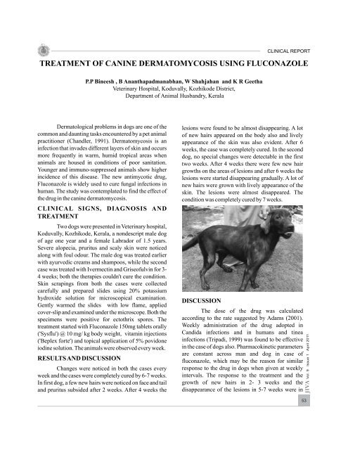
Two dogs were presented in Veterinary hospital,<br />
Koduvally, Kozhikode, Kerala, a nondescript male dog<br />
<strong>of</strong> age one year and a female Labrador <strong>of</strong> 1.5 years.<br />
Severe alopecia, pruritus and scaly skin were noticed<br />
along with foul odour. The male dog was treated earlier<br />
with ayurvedic creams and shampoos, while the second<br />
case was treated with Ivermectin and Grise<strong>of</strong>ulvin for 3-<br />
4 weeks; both the therapies couldn't cure the condition.<br />
Skin scrapings from both the cases were collected<br />
carefully and prepared slides <strong>using</strong> 20% potassium<br />
hydroxide solution for microscopical examination.<br />
Gently warmed the slides with low flame, applied<br />
cover-slip and examined under the microscope. Both the<br />
specimens were positive for ectothrix spores. The<br />
<strong>treatment</strong> started with Fluconazole 150mg tablets orally<br />
('Sysflu') @ 10 mg/ kg body weight, vitamin injections<br />
('Beplex forte') and topical application <strong>of</strong> 5% povidone<br />
iodine solution. The animals were observed every week.<br />
RESULTS AND DISCUSSION<br />
Changes were noticed in both the cases every<br />
week and the cases were completely cured by 6-7 weeks.<br />
In first dog, a few new hairs were noticed on face and tail<br />
and pruritus subsided after 2 weeks. After 4 weeks the<br />
lesions were found to be almost disappearing. A lot<br />
<strong>of</strong> new hairs appeared on the body also and lively<br />
appearance <strong>of</strong> the skin was also evident. After 6<br />
weeks, the case was completely cured. In the second<br />
dog, no special changes were detectable in the first<br />
two weeks. After 4 weeks there were few new hair<br />
growths on the areas <strong>of</strong> lesions and after 6 weeks the<br />
lesions were started disappearing gradually. A lot <strong>of</strong><br />
new hairs were grown with lively appearance <strong>of</strong> the<br />
skin. The lesions were almost disappeared. The<br />
condition was completely cured by 7 weeks.<br />
DISCUSSION<br />
The dose <strong>of</strong> the drug was calculated<br />
according to the rate suggested by Adams (2001).<br />
Weekly administration <strong>of</strong> the drug adopted in<br />
Candida infections and in humans and tinea<br />
infections (Tripadi, 1999) was found to be effective<br />
in the case <strong>of</strong> dogs also. Pharmacoki<strong>net</strong>ic parameters<br />
are constant across man and dog in case <strong>of</strong><br />
<strong>fluconazole</strong>, which may be the reason for similar<br />
response to the drug in dogs when given at weekly<br />
intervals. The response to the <strong>treatment</strong> and the<br />
growth <strong>of</strong> new hairs in 2- 3 weeks and the<br />
disappearance <strong>of</strong> the lesions in 5-7 weeks were in<br />
JIVA Vol. 9 Issue 1 April 2011<br />
53
CLINICAL REPORT<br />
accordance with the findings <strong>of</strong> Or (2000). There<br />
were no toxicity symptoms during the <strong>treatment</strong><br />
period. The effective response to the <strong>treatment</strong> was<br />
obtained probably due to drug's complete absorption<br />
from the gastrointestinal tract, increased aqueous<br />
solubility which is acid independent and the<br />
maintenance <strong>of</strong> the concentration in the skin similar<br />
to plasma (Adams 2001).The failure <strong>of</strong> the<br />
grise<strong>of</strong>ulvin <strong>treatment</strong> can be attributed to the<br />
inefficient absorption <strong>of</strong> the drug from digestive tract<br />
and the delayed accomplishment <strong>of</strong> the desired<br />
concentration <strong>of</strong> the drug in the skin (Greene 1998).<br />
The usage <strong>of</strong> drug in the <strong>dermatomycosis</strong> cases was<br />
found to be very effective and economical.<br />
REFERENCES<br />
Adams, H.R. (2001) Veterinary pharmacology and<br />
therapeutics, 8th ed., Iowa State University Press,<br />
Ames, Iowa.p930.<br />
Chandler E A, Thompson D J, Sutton J B and Price C J (1991)<br />
rd<br />
Canine medicine and therapeutics, 3 ed, Blackwell<br />
Scientific publications, pp-382-383.<br />
nd<br />
Green C E (1998) Infectious diseases <strong>of</strong> dog and cat, 2 ed.<br />
WB Saunders company, Philadelphia, London.<br />
Or E,Dodurka T and Tan H (2000)Clinical use <strong>of</strong> the oral<br />
antimycotic <strong>fluconazole</strong> for the detrmatophytosis<br />
therapy <strong>of</strong> the dog. VeterinerFakultesiDergisi(Istanbul)<br />
26(1):215-221<br />
th<br />
Tripadi K D (1999)Essentails <strong>of</strong> Medical Pharmaclogy, 4 ed.<br />
Jaypeepublicastions, p-777.<br />
CLINICAL REPORT<br />
RENAL CALCULI IN A COW<br />
P. Biju<br />
Veterinary Dispensary, Parathodu, Kottayam District<br />
Department <strong>of</strong> Animal Husbandry, Kerala<br />
JIVA Vol. 9 Issue 1 April 2011<br />
54<br />
A cross-bred cow aged 2 years was presented<br />
with a history <strong>of</strong> dysuria and anorexia. No improvement<br />
was noticed upon <strong>treatment</strong>. The animal died after 1<br />
week. On postmortem examination, the kidneys, ureter,<br />
and the bladder revealed calculi <strong>of</strong> varying sizes ranging<br />
from 2 mm to 6.5 cm (fig.). The kidneys appeared pale<br />
and s<strong>of</strong>t. All the calyces contained calculi. The largest<br />
calculi was recovered from the pelvis <strong>of</strong> the right kidney.<br />
Both the ureters were occluded with calculi. The<br />
bladder also contained calculi <strong>of</strong> varying sizes. The<br />
calculi were <strong>of</strong> different shapes with sharp edges. The<br />
chemical examination revealed the calculi were <strong>of</strong><br />
hippuric acid. No references could be traced out<br />
regarding the observation <strong>of</strong> a kidney stone as big as<br />
this.


