morphogenesis of medulla oblongata * in goat ... - Jivaonline.net
morphogenesis of medulla oblongata * in goat ... - Jivaonline.net
morphogenesis of medulla oblongata * in goat ... - Jivaonline.net
You also want an ePaper? Increase the reach of your titles
YUMPU automatically turns print PDFs into web optimized ePapers that Google loves.
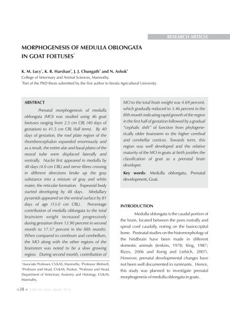
RESEARCH ARTICLEMORPHOGENESIS OF MEDULLA OBLONGATA*IN GOAT FOETUSES1 2 3 4K. M. Lucy , K. R. Harshan , J. J. Chungath and N. AshokCollege <strong>of</strong> Veter<strong>in</strong>ary and Animal Sciences, Mannuthy.*Part <strong>of</strong> the PhD thesis submitted by the first author to Kerala Agricultural University.ABSTRACTPrenatal <strong>morphogenesis</strong> <strong>of</strong> <strong>medulla</strong><strong>oblongata</strong> (MO) was studied us<strong>in</strong>g 46 <strong>goat</strong>foetuses rang<strong>in</strong>g from 2.5 cm CRL (40 days <strong>of</strong>gestation) to 41.5 cm CRL (full term). By 40days <strong>of</strong> gestation, the ro<strong>of</strong> plate region <strong>of</strong> therhombencephalon expanded enormously andas a result, the entire alar and basal plates <strong>of</strong> theneural tube were displaced laterally andventrally. Nuclei first appeared <strong>in</strong> <strong>medulla</strong> by48 days (4.0 cm CRL) and nerve fibres cross<strong>in</strong>g<strong>in</strong> different directions broke up the graysubstance <strong>in</strong>to a mixture <strong>of</strong> gray and whitemater, the reticular formation. Trapezoid bodystarted develop<strong>in</strong>g by 48 days. Medullarypyramids appeared on the ventral surface by 81days <strong>of</strong> age (13.0 cm CRL).Percentagecontribution <strong>of</strong> <strong>medulla</strong> <strong>oblongata</strong> to the totalbra<strong>in</strong>stem weight <strong>in</strong>creased progressivelydur<strong>in</strong>g gestation (from 13.90 percent <strong>in</strong> secondmonth to 17.57 percent <strong>in</strong> the fifth month).When compared to cerebrum and cerebellum,the MO along with the other regions <strong>of</strong> thebra<strong>in</strong>stem was noted to be a slow grow<strong>in</strong>gregion. Dur<strong>in</strong>g second month, contribution <strong>of</strong>¹Associate Pr<strong>of</strong>essor, CV&AS, Mannuthy, ²Pr<strong>of</strong>essor (Retired),4³Pr<strong>of</strong>essor and Head, CV&AS, Pookot, Pr<strong>of</strong>essor and Head,Department <strong>of</strong> Veter<strong>in</strong>ary Anatomy and Histology, CV&AS,Mannuthy.MO to the total bra<strong>in</strong> weight was 4.69 percent,which gradually reduced to 3.46 percent <strong>in</strong> thefifth month <strong>in</strong>dicat<strong>in</strong>g rapid growth <strong>of</strong> the region<strong>in</strong> the first half <strong>of</strong> gestation followed by a gradual“cephalic shift” <strong>of</strong> function from phyloge<strong>net</strong>icallyolder bra<strong>in</strong>stem to the higher cerebraland cerebellar cortices. Towards term, thisregion was well developed and the relativematurity <strong>of</strong> the MO <strong>in</strong> <strong>goat</strong>s at birth justifies theclassification <strong>of</strong> <strong>goat</strong> as a prenatal bra<strong>in</strong>developer.Key words: Medulla <strong>oblongata</strong>, Prenataldevelopment, Goat.INTRODUCTIONMedulla <strong>oblongata</strong> is the caudal portion <strong>of</strong>the bra<strong>in</strong>, located between the pons rostrally andsp<strong>in</strong>al cord caudally, rest<strong>in</strong>g on the basioccipitalbone. Postnatal studies on the histomorphology <strong>of</strong>the h<strong>in</strong>dbra<strong>in</strong> have been made <strong>in</strong> differentdomestic animals (Jenk<strong>in</strong>s, 1978; K<strong>in</strong>g, 1987;Rizzo, 2006 and Konig and Liebich, 2007).However, prenatal developmental changes havenot been well documented <strong>in</strong> rum<strong>in</strong>ants. Hence,this study was planned to <strong>in</strong>vestigate prenatal<strong>morphogenesis</strong> <strong>of</strong> <strong>medulla</strong> <strong>oblongata</strong> <strong>in</strong> <strong>goat</strong>s.28 J. Ind. Vet. Assoc., Kerala. 10 (3)
MATERIALS AND METHODSPrenatal <strong>morphogenesis</strong> <strong>of</strong> <strong>medulla</strong><strong>oblongata</strong> (MO) <strong>in</strong> <strong>goat</strong>s was studied us<strong>in</strong>g 46 <strong>goat</strong>foetuses rang<strong>in</strong>g from 2.5 cm CRL (40 days <strong>of</strong>gestation) to 41.5 cm CRL (full term). The materialavailable <strong>in</strong> the Department <strong>of</strong> Anatomy and thosecollected from the farms and cl<strong>in</strong>ics were used forthe study. Body weight, body parameters and skullparameters <strong>of</strong> the subjects were recorded. Theage <strong>of</strong> the foetuses was calculated from the0.33formula, W = 0.096 (t-30) derived by S<strong>in</strong>ghet.al. (1979) for the <strong>goat</strong> foetuses, where 'W' is thebody weight <strong>of</strong> the foetus <strong>in</strong> g and 't' is the age <strong>of</strong> thefoetus <strong>in</strong> days. Based on the age, the foetuses weredivided <strong>in</strong>to four groups represent<strong>in</strong>g second,third, fourth and fifth months <strong>of</strong> gestation. Theheads were separated at the occipito-atlantaljunction and the bra<strong>in</strong> was then carefully dissectedout and fixed <strong>in</strong> 10 percent neutral bufferedformal<strong>in</strong>. After record<strong>in</strong>g the whole bra<strong>in</strong>parameters, the MO was separated at the caudalborder <strong>of</strong> the pons (rostral boundary) and therostral limit <strong>of</strong> orig<strong>in</strong> <strong>of</strong> first pair <strong>of</strong> cervical sp<strong>in</strong>alnerves (caudal boundary). Measurements weretaken and the data were analysed statistically(Snedecor and Cochran, 1985). Standardprocedures were adopted for histoarchitecturalstudies (Luna, 1968).RESULTS AND DISCUSSIONDevelopment <strong>in</strong> the Second MonthMedulla <strong>oblongata</strong>, the caudal mostsegment <strong>of</strong> the bra<strong>in</strong>stem, extended from the level<strong>of</strong> first pair <strong>of</strong> cervical sp<strong>in</strong>al nerves to the caudaledge <strong>of</strong> the pons (Fig. 1). It lay on the unossifiedbasioccipital and this cartilag<strong>in</strong>ous skeletondeveloped <strong>in</strong> the sixth week <strong>of</strong> gestation.Measurements <strong>of</strong> <strong>medulla</strong> <strong>oblongata</strong> at differentstages <strong>of</strong> gestation are given <strong>in</strong> table 1. By 40 days,the ro<strong>of</strong> plate region <strong>of</strong> the embryonicrhombencephalon expanded enormously. As aresult, the entire alar and basal plates <strong>of</strong> the neuraltube were displaced laterally and ventrally. Arey(1957) compared this to an opened book whoseh<strong>in</strong>ge was the floor plate. The lumen became thefourth ventricle covered dorsally by the th<strong>in</strong>, s<strong>in</strong>glelayer <strong>of</strong> ependyma, the ro<strong>of</strong> plate. This constitutedthe anterior and posterior <strong>medulla</strong>ry vela (Figs. 2and 3). These vela were cont<strong>in</strong>uous with thecerebellum cranially and the ro<strong>of</strong> <strong>of</strong> the centralcanal <strong>of</strong> sp<strong>in</strong>al cord caudally. The sulcus limitanspresent on the ventrolateral wall <strong>of</strong> the fourthventricle provided the plane <strong>of</strong> division <strong>of</strong> the<strong>medulla</strong> <strong>in</strong>to a ventromedial basal plate and adorsolateral alar plate. Similar observations weremade <strong>in</strong> dog foetuses by Jenk<strong>in</strong>s (1978). Thelumen was filled with CSF. Nuclei appeared by 48days and nerve fibres cross<strong>in</strong>g <strong>in</strong> differentdirections broke up the gray substance <strong>in</strong>to amixture <strong>of</strong> gray and white known as the reticularformation. The trapezoid body started develop<strong>in</strong>gat 48 days (4 cm CRL). The po<strong>in</strong>t <strong>of</strong> emergence <strong>of</strong>the facial nerve from the <strong>medulla</strong> is illustrated <strong>in</strong>figure 4. The endolymphatic duct also could beseen with<strong>in</strong> the petrous temporal.Vascular mesenchyme occupied theependymal ro<strong>of</strong>; the comb<strong>in</strong>ed membrane, thetela choroidea, <strong>in</strong>folded as vascular tufts <strong>in</strong>to thecavity <strong>of</strong> the myelencephalon constitut<strong>in</strong>g thechoroid plexus <strong>of</strong> the fourth ventricle. Thisdeveloped <strong>in</strong> the <strong>goat</strong> foetus <strong>in</strong> the sixth week <strong>of</strong>gestation. Keith (1947) reported that the choroidvilli developed on the ventricular surface <strong>of</strong> thecaudal <strong>medulla</strong>ry velum at eight weeks <strong>in</strong> thehuman foetus and CSF was be<strong>in</strong>g produced dur<strong>in</strong>gVol. 10 Issue 3 December 2012 JIVA 29
the third month. Later dur<strong>in</strong>g the seventh week,the ependymal ro<strong>of</strong> plate became broad and th<strong>in</strong>.The cavity <strong>of</strong> the rhombencephalon thusexpanded to the sides, flattened dorsoventrallyand was filled with CSF. The rhombic lip, ridgewhere the tela jo<strong>in</strong>ed the alar plate was made up <strong>of</strong>three to four layers <strong>of</strong> cells. Harrison (1978)reported that the cells <strong>of</strong> rhombic lip were activelymitotic and provided large number <strong>of</strong> neuroblasts,which migrated cephalad <strong>in</strong>to the ventral aspect <strong>of</strong>the h<strong>in</strong>dbra<strong>in</strong> where they formed the pont<strong>in</strong>enuclei and the olivary nuclear complex.Caudally the <strong>medulla</strong> <strong>oblongata</strong> wascont<strong>in</strong>uous with the sp<strong>in</strong>al cord. The fourthventricle narrowed posteriorly to be cont<strong>in</strong>ued asthe central canal <strong>of</strong> sp<strong>in</strong>al cord. Dorsal wall <strong>of</strong> thisregion showed thickened epithelium constitut<strong>in</strong>gthe circumventricular organ. Medulla <strong>oblongata</strong>contributed 4.69 percent <strong>of</strong> the bra<strong>in</strong> weight and13.90 percent <strong>of</strong> the bra<strong>in</strong>stem weight at this stage.Fig. 1 Dorsal surface <strong>of</strong> bra<strong>in</strong> (55 days)1. Cerebrum; 2. Corpora quadrigem<strong>in</strong>a;3. Cerebellum; 4. Medulla <strong>oblongata</strong>Fig. 2 C.S. <strong>of</strong> the <strong>medulla</strong> <strong>oblongata</strong> at the level<strong>of</strong> rostral <strong>medulla</strong>ry velum (48 days). H&E. x 1001. Medulla <strong>oblongata</strong>; 2. Nuclear aggregation;3. Fourth ventricle with CSF; 4. Rostral <strong>medulla</strong>ry velum;5. Body wall; 6. Internal glial limit<strong>in</strong>g membrane30 J. Ind. Vet. Assoc., Kerala. 10 (3)
Development In The Third MonthRESEARCH ARTICLEFig. 3 C.S. <strong>of</strong> the <strong>medulla</strong> <strong>oblongata</strong> at the level <strong>of</strong>caudal <strong>medulla</strong>ry velum (48 days). H&E. x 1001. Caudal <strong>medulla</strong>ry velum; 2. Choroid plexus;3. Fourth ventricle with CSF; 4. Medulla <strong>oblongata</strong>;5. Nuclear aggregation; 6. Median sulcus;7. Sulcus limitansFig. 4 C.S. <strong>of</strong> the <strong>medulla</strong> <strong>oblongata</strong> show<strong>in</strong>g thefacial nerve and endolymph duct (48 days). H&E.x1001. Medulla <strong>oblongata</strong>; 2. Facial nerve;3. Endolymph duct; 4. Petrous temporal boneFig. 5 C.S. <strong>of</strong> the <strong>medulla</strong> <strong>oblongata</strong> show<strong>in</strong>gforamen <strong>of</strong> Lushcka and caudal <strong>medulla</strong>ry velum.(76 days). H&E. x 100 1. Foramen <strong>of</strong> Lushcka;2. Caudal <strong>medulla</strong>ry velum; 3. Choroid plexus;4. Sulcus limitansThe <strong>medulla</strong>ry pyramids could bedist<strong>in</strong>guished from 81 days (13 cm CRL) and were<strong>in</strong> the form <strong>of</strong> longitud<strong>in</strong>al ridges on either side <strong>of</strong>the ventral median fissure but they were notwidely separated <strong>in</strong> the rostral portion. Theseagree with the f<strong>in</strong>d<strong>in</strong>gs <strong>of</strong> Dellmann and Mc Clure(1975) <strong>in</strong> small rum<strong>in</strong>ants. However, <strong>in</strong> cattle thepyramids were widely separated at the po<strong>in</strong>t <strong>of</strong>emergence from the caudal aspect <strong>of</strong> pons.The dorsal surface <strong>of</strong> the <strong>medulla</strong> formedthe floor <strong>of</strong> fourth ventricle as noted <strong>in</strong> the secondmonth. The caudal <strong>medulla</strong>ry velum projectedfrom the dorso-medial angle <strong>of</strong> <strong>medulla</strong><strong>oblongata</strong>. The fourth ventricle communicatedwith the subarachnoid space by the foramen <strong>of</strong>Lushcka (Fig. 5). Floor <strong>of</strong> the ventricle was markedby a deep median sulcus, which became shallowerrostrally. On each side <strong>of</strong> the median sulcus was acont<strong>in</strong>uous ridge, the medial em<strong>in</strong>ence, boundedlaterally by the sulcus limitans as observed by Truexand Carpenter (1969) <strong>in</strong> man and Dyce et al.(1996) <strong>in</strong> domestic animals. The medialem<strong>in</strong>ence, or trigonum hypoglossi was formed bythe nucleus <strong>of</strong> hypoglossal nerve. The lateralem<strong>in</strong>ence was occupied by the caudal poles <strong>of</strong> themedial and <strong>in</strong>ferior vestibular nuclei. In betweenthe medial and lateral em<strong>in</strong>ences was the<strong>in</strong>termediate em<strong>in</strong>ence, the trigonum vagi.Mean weight <strong>of</strong> <strong>medulla</strong> <strong>oblongata</strong> was0.220 ± 0.032g dur<strong>in</strong>g third month. Width <strong>of</strong><strong>medulla</strong> <strong>oblongata</strong> was more than its heightthroughout gestation. Average length and width <strong>of</strong><strong>medulla</strong>ry pyramids were 0.805 ± 0.031cm and0.230 ± 0.013cm, respectively. The trapezoidbody was clearly demarcated and the meanrostrocaudal distance was 0.117 ± 0.005cm.Vol. 10 Issue 3 December 2012 JIVA 31
Dellmann and Mc Clure (1975) reported that thetrapezoid body was more clearly demarcated <strong>in</strong>small rum<strong>in</strong>ants than <strong>in</strong> cattle.Development In The Fourth MonthMedulla <strong>oblongata</strong> contributed 17.57percent <strong>of</strong> the bra<strong>in</strong>stem weight. Percentagecontribution <strong>of</strong> <strong>medulla</strong> <strong>oblongata</strong> to the totalbra<strong>in</strong>stem weight <strong>in</strong>creased progressively dur<strong>in</strong>ggestation. Dur<strong>in</strong>g the fourth month, mean length,width and thickness <strong>of</strong> the <strong>medulla</strong> <strong>oblongata</strong> were1.183 ± 0.027cm, 0.855 ± 0.019cm and 0.682 ±0.011cm, respectively. Unlike <strong>in</strong> the pons region,maximum width <strong>of</strong> <strong>medulla</strong> <strong>oblongata</strong> exceededthe maximum height. Morphological features didnot change much dur<strong>in</strong>g the fourth month. Lengthand width <strong>of</strong> <strong>medulla</strong>ry pyramids <strong>in</strong>creased 15.03and 93.04 percent, respectively from third t<strong>of</strong>ourth month. More <strong>in</strong>crease <strong>in</strong> widthcorresponded to growth <strong>of</strong> cerebrum s<strong>in</strong>ce thesefibres have their orig<strong>in</strong> <strong>in</strong> the cerebral cortex.Development In The Fifth MonthTrapezoid body was clearly demarcatedfrom the pons. Cranio-caudal length <strong>of</strong> trapezoidbody was 0.298 ± 0.003cm. Mean weight <strong>of</strong><strong>medulla</strong> <strong>oblongata</strong> <strong>in</strong>creased three-fold dur<strong>in</strong>gfifth month. Correspond<strong>in</strong>g changes were alsonoticed <strong>in</strong> the length, height and width <strong>of</strong> <strong>medulla</strong><strong>oblongata</strong> (Table. 1). Dur<strong>in</strong>g second monthcontribution <strong>of</strong> <strong>medulla</strong> <strong>oblongata</strong> to the totalbra<strong>in</strong> weight was 4.69 percent which graduallyreduced to 3.46 percent <strong>in</strong> the fifth month<strong>in</strong>dicat<strong>in</strong>g rapid growth <strong>of</strong> the region <strong>in</strong> the first half<strong>of</strong> gestation followed by a gradual “cephalic shift”<strong>of</strong> function from phyloge<strong>net</strong>ically older bra<strong>in</strong>stemto the higher cerebral and cerebellar cortices.When compared to other divisions <strong>of</strong> bra<strong>in</strong>, the<strong>medulla</strong> <strong>oblongata</strong> along with other regions <strong>of</strong>bra<strong>in</strong>stem was noted to be a slow grow<strong>in</strong>g region.Ventral median fissure was flanked by thepyramids. Mean length and width <strong>of</strong> <strong>medulla</strong>rypyramids were 1.585 ± 0.088 cm and 0.656 ±0.008 cm, respectively. Grossly, <strong>medulla</strong> <strong>oblongata</strong>was adult-like dur<strong>in</strong>g the fifth month (Fig. 6).Fig. 6 Dorsal view <strong>of</strong> bra<strong>in</strong>stem (124 days)1. Thalamus; 2. P<strong>in</strong>eal gland; 3. Rostral colliculus;4. Caudal colliculus; 5. Fourth ventricle;6. Medulla <strong>oblongata</strong>The <strong>medulla</strong> <strong>oblongata</strong> is a greatsuprasegmental conveyor and co-ord<strong>in</strong>ator forpathways and nuclei <strong>in</strong>volved with vital regulatoryand protective processes which affect the wholebody. Towards term, this region was welldeveloped and the relative maturity <strong>of</strong> the MO <strong>in</strong><strong>goat</strong>s at birth justifies the classification <strong>of</strong> <strong>goat</strong> as aprenatal bra<strong>in</strong> developer. Foetal bra<strong>in</strong> is mostvulnerable when it is grow<strong>in</strong>g rapidly andnutritional deficiencies and diseases dur<strong>in</strong>g thegrow<strong>in</strong>g period can cause permanent damage.REFERENCESthArey, L.B. 1957. Developmental Anatomy. 6 edn,W.B.Saunders Company, Philadelphia. pp.454-501.32 J. Ind. Vet. Assoc., Kerala. 10 (3)
Dellmann, H.D and Mc Clure, R.G. 1975. Centralnervous system. Sisson and Grossman'sthThe Anatomy <strong>of</strong> the Domestic Animals. 5edn, (Ed.) Getty R. W.B. SaundersCompany, Philadelphia. pp.1065-80.Dyce, K.M., Sack, W.O. and Wens<strong>in</strong>g, C.J.G.1996. Textbook <strong>of</strong> Veter<strong>in</strong>ary Anatomy.nd2 edn, W.B. Saunders Company,Philadelphia. pp. 259-324.Harrison, R.G. 1978. Cl<strong>in</strong>ical Embryology.Academic Press, London. p. 250.Jenk<strong>in</strong>s, T.W. 1978. Functional MammalianndNeuroanatomy. 2 edn, Lea and Febiger,Philadelphia. p. 480.Keith, A. 1947. Human Embryology andthMorphology. 6 edn, Williams andWilk<strong>in</strong>s, Baltimore. pp: 117-53.K<strong>in</strong>g, A.S. 1987. Physiological and Cl<strong>in</strong>icalAnatomy <strong>of</strong> the Domestic Mammals.Oxford University Press, New York. p.325.Konig, H.E. and Liebich, H.G. 2007. Veter<strong>in</strong>aryAnatomy <strong>of</strong> Domestic Animals, Text Bookrdand Colour Atlas. 3 edn, Schattauer, NewYork. pp: 40-48.Luna, L.G. 1968. Manual<strong>of</strong> HistologicalSta<strong>in</strong><strong>in</strong>g Methods <strong>of</strong> the Armed ForcesrdInstitute <strong>of</strong> Pathology. 3 edn, Mc Graw-Hill Book Company, New York. p.258.Rizzo, D.C. 2006. Fundamentals <strong>of</strong> Anatomy andndPhysiology. 2 edn, Thomson DelmarLearn<strong>in</strong>g, Australia. pp: 244-69.S<strong>in</strong>gh, Y., Sharma, D.N. and Dh<strong>in</strong>gra, L.D. 1979.Morphogenesis <strong>of</strong> the testis <strong>in</strong> <strong>goat</strong>. IndianJ. Anim. Sci. 49: 925-31.Snedecor, G.W. and Cochran, W.G. 1985.thStatistical Methods. 7 edn, The Iowa StateUniversity Press, USA. p. 313.Truex, R.C. and Carpenter, M.B. 1969. HumanthNeuroanatomy. 6 edn, The Williams andWilk<strong>in</strong>s, Baltimore. p. 673.Vol. 10 Issue 3 December 2012 JIVA 33


