What is synchrotron radiation?
What is synchrotron radiation?
What is synchrotron radiation?

You also want an ePaper? Increase the reach of your titles
YUMPU automatically turns print PDFs into web optimized ePapers that Google loves.
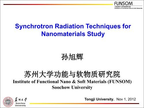
Synchrotron Radiation Techniques for<br />
Nanomaterials Study<br />
孙 旭 辉<br />
苏 州 大 学 功 能 与 软 物 质 研 究 院<br />
Institute of Functional Nano & Soft Materials (FUNSOM)<br />
Soochow University<br />
Tongji University, Nov 1, 2012
• SR techniques and its advantages in nano studies<br />
• Nanomaterials study by SR techniques<br />
o Electronic structure of nanomaterials (XAS, XES, STXM)<br />
o Optical property of nanomaterials (XEOL)<br />
o Crystal structure of nanomaterials (XRD)<br />
o Development of in-situ XAS techniques<br />
• Summary
<strong>What</strong> <strong>is</strong> <strong>synchrotron</strong> <strong>radiation</strong><br />
When an electron traveling at a speed close to the speed of light<br />
in an orbit, it emits a continuum of electromagnetic <strong>radiation</strong><br />
tangential to the orbit, which <strong>is</strong> Synchrotron light.<br />
3
Then they pass into the<br />
booster ring where they are<br />
accelerated to 99.9999% of<br />
the speed of light<br />
Electrons are<br />
generated here<br />
Creating the light<br />
And are finally transferred into the<br />
storage ring<br />
And initially<br />
accelerated in<br />
the LINAC<br />
Scheme of <strong>synchrotron</strong> <strong>radiation</strong> facility
NSRFII<br />
Diamond Light Source<br />
Shanghai Synchrotron Radiation Facility
Interior of the Canadian Light Source
Synchrotron Radiation Properties<br />
• Tunability (IR to hard x-rays, element specific)<br />
• Brightness (highly collimated, micro/nano beam)<br />
• Polarization (linear, circular, tunable, dichro<strong>is</strong>m)<br />
• Time structure (short pulse, dynamics)
Techniques of SR<br />
The fundamental parameters necessary for perception of physical world:<br />
• Energy (spectroscopy, state of matter)<br />
• Momentum (scattering)<br />
• Position (imaging, spatial d<strong>is</strong>tribution)<br />
• Time (dynamics)<br />
numerous techniques of SR<br />
Spectroscopy Scattering Imaging<br />
01 Low-Energy Spectroscopy 05 Hard X-Ray Diffraction 09 Hard X-Ray Imaging<br />
02 Soft X-Ray Spectroscopy 06 Macromolecular<br />
10 Soft X-Ray Imaging<br />
Crystallography<br />
03 Hard X-Ray Spectroscopy 07 Hard X-Ray Scattering 11 Infrared Imaging<br />
04 Optics/Calibration/Metrology 08 Soft X-Ray Scattering 12 Lithography
A Single Storage Ring Serves Many Scientific User Groups
Synchrotron Radiation Techniques at SSRF<br />
BL08U-A – Soft X-ray Spectromicroscopy (STXM)<br />
BL08U-B – X-ray Interference Lithography (XIL)<br />
BL14W – X-ray Absorption Fine Structure (XAFS)<br />
BL14B – X-ray Diffraction (XRD)<br />
BL15U – Hard X-ray Micro-focus<br />
BL16B – Small Angle X-ray Scattering (SAXS)<br />
BL10W – X-ray Imaging and Biomedical Application<br />
BL17U – Macromolecular Crystallography (MC)
<strong>What</strong> <strong>is</strong> X-ray absorption fine structures (XAFS) <br />
• As core electron <strong>is</strong> excited with hv the threshold<br />
(E o ), it <strong>is</strong> excited to a final state defined by the<br />
chemical environment, which modulates the<br />
absorption coefficient relative to that of a free<br />
atom. Th<strong>is</strong> modulation <strong>is</strong> known the XAFS,<br />
• XAFS contains all the information about the local<br />
structure and bonding of the absorbing atom<br />
• XAFS study requires a tunable X-ray source –<br />
<strong>synchrotron</strong> <strong>radiation</strong><br />
11
t<br />
<strong>What</strong> does XAFS look like<br />
Near edge<br />
XANES<br />
(NEXAFS)<br />
1s to LUMO<br />
(t3)<br />
EXAFS<br />
Si(CH3)4<br />
C<br />
Si<br />
Si K-edge<br />
12
Synchrotron Probe<br />
Spectroscopy<br />
Structure<br />
Valence<br />
Electrons<br />
Core Electrons<br />
Photon<br />
Energy<br />
Wavelength<br />
10eV 100eV 1keV 10keV<br />
100nm 10nm<br />
1nm 1Å<br />
Lithography<br />
Nanostructures<br />
Proteomics<br />
Protein<br />
Crystallography
Advantage of SR Techniques in Nano Study<br />
Comprehensive techniques (electron, ion,<br />
photon), various information<br />
New techniques: XAFS,<br />
XEOL, XES, RIXS,<br />
STXM, etc<br />
Individual nanomaterial<br />
characterization<br />
SR<br />
Techniques<br />
High Signal/No<strong>is</strong>e<br />
Ambient Conditions<br />
In-situ, dynamic<br />
Depth profile with SR X-ray<br />
(Surface & Interface)<br />
Less sample damage,<br />
reliable data
Absorption Techniques for Nano Studies<br />
• XAFS (X-ray Absorption Fine Structures)<br />
Measurement: Absorption coefficient above an edge<br />
Information: Conduction band, Local structure and<br />
bonding<br />
• XES (X-ray Em<strong>is</strong>sion Spectroscopy)<br />
Measurement: X-rays in, x-rays out<br />
Information: Valence band electron d<strong>is</strong>tribution<br />
• XEOL (X-ray Excited Optical Luminescence)<br />
Measurement: X-rays in, optical photons out<br />
Information: origin of luminescence, time dependent<br />
events (dynamics)
XANES<br />
XAFS, XEOL and XES<br />
E<br />
(III)<br />
PLY<br />
XEOL<br />
hv ex<br />
(I)<br />
Abs.<br />
CB<br />
VB<br />
(II)<br />
E F<br />
core<br />
Mono<br />
hv op<br />
hv f<br />
FLY<br />
Mono<br />
Channel<br />
Plate<br />
200 850<br />
Wavelength (nm)<br />
VB<br />
XES<br />
Energy (eV) E F
XANES and XES of Zn nanostructures<br />
500 nm
XEOL - Conversion of X-ray energy into optical em<strong>is</strong>sion<br />
X-ray phosphor<br />
thermalization<br />
hv >E o<br />
Recombination via<br />
exciton<br />
UV<br />
v<strong>is</strong>.<br />
Elliot & Gibson<br />
Solid State<br />
Physics Haper &<br />
Row, 1974<br />
o<br />
o<br />
hv ~ E o<br />
Core level<br />
Recombination via bound exciton,<br />
impurity and defect states
XEOL Techniques<br />
Energy Domain<br />
XEOL: Luminescence induced by selected excitation<br />
photon energy (usually across an absorption edge)<br />
Optical XAFS: Absorption across an edge monitored with<br />
the optical signal (photoluminescence yield )<br />
Time Domain<br />
Lifetime: Synchrotron pulse as the start/stop<br />
Time-gated XEOL: Luminescence within a selected time<br />
window between pulses<br />
Time-gated Optical XAFS: Photoluminescence yield<br />
with a time window
TRXEOL<br />
Photon Energy (eV)<br />
CLS-July 16, 2012<br />
Optical XAFS<br />
(time & wavelength selected)<br />
imaging
Normalized Intensity<br />
CdSe -Si Heteronanostructure<br />
Si<br />
CdSe<br />
CdSe-Si 1100 eV<br />
0.007<br />
0.006<br />
total<br />
Si CdSe<br />
0.005<br />
0.004<br />
0.003<br />
0.002<br />
slow<br />
(20-150 ns)<br />
fast<br />
(0-20 ns)<br />
0.001<br />
0.000<br />
300 400 500 600 700 800 900<br />
Wavelength (eV)<br />
hv = 1100 eV<br />
J. Phys. Chem. C, 111, 8475 (2007)
CdSe-Si Heterostructure: Se L 3,2 - edge<br />
Appl. Phys. Lett., 89, 243102 (2006)
• Advantages of SR techniques in nano studies<br />
• Nanomaterials study by SR techniques<br />
o Electronic structure of nanomaterials (XAS, XES, STXM)<br />
o Optical property of nanomaterials (XEOL)<br />
o Crystal structure of nanomaterials (XRD)<br />
o Development of in-situ XAS techniques<br />
• Summary
Electronic structure of nanomaterials<br />
by XAS, XES, and STXM
Probing solid state N-doping in graphene by XAS<br />
C K-edge<br />
GO: Graphene oxide<br />
GRA-x: Graphene-urea annealed<br />
at x degree<br />
A: C-C π* excitation<br />
B1: Amino C-N<br />
B2: C-O or C=O<br />
B3: Urea C=O π* excitation<br />
C: C-C σ* excitation<br />
Carbon 2012, 50, 321–341.
N K-edge<br />
GRA-x: Graphene-urea annealed<br />
at x degree<br />
N1: Pyridinic C-N<br />
N2: Amino C-N<br />
N3: Graphitic C-N<br />
N4: Urea C-N<br />
N5: C-N σ* excitation
Intensity(a.u.)<br />
Unpubl<strong>is</strong>hed<br />
Carbon nanocages (CNCs) studied by XAS and XES<br />
P-CNC<br />
N-CNC<br />
Pure CNCs<br />
282<br />
284<br />
286<br />
Photon Energy(eV)<br />
288<br />
290
Electronic Structure of Graphdiyne Probed by XAS and STXM<br />
JPCC, submitted
STXM and XANES study of N-doped CNTs sealed with N 2 gas<br />
J. Appl. Phys. 111, 124318 (2012)
Optical property of nanomaterials by XEOL
XEOL Study of PL Mechan<strong>is</strong>m of Nanostructures<br />
PL of Single Crystalline GeO2 Nanowires<br />
Ge:O≈1:1.8<br />
J. Phys. Chem. C, 115, 11420 (2011)
XAFS and XEOL of GeO2 Nanowires
Crystal structure of nanomaterials by XRD
SR-XRD study of In 2 Se 3 nanowires<br />
In:Se≈2:3<br />
TEM image and corresponding EDS spectra of an individual In 2 Se 3<br />
nanowire. Scare bar <strong>is</strong> 100 nm<br />
Appl. Phys. Lett., 89 (2006) 233121
SR-XRD of In 2 Se 3 nanowires<br />
JCPDS PDF 341279<br />
J. Mater. Chem., 21 (2011) 6944
In2Se3 Nanowires: HR-TEM Image<br />
(a)<br />
(b)<br />
α-phase<br />
κ-phase
In-situ phase transition of In 2 Se 3 nanowires
Electrical Res<strong>is</strong>tances of In 2 Se 3 Nanowires<br />
I-V character<strong>is</strong>tics of two devices fabricated with<br />
nanowires of as- synthesized and after annealing
Summary<br />
Synchrotron <strong>radiation</strong> techniques including absorption<br />
spectroscopy such as XAFS, (XANES and EXFAS), deexcitation<br />
spectroscopy such as XEOL, TRXEOL, and XES<br />
(RIXS), XRD and spectro-microscoy (STXM) have<br />
provided unprecedented research opportunities in probing<br />
the structure and electronic properties of nanomaterials.
Acknowledgements:<br />
Soochow-Western Joint Center for Synchrotron Radiation Research<br />
SR Team @ FUNSOM, Soochow University<br />
Prof. Suidong Wang<br />
Prof. Youyong Li<br />
Dr. Jun Zhong<br />
Dr. Xiujuan Zhang<br />
Prof. Steffen Duhm<br />
Dr. Keith Gilmore<br />
University of Western Ontario & Canadian Light Source<br />
Profs. T. K. Sham, X. L. Sun, Y. Song<br />
Dr. Yongfeng Hu, Tom Reger<br />
Shanghai Synchrotron Radiation Facility<br />
Dr. Xingtai Zhou<br />
Dr. Renzhong Tai<br />
Dr. Zheng Jiang<br />
Dr. Wen Wen<br />
Advanced Light Source, Berkeley Lawrence<br />
National Lab<br />
Dr. Jinghua Guo, Dr. Zhi Liu, Dr. Xiaosong Liu<br />
National Synchrotron Radiation Lab (Hefei)<br />
Prof. Ziyu Wu,Dr. Zhiyun Pan
Thank you for your attention!
Acknowledgements: