69-72 - Polskie Stowarzyszenie BiomateriaÅów
69-72 - Polskie Stowarzyszenie BiomateriaÅów
69-72 - Polskie Stowarzyszenie BiomateriaÅów

Create successful ePaper yourself
Turn your PDF publications into a flip-book with our unique Google optimized e-Paper software.
20 Wykonano trzy próby według schematu przedstawionego its original position. In figure 1, the heating elements are<br />
represented as spirals. The specimen was removed from the<br />
furnace at a temperature of approximately 200°C. TABLE 2<br />
depicts the parameters of the three test runs.<br />
w tabeli 2. Proszek z Ti-6Al-4V umieszczono w formie<br />
ze szkła kwarcowego (Rys.1b) i wprowadzono do pieca<br />
z atmosferą ochronną (Rys.1c). Próbę umieszczono<br />
w piecu kwarcowym ze stałą temperaturą i atmosferą<br />
gazu ochronnego. Jako gazu ochronnego użyto argonu.<br />
W trakcie eksperymentu kilkakrotnie zmieniano położenie<br />
formy przesuwając ją w kierunku otworu wyjściowego<br />
pieca a tym samym obniżając temperaturę spieku próbki,<br />
by następnie skierować ją w miejsce zapoczątkowania<br />
eksperymentu. Na Rys.1c zaznaczono kręgi przedstawiające<br />
elementy grzewcze. Próbkę wyjęto przy temperaturze<br />
spieku około 200 °C.<br />
Wyniki i dyskusja<br />
Na ogół spiekanie metali przeprowadza się w temperaturze<br />
o wartości około 3/4 temperatury topnienia stopu,<br />
aktywując termicznie ruch wiązań pomiędzy atomami [4,5].<br />
W przypadku przeprowadzonego doświadczenia celem<br />
szczegółowego poznania mechanizmów aktywujących<br />
wiązanie metaliczne pomiędzy poszczególnymi strukturami<br />
kulistymi przeprowadzono porównawczo dwie próby<br />
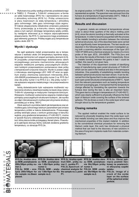
z temperaturami znacznie niższymi. Wyniki w postaci zdjęć<br />
z mikroskopu skaningowego LEO 1455 VP wraz ze spektrum<br />
analizy chemicznej wykonanym mikrosondą JEOL<br />
JXA-8900R przedstawiono dla próby numer 2 na rys.2a-f<br />
oraz dla próby numer 3 na Rys.3 a-c. Dla próby numer 1<br />
nie wykazano wiązania metalicznego i nie przeprowadzono<br />
badań.<br />
Istotą doświadczenia było wykazanie możliwości wytworzenia<br />
struktury otwartoporowatej na bazie stopu tytanu<br />
Ti-6Al-4V używanego w medycynie i transporcie lotniczym.<br />
Wykazano możliwość wytworzenia wiązania metalicznego<br />
pomiędzy poszczególnymi strukturami kulistymi. Jak wynika<br />
ze zdjęć mikroskopowych możliwe jest otrzymanie struktury<br />
porowatej już w temp. 900 o C.<br />
Obok ważnych czynników takich jak temperatura oraz atmosfera<br />
gazu ochronnego istotnym parametrem jest zmiana<br />
temperatury próbki w trakcie doświadczenia. Przesuwając<br />
próbkę wewnątrz pieca z centrum grzewczego w kierunku<br />
wyjścia, przy gradiencie temperatury ∆T=(40÷60)°C, można<br />
w sposób fizyczny oddziaływać na powierzchnię spiekanej<br />
kuli, którą otacza ściśle przylegający tlenek tytanu. Powoduje<br />
to pęknięcie skorupy tlenku wskutek działania gradientu<br />
temperatury, co obrazuje rys.2e.<br />
Results and discussion<br />
In general, metals are sintered at temperatures whose<br />
value is about three quarters of the alloy’s melting point<br />
[4-5], since the atomic bonding is thermally activated at this<br />
temperature. In order to become acquainted with the process<br />
of the metallic bonding’s activation between the individual<br />
spherical structures, two of the described tests were carried<br />
out at significantly lower temperatures. The test results are<br />
depicted in the following figures and were investigated using<br />
both a scanning electron microscope of the type LEO<br />
1455 VP SEM and a chemical analysis by means of a micro<br />
probe of the type JEOL JXA-8900R. The FIGs.2a-e and<br />
3a-c show the results of the tests 2 and 3 respectively. Since<br />
no metallic bonding between the grains in test 1 could be<br />
verified, this result is not given here.<br />
The intention of this experiment consists of identifying<br />
ways of manufacturing open pored structures of Ti-6Al-4V<br />
which can be used in the medical and also in the aerospace<br />
industries. One possibility of establishing metallic bonding<br />
between spherical structures has been shown. It can be discerned<br />
from the figures that it is also possible to manufacture<br />
such open pored structures at temperatures of 900°C. Apart<br />
from the relevant parameters such as heating temperature<br />
and inert scavenging gas, it transpired that the temperature<br />
change effected by translating the specimen towards the<br />
furnace door during the test, is also an important factor.<br />
This gave rise to a change in temperature of ∆T=(40÷60)°C,<br />
which was clearly sufficient to physically influence the thick<br />
layer of titanium oxide covering the surface of the sintered<br />
spheres. FIG.2e shows a crack in the oxide layer which was<br />
brought about by the temperature gradient.<br />
Closing remarks<br />
The applied method enabled the metal surface to be<br />
reduced by physically breaking down the oxide layer such<br />
that metallic bonding can take place and thus improve the<br />
mechanical properties of the implant material. In addition<br />
to the conventional chemical procedure for removing the<br />
titanium oxide layer, this work has introduced a physical<br />
method that can lead to the discovery of new solutions in<br />
the area of long term implants made from materials containing<br />
titanium.<br />
RYS.2a. Analiza obrazu<br />
SEM, powiększenie<br />
200x100µm próby numer<br />
2.<br />
FIG.2a. SEM – analysis<br />
200x100µm for<br />
the sample no.2been<br />
placed.<br />
RYS.2b. Analiza obrazu<br />
SEM, powiększenie<br />
1.00x10µm próby<br />
numer 2.<br />
FIG.2b. SEM – analysis<br />
1.00x10µm for the<br />
sample no.2.<br />
RYS.2c. Analiza spektrum<br />
EDX , próby numer<br />
2 , punktu P1.<br />
FIG.2c.Spectrum of<br />
chemical analysis for<br />
the sample no.3, point<br />
P1.<br />
RYS.2d. Analiza obrazu<br />
SEM, powiększenie<br />
50x100µm próby<br />
numer 2.<br />
FIG.2d. SEM –analysis<br />
50x100µm for the<br />
sample no.2.<br />
RYS.2e. Obraz pęknięcia<br />
powłoki tlenkowej<br />
na powierzchni<br />
sferycznej cząstki<br />
spieku.<br />
FIG.2e. Image of a<br />
crack of the oxide<br />
layer on the surface<br />
of the spherical sinter<br />
particle.