THE JOURNAL OF TEHRAN UNIVERSITY HEART CENTER
THE JOURNAL OF TEHRAN UNIVERSITY HEART CENTER
THE JOURNAL OF TEHRAN UNIVERSITY HEART CENTER

Create successful ePaper yourself
Turn your PDF publications into a flip-book with our unique Google optimized e-Paper software.
The Journal of Tehran University Heart Center<br />
Ali Kazemisaeid et al.<br />
artery giving rise to it.<br />
Statistical analysis was performed using SPSS version<br />
15.0 software. Results for the quantitative variables were<br />
reported as mean ± standard deviation and as frequencies for<br />
the qualitative variables. The mean values of the quantitative<br />
variables of the two study groups were compared using the<br />
Student t-test. The frequency distributions of the qualitative<br />
variables, obtained from the two study groups, were<br />
compared using the chi-square test. A p value ≤ 0.05 was<br />
considered a statistically significant difference.<br />
Results<br />
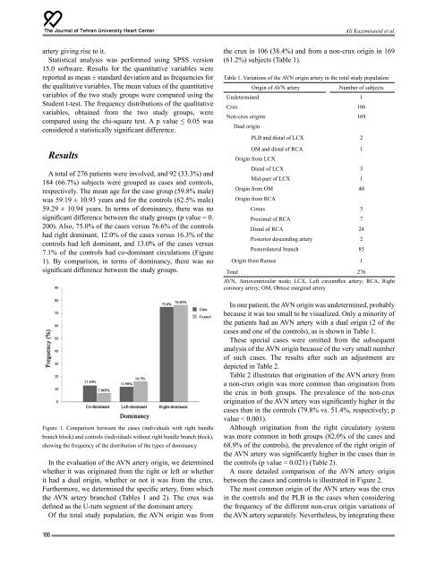
A total of 276 patients were involved, and 92 (33.3%) and<br />
184 (66.7%) subjects were grouped as cases and controls,<br />
respectively. The mean age for the case group (59.8% male)<br />
was 59.19 ± 10.93 years and for the controls (62.5% male)<br />
59.29 ± 10.94 years. In terms of dominancy, there was no<br />
significant difference between the study groups (p value = 0.<br />
200). Also, 75.0% of the cases versus 76.6% of the controls<br />
had right dominant, 12.0% of the cases versus 16.3% of the<br />
controls had left dominant, and 13.0% of the cases versus<br />
7.1% of the controls had co-dominant circulations (Figure<br />
1). By comparison, in terms of dominancy, there was no<br />
significant difference between the study groups.<br />
Figure 1. Comparison between the cases (individuals with right bundle<br />
branch block) and controls (individuals without right bundle branch block),<br />
showing the frequency of the distribution of the types of dominancy<br />
In the evaluation of the AVN artery origin, we determined<br />
whether it was originated from the right or left or whether<br />
it had a dual origin, whether or not it was from the crux.<br />
Furthermore, we determined the specific artery, from which<br />
the AVN artery branched (Tables 1 and 2). The crux was<br />
defined as the U-turn segment of the dominant artery.<br />
Of the total study population, the AVN origin was from<br />
the crux in 106 (38.4%) and from a non-crux origin in 169<br />
(61.2%) subjects (Table 1).<br />
Table 1. Variations of the AVN origin artery in the total study population<br />
Origin of AVN artery<br />
Number of subjects<br />
Undetermined 1<br />
Crux 106<br />
Non-crux origins 169<br />
Dual origin<br />
PLB and distal of LCX 2<br />
OM and distal of RCA 1<br />
Origin from LCX<br />
Distal of LCX 3<br />
Mid-part of LCX 1<br />
Origin from OM 40<br />
Origin from RCA<br />
Conus 3<br />
Proximal of RCA 7<br />
Distal of RCA 24<br />
Posterior descending artery 2<br />
Posterolateral branch 85<br />
Origin from Ramus 1<br />
Total 276<br />
AVN, Atrioventricular node; LCX, Left circumflex artery; RCA, Right<br />
coronary artery; OM, Obtuse marginal artery<br />
In one patient, the AVN origin was undetermined, probably<br />
because it was too small to be visualized. Only a minority of<br />
the patients had an AVN artery with a dual origin (2 of the<br />
cases and one of the controls), as is shown in Table 1.<br />
These special cases were omitted from the subsequent<br />
analysis of the AVN origin because of the very small number<br />
of such cases. The results after such an adjustment are<br />
depicted in Table 2.<br />
Table 2 illustrates that origination of the AVN artery from<br />
a non-crux origin was more common than origination from<br />
the crux in both groups. The prevalence of the non-crux<br />
origination of the AVN artery was significantly higher in the<br />
cases than in the controls (79.8% vs. 51.4%, respectively; p<br />
value < 0.001).<br />
Although origination from the right circulatory system<br />
was more common in both groups (82.0% of the cases and<br />
68.9% of the controls), the prevalence of the right origin of<br />
the AVN artery was significantly higher in the cases than in<br />
the controls (p value = 0.021) (Table 2).<br />
A more detailed comparison of the AVN artery origin<br />
between the cases and controls is illustrated in Figure 2.<br />
The most common origin of the AVN artery was the crux<br />
in the controls and the PLB in the cases when considering<br />
the frequency of the different non-crux origin variations of<br />
the AVN artery separately. Nevertheless, by integrating these<br />
166