THE JOURNAL OF TEHRAN UNIVERSITY HEART CENTER
THE JOURNAL OF TEHRAN UNIVERSITY HEART CENTER
THE JOURNAL OF TEHRAN UNIVERSITY HEART CENTER

Create successful ePaper yourself
Turn your PDF publications into a flip-book with our unique Google optimized e-Paper software.
The Journal of Tehran University Heart Center<br />
Mozhgan Parsaee et al.<br />
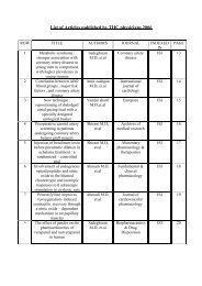
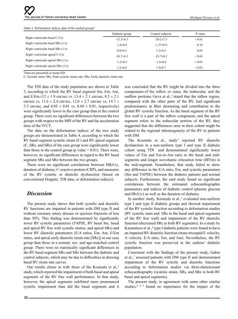
Table 4. Deformation indices data of the studied groups * Diabetic group Control subjects P value<br />
Right ventricular basal Є (%)<br />
Right ventricular basal SRs (1/s)<br />
Right ventricular basal SRe (1/s)<br />
Right ventricular apical Є (%)<br />
Right ventricular apical SRs (1/s)<br />
Right ventricular apical SRe (1/s)<br />
*<br />
Data are presented as mean±SD<br />
Є, Systolic stain; SRs, Peak systolic strain rate; SRe, Early diastolic strain rate<br />
-13.3±4.3 -20.2±7.3 < 0.01<br />
-1.0±0.5 -1.37±0.9 0.18<br />
0.9±0.3 1.2±0.5 0.05<br />
-18.7±4.3 -25.7±8.2 < 0.01<br />
-1.2±0.3 -1.6±0.6 < 0.01<br />
1.2±0.4 1.9±0.7 < 0.01<br />
The TDI data of the study population are shown in Table<br />
3, according to which the RV basal segment Sm, Em, Am,<br />
and E/Em (12 ± 1.9 cm/sec vs. 13.4 ± 2.1 cm/sec, 8.5 ± 2.1<br />
cm/sec vs. 11.6 ± 2.4 cm/sec, 12.6 ± 2.7 cm/sec vs. 14.7 ±<br />
3.3 cm/sec, and 0.05 ± 0.01 vs. 0.04 ± 0.01, respectively)<br />
were significantly lower in the case group than in the control<br />
group. There were no significant differences between the two<br />
groups with respect to the MPI of the RV and the acceleration<br />
time of the IVCT.<br />
The data on the deformation indices of the two study<br />
groups are demonstrated in Table 4, according to which the<br />
RV basal segment systolic strain (Є) and RV apical segment<br />
(Є, SRs, and SRe) of the case group were significantly lower<br />
than those in the control group (p value < 0.01). There were,<br />
however, no significant differences in regard to the RV basal<br />
segment SRs and SRe between the two groups.<br />
There were no significant correlations between HBA1c,<br />
duration of diabetes, C-reactive protein (CRP), and measures<br />
of the RV systolic or diastolic dysfunction (based on<br />
conventional Doppler, TDI data, or deformation indices).<br />
Discussion<br />
The present study shows that both systolic and diastolic<br />
RV functions are impaired in patients with DM type II and<br />
without coronary artery disease or ejection fractions of less<br />
than 50%. This finding was demonstrated by significantly<br />
lower RV systolic parameters (TAPSE, RV basal Sm, basal<br />
and apical RV free wall systolic strains, and apical SRs) and<br />
lower RV diastolic parameters (E/A ratios, Em, Am, E/Em<br />
ratios, and apical early diastolic strain rate [SRe]) in our case<br />
group than those in a normal, sex- and age-matched control<br />
group. There were no statistically significant differences in<br />
the RV basal segment SRs and SRe between the diabetic and<br />
control subjects, which may be due to difficulties in drawing<br />
basal RV strain rate curves.<br />
Our results chime in with those of the Kosmala et al., 4<br />
study, which reported the impairment of both basal and apical<br />
segments of the RV free wall performance. In that study,<br />
however, the apical segments exhibited more pronounced<br />
systolic impairment than did the basal segments and it<br />
was concluded that the RV might be divided into the three<br />
components of the inflow or sinus, the trabecular, and the<br />
outflow portions. Geva et al., 5 stated that the inflow region,<br />
compared with the other parts of the RV, had significant<br />
predominance in fiber shortening and contribution to the<br />
global RV systolic function. As the basal segment of the RV<br />
free wall is a part of the inflow component, and the apical<br />
segment refers to the trabecular portion of the RV, they<br />
suggested that the differences seen in their cohort might be<br />
related to the regional inhomogeneity of the RV in patients<br />
with DM.<br />
The Kosmala et al., study 4 reported RV diastolic<br />
dysfunction in a non-uniform type I and type II diabetic<br />
cohort using TDI and demonstrated significantly lower<br />
values of Em and Em-to-Am ratio in the basal and midsegments<br />
and longer isovolumic relaxation time (IRTm) in<br />
the mid-segment. Nonetheless, that study failed to show<br />
any difference in the E/A ratio, Em, and systolic parameters<br />
(Sm and TAPSE) between the diabetic patients and normal<br />
subjects. Furthermore, the said study found no significant<br />
correlations between the estimated echocardiographic<br />
parameters and indices of diabetic control (plasma glucose<br />
and HbA1c) as well as the duration of diabetes.<br />
In another study, Kosmala et al., 4 evaluated non-uniform<br />
type I and type II diabetic groups and showed impairment<br />
of the RV systolic function according to deformation studies<br />
(RV systolic stain and SRs in the basal and apical segments<br />
of the RV free wall) and impairment of the RV diastolic<br />
function (decreased SRe in both RV segments). In a study by<br />
Karamitsos et al., 6 type I diabetic patients were found to have<br />
an impaired RV diastolic function (trans-tricuspid E velocity,<br />
A velocity, E/A ratio, Em, and Am). Nevertheless, the RV<br />
systolic function was preserved in the authors’ diabetic<br />
population.<br />
Consistent with the findings of the present study, Gaber<br />
et al., 7 assessed patients with DM type II and demonstrated<br />
impairment of the RV systolic and diastolic functions<br />
according to deformation studies via three-dimensional<br />
echocardiography (systolic strain, SRs, and SRe in both RV<br />
basal and apical segments).<br />
The present study, in agreement with some other similar<br />
studies, 4, 8, 9 found no importance for the impact of the<br />
180