Journal of Tehran University Heart Center
Journal of Tehran University Heart Center
Journal of Tehran University Heart Center

You also want an ePaper? Increase the reach of your titles
YUMPU automatically turns print PDFs into web optimized ePapers that Google loves.
Zohair Yousef Al-halees, MD , FRCSC, FACS<br />
King Faisal <strong>Heart</strong> Institute, Saudi Arabia<br />
Yadolah Dodge, PhD<br />
<strong>University</strong> <strong>of</strong> Neuchâtel, Switzerland<br />
Ali Dodge-Khatami, MD, PhD<br />
<strong>University</strong> <strong>of</strong> Hamburg-Eppendorf School <strong>of</strong> Medicne, Germany<br />
Iradj Gandjbakhch, MD<br />
Hopital Pitie, France<br />
Omer Isik, MD<br />
Yeditepe <strong>University</strong>, School <strong>of</strong> Medicine, Turkey<br />
Sami S. Kabbani, MD<br />
Damascus <strong>University</strong> Cardiovascular Surgical <strong>Center</strong>, Syria<br />
Kayvan Kamalvand, MD, FRCP, FACC<br />
William Harvey Hospital, United Kingdom<br />
Jean Marco, MD, FESC<br />
Centre Cardio- Thoracique de Monaco, France<br />
Ali Massumi, MD<br />
Texas <strong>Heart</strong> Institute, U. S. A<br />
Carlos-A. Mestres, MD<br />
<strong>University</strong> <strong>of</strong> Barcelona, Spain<br />
Hossien Ahmadi, MD<br />
<strong>Tehran</strong> <strong>University</strong> <strong>of</strong> Medical Sciences<br />
Shahin Akhondzadeh, PhD<br />
<strong>Tehran</strong> <strong>University</strong> <strong>of</strong> Medical Sciences<br />
Mohammad Alidoosti, MD<br />
<strong>Tehran</strong> <strong>University</strong> <strong>of</strong> Medical Sciences<br />
Mohammad Ali Boroumand, MD<br />
<strong>Tehran</strong> <strong>University</strong> <strong>of</strong> Medical Sciences<br />
Ahmad Reza Dehpour, PhD<br />
<strong>Tehran</strong> <strong>University</strong> <strong>of</strong> Medical Sciences<br />
Abbasali Karimi, MD<br />
<strong>Tehran</strong> <strong>University</strong> <strong>of</strong> Medical Sciences<br />
Seyed Ebrahim Kassaian, MD<br />
<strong>Tehran</strong> <strong>University</strong> <strong>of</strong> Medical Sciences<br />
Davood Kazemi Saleh, MD<br />
Baghiatallah <strong>University</strong> <strong>of</strong> Medical Sciences<br />
Majid Maleki, MD<br />
Iran <strong>University</strong> <strong>of</strong> Medical Sciences<br />
Mehrab Marzban, MD<br />
<strong>Tehran</strong> <strong>University</strong> <strong>of</strong> Medical Sciences<br />
Mansor Moghadam, MD<br />
<strong>Tehran</strong> <strong>University</strong> <strong>of</strong> Medical Sciences<br />
Sina Moradmand Badie, MD<br />
<strong>Tehran</strong> <strong>University</strong> <strong>of</strong> Medical Sciences<br />
Seyed Mahmood Mirhoseini, MD, DSc, FACC, FAES<br />
<strong>Tehran</strong> <strong>University</strong> <strong>of</strong> Medical Sciences<br />
THE<br />
JOURNAL OF TEHRAN<br />
UNIVERSITY HEART<br />
CENTER<br />
Editor-in-Chief<br />
ABBASALI KARIMI, MD<br />
PROFESSOR OF CARDIAC SURGERY<br />
TEHRAN UNIVERSITY OF MEDICAL SCIENCES<br />
Managing Editor<br />
SEYED HESAMEDDIN ABBASI, MD<br />
TEHRAN HEART CENTER<br />
TEHRAN UNIVERSITY OF MEDICAL SCIENCES<br />
International Editors<br />
Editorial Board<br />
Fred Morady, MD<br />
<strong>University</strong> <strong>of</strong> Michigan, U. S. A<br />
Mohammed T. Numan, MD<br />
<strong>University</strong> <strong>of</strong> Texas, U. S. A<br />
Ahmand S. Omran, MD, FACC, FASE<br />
King Abdulaziz Cardiac <strong>Center</strong>, Saudi Arabia<br />
Fausto J. Pinto, MD, PhD, FESC, FACC, FASA, FSCAI,<br />
FASE<br />
Lisbon <strong>University</strong>, Portugal<br />
Mehrdad Rezaee, MD, PhD<br />
Stanford <strong>University</strong>, School <strong>of</strong> Medicine, U. S. A<br />
Gregory S. Thomas, MD, MPH, FACC, FACP, FASNC<br />
<strong>University</strong> <strong>of</strong> California, U. S. A<br />
Lee Samuel Wann, MD<br />
Wisconsin <strong>Heart</strong> Hospital, U. S. A<br />
Hein J. Wellens, MD<br />
Cardiovascular Research Institute, Maastricht, The Netherlands<br />
Douglas P. Zipes, MD<br />
Indiana <strong>University</strong>, School <strong>of</strong> medicine, U. S. A<br />
Seyed Rasoul Mirsharifi, MD<br />
<strong>Tehran</strong> <strong>University</strong> <strong>of</strong> Medical Sciences<br />
Ahmad Mohebi, MD<br />
Iran <strong>University</strong> <strong>of</strong> Medical Sciences<br />
Mohammad-Hasan Namazi<br />
Shaheed beheshti <strong>University</strong> <strong>of</strong> Medical Sciences<br />
Ebrahim Nematipour, MD<br />
<strong>Tehran</strong> <strong>University</strong> <strong>of</strong> Medical Sciences<br />
Rezayat Parvizi, MD<br />
Tabriz <strong>University</strong> <strong>of</strong> Medical Sciences<br />
Masoud Pezeshkian<br />
Tabriz <strong>University</strong> <strong>of</strong> Medical Sciences<br />
Hassan Radmehr, MD<br />
<strong>Tehran</strong> <strong>University</strong> <strong>of</strong> Medical Sciences<br />
Saeed Sadeghian, MD<br />
<strong>Tehran</strong> <strong>University</strong> <strong>of</strong> Medical Sciences<br />
Mojtaba Salarifar, MD<br />
<strong>Tehran</strong> <strong>University</strong> <strong>of</strong> Medical Sciences<br />
Nizal Sarraf –Zadegan, MD<br />
Isfahan <strong>University</strong> <strong>of</strong> Medical Sciences<br />
Ahmad Yaminisharif, MD<br />
<strong>Tehran</strong> <strong>University</strong> <strong>of</strong> Medical Sciences<br />
Mohammad Reza Zafarghandi, MD<br />
<strong>Tehran</strong> <strong>University</strong> <strong>of</strong> Medical Sciences<br />
Aliakbar Zeinaloo, MD<br />
<strong>Tehran</strong> <strong>University</strong> <strong>of</strong> Medical Sciences
Advisory Board<br />
Kiyomars Abbasi, MD<br />
<strong>Tehran</strong> <strong>University</strong> <strong>of</strong> Medical Sciences<br />
Seifollah Abdi, MD<br />
Iran <strong>University</strong> <strong>of</strong> Medical Sciences<br />
Alireza Amirzadegan, MD<br />
<strong>Tehran</strong> <strong>University</strong> <strong>of</strong> Medical Sciences<br />
Naser Aslanabadi, MD<br />
Tabriz <strong>University</strong> <strong>of</strong> Medical Sciences<br />
Sirous Darabian, MD<br />
<strong>Tehran</strong> <strong>University</strong> <strong>of</strong> Medical Sciences<br />
Gholamreza Davoodi, MD<br />
<strong>Tehran</strong> <strong>University</strong> <strong>of</strong> Medical Sciences<br />
Saeed Davoodi, MD<br />
<strong>Tehran</strong> <strong>University</strong> <strong>of</strong> Medical Sciences<br />
Iraj Firoozi, MD<br />
Iran <strong>University</strong> <strong>of</strong> Medical Sciences<br />
Seyed Khalil Foroozannia, MD<br />
Shaheed Sadoghi <strong>University</strong> <strong>of</strong> Medical Sciences<br />
Armen Gasparyan MD, PhD<br />
Armenia<br />
Ali Ghaemian, MD<br />
Mazandaran <strong>University</strong> <strong>of</strong> Medical Sciences<br />
Namvar Ghasemi Movahedi, MD<br />
<strong>Tehran</strong> <strong>University</strong> <strong>of</strong> Medical Sciences<br />
Abbas Ghiasi, MD<br />
<strong>Tehran</strong> <strong>University</strong> <strong>of</strong> Medical Sciences<br />
Ali Kazemi Saeed, MD<br />
<strong>Tehran</strong> <strong>University</strong> <strong>of</strong> Medical Sciences<br />
Ali Mohammad Haji Zeinali, MD<br />
<strong>Tehran</strong> <strong>University</strong> <strong>of</strong> Medical Sciences<br />
Mohammad Jafar Hashemi, MD<br />
Iran <strong>University</strong> <strong>of</strong> Medical Sciences<br />
Mahmood Sheikh Fathollahi<br />
Pedram Amouzadeh<br />
Fatemeh Esmaeili Darabi<br />
Seyed Kianoosh Hoseini<br />
<strong>Tehran</strong> <strong>University</strong> <strong>of</strong> Medical Sciences<br />
Elise Langdon- Neuner<br />
The editor <strong>of</strong> The Write Stuff, Austria<br />
Jalil Majd Ardekani, MD<br />
<strong>Tehran</strong> <strong>University</strong> <strong>of</strong> Medical Sciences<br />
Fardin Mirbolook, MD<br />
Gilan <strong>University</strong> <strong>of</strong> Medical Sciences<br />
Mehdi Najafi, MD<br />
<strong>Tehran</strong> <strong>University</strong> <strong>of</strong> Medical Sciences<br />
Younes Nozari, MD<br />
<strong>Tehran</strong> <strong>University</strong> <strong>of</strong> Medical Sciences<br />
Hamid Reza Pour Hosseini, MD<br />
<strong>Tehran</strong> <strong>University</strong> <strong>of</strong> Medical Sciences<br />
Hakimeh Sadeghian, MD<br />
<strong>Tehran</strong> <strong>University</strong> <strong>of</strong> Medical Sciences<br />
Mohammad Saheb Jam, MD<br />
<strong>Tehran</strong> <strong>University</strong> <strong>of</strong> Medical Sciences<br />
Abbas Salehi Omran, MD<br />
<strong>Tehran</strong> <strong>University</strong> <strong>of</strong> Medical Sciences<br />
Mahmood Shabestari, MD<br />
Mashhad <strong>University</strong> <strong>of</strong> Medical Sciences<br />
Shapour Shirani, MD<br />
<strong>Tehran</strong> <strong>University</strong> <strong>of</strong> Medical Sciences<br />
Abbas Soleimani, MD<br />
Kerman <strong>University</strong> <strong>of</strong> Medical Sciences<br />
Seyed Abdolhosein Tabatabaei, MD<br />
<strong>Tehran</strong> <strong>University</strong> <strong>of</strong> Medical Sciences<br />
Murat Ugurlucan, MD<br />
Rostock <strong>University</strong> Medical Faculty<br />
Arezou Zoroufian, MD<br />
<strong>Tehran</strong> <strong>University</strong> <strong>of</strong> Medical Sciences<br />
Statistical Consultant<br />
Technical Editors<br />
Graphic Design & Office<br />
The <strong>Journal</strong> <strong>of</strong> <strong>Tehran</strong> <strong>University</strong> <strong>Heart</strong> <strong>Center</strong> is indexed in Scopus, EMBASE, CAB Abstracts, Global Health,<br />
Chemical Abstract Service, Cinahl, Google Scholar, DOAJ, Geneva Foundation for Medical Education and<br />
Research, Index Copernicus, Index Medicus for the WHO Eastern Mediterranean Region (IMEMR), ISC, SID,<br />
Iranmedex and Magiran<br />
Address<br />
North Kargar Street, <strong>Tehran</strong> <strong>Heart</strong> <strong>Center</strong>, <strong>Tehran</strong>, Iran 1411713138. Tel: +98-21-88029720. Fax: +98-21-88029702.<br />
Web Site: http://jthc.tums.ac.ir. E-mail: jthc@tums.ac.ir.
TEHRAN HEART CENTER<br />
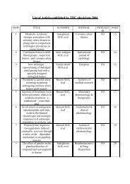
Content<br />
Volume: 6 Number: 2 Spring 2011<br />
The <strong>Journal</strong> <strong>of</strong> <strong>Tehran</strong> <strong>University</strong> <strong>Heart</strong> <strong>Center</strong><br />
Review Article<br />
Cardiovascular Effects <strong>of</strong> Saffron: An Evidence-Based Review<br />
Maryam Kamalipour, Shahin Akhondzadeh …………….................................................................................................................................................... 59<br />
Original Articles<br />
Demographics and Angiographic Findings in Patients under 35 Years <strong>of</strong> Age with Acute ST Elevation Myocardial<br />
Infarction<br />
Seyed Kianoosh Hosseini, Abbas Soleimani, Mojtaba Salarifar, Hamidreza Pourhoseini, Ebrahim Nematipoor, Seyed Hesameddin Abbasi, Ali Abbasi ...... 62<br />
Signal-Averaged Electrocardiography in Patients with Advanced <strong>Heart</strong> Failure: A Better Indicator <strong>of</strong> Left<br />
Ventricular Enlargement Compared with Conventional Electrocardiography<br />
Mohammad Alasti, Majid Haghjoo, Abolfath Alizadeh, Mohammad Hossein Nikoo, Hamid Reza Bonakdar, Bita Omidvar .......................................... 68<br />
Carotid Artery Wall Motion Estimation from Consecutive Ultrasonic Images: Comparison <strong>of</strong> Block Matching and<br />
Maximum Gradient Algorithms<br />
Effat Soleimani, Manijhe Mokhtari-Dizaji, Hajir Saberi .................................................................................................................................................... 72<br />
Transcatheter Closure <strong>of</strong> Atrial Septal Defect with Amplatzer Septal Occluder in Adults: Immediate, Short, and<br />
Intermediate-Term Results<br />
Mostafa Behjati, Mansour Rafiei, Mohammad Hossein Soltani, Mahmoud Emami, Majid Dehghani ................................................................ 79<br />
Case Reports<br />
Incidental Finding <strong>of</strong> Cor Triatriatum Sinistrum in a Middle-Aged Man Candidated for Coronary Bypass Grafting<br />
(with Three-D Imaging)<br />
Afsoon Fazlinezhad, Farveh Vakilian, Asadollah Mirzaei, Azadeh Fallah Rastegar ……................................................................................................... 85<br />
Segmented Coronary Artery Aneurysms and Kawasaki Disease<br />
Mohammad Yoosef Aarabi Moghadam, Hojat Mortazaeian, Mehdi Ghaderian, Hamid Reza Ghaemi ……..…................................................................ 89<br />
Traumatic Left Anterior Descending Coronary Artery-Right Ventricle Fistula: A Case Report<br />
Mohammad Ali Sheikhi, Mehdi Asgari, Mehdi Dehghani Firouzabadi, Mohammad Reza Zeraati, Alireza Rezaee...……….............................……..… 92<br />
Letter to the Editor<br />
Percutaneous Revascularization <strong>of</strong> Patients with History <strong>of</strong> Coronary Artery Bypass Grafting<br />
Seyed Kianoosh Hoseini, Fatemeh Behboudi ………………………………………………..……………….......................................……....….....…… 95<br />
The <strong>Journal</strong> <strong>of</strong> <strong>Tehran</strong> <strong>University</strong> <strong>Heart</strong> <strong>Center</strong>
TEHRAN HEART CENTER<br />
Review Article<br />
Cardiovascular Effects <strong>of</strong> Saffron: An Evidence-Based<br />
Review<br />
Maryam Kamalipour, MSc, Shahin Akhondzadeh, PhD, FBPharmacolS *<br />
Roozbeh Psychiatric Hospital, <strong>Tehran</strong> <strong>University</strong> <strong>of</strong> Medical Sciences, <strong>Tehran</strong>, Iran.<br />
Received 15 January 2011; Accepted 28 March 2011<br />
Abstract<br />
Herbal medicine can be a valuable source <strong>of</strong> assistance for traditional medicine. There are a number <strong>of</strong> herbs that can be<br />
used in conjunction with modern medicine. Herbs can also be taken to aid recovery from serious diseases. Although one should<br />
never aim to treat diseases such as cardiovascular disease solely with herbal medicine, the value <strong>of</strong> herbs used in tandem with<br />
modern medicine cannot be ignored. Saffron has been reported to help lower cholesterol and keep cholesterol levels healthy.<br />
Animal studies have shown saffron to lower cholesterol by as much as 50%. Saffron has antioxidant properties; it is, therefore,<br />
helpful in maintaining healthy arteries and blood vessels. Saffron is also known to have anti-inflammatory properties, which<br />
are beneficial to cardiovascular health. The people <strong>of</strong> Mediterranean countries, where saffron use is common, have lower<br />
than normal incidence <strong>of</strong> heart diseases. From saffron's cholesterol lowering benefits to its anti inflammatory properties,<br />
saffron may be one <strong>of</strong> the best supplements for cardiac health. This paper reviews the studies regarding the beneficial effects<br />
<strong>of</strong> saffron in cardiovascular health.<br />
J Teh Univ <strong>Heart</strong> Ctr 2011;6(2):59-61<br />
This paper should be cited as: Kamalipour M, Akhondzadeh S. Cardiovascular Effects <strong>of</strong> Saffron: An Evidence-Based Review. J<br />
Teh Univ <strong>Heart</strong> Ctr 2011;6(2):59-61.<br />
Keywords: Anti-inflammatory agents, non-steriodal • Cardiovascular agents • Lipid regulating agents • Crocus sativus<br />
Introduction<br />
The role <strong>of</strong> alternative medicine in general and<br />
phytotherapy in various diseases in particular has been <strong>of</strong><br />
extreme interest to various scientific and non-scientific<br />
communities throughout the world. Phytotherapy is broadly<br />
defined as the use <strong>of</strong> natural therapeutic agents derived from<br />
plants or crude herbal drugs. Herbal medicine has a long and<br />
respected history and holds a valuable place in the treatment<br />
<strong>of</strong> cardiovascular diseases as well as the vast majority <strong>of</strong><br />
health problems. Utilizing the leaves, flowers, stems, berries,<br />
and roots <strong>of</strong> plants to both prevent and treat illness, herbal<br />
medicine not only helps to alleviate symptoms but also helps<br />
to treat the underlying problem, as well as strengthen the<br />
overall functioning <strong>of</strong> a particular organ or body system. 1, 2<br />
Cardiovascular diseases are now considered a major cause<br />
<strong>of</strong> mortality not only in the developed world but also in the<br />
developing countries. In the age <strong>of</strong> genomics, nanotechnology,<br />
and proteomics, cardiovascular diseases continue to remain<br />
a major challenge to therapeutically manage; and the search<br />
for a viable evidence-based alternative continues.<br />
Saffron (Crocis sativus) is a spice derived from the flower<br />
<strong>of</strong> the saffron crocus (Crocus sativus), a species <strong>of</strong> crocus in<br />
the family Iridaceae. The flower has three stigmas, which<br />
are the distal ends <strong>of</strong> the plant’s carpels. Together with its<br />
style, the stalk connecting the stigmas to the rest <strong>of</strong> the plant,<br />
*<br />
Corresponding Author: Shahin Akhondzadeh, Pr<strong>of</strong>essor <strong>of</strong> Neuroscience, Psychiatric Research <strong>Center</strong>, Roozbeh Psychiatric Hospital, <strong>Tehran</strong><br />
<strong>University</strong> <strong>of</strong> Medical Sciences, South Kargar Street, <strong>Tehran</strong>, Iran. 13337. Tel: +98 21 88281866, Fax: +98 21 55419113. E-mail: s.akhond@neda.net.<br />
The <strong>Journal</strong> <strong>of</strong> <strong>Tehran</strong> <strong>University</strong> <strong>Heart</strong> <strong>Center</strong> 59
The <strong>Journal</strong> <strong>of</strong> <strong>Tehran</strong> <strong>University</strong> <strong>Heart</strong> <strong>Center</strong><br />
Shahin Akhondzadeh et al.<br />
these components are <strong>of</strong>ten dried and used in cooking as a<br />
seasoning and coloring agent. Saffron, which has for decades<br />
been the world’s most expensive spice by weight, is native to<br />
Iran; it was first cultivated in the Persian Empire. 2, 3 Saffron<br />
is characterized by a bitter taste and an iod<strong>of</strong>orm- or haylike<br />
fragrance; these are caused by the chemicals picrocrocin<br />
and safranal. It also contains a carotenoid dye, crocin, which<br />
gives food a rich golden-yellow hue. These traits make<br />
saffron a much-sought ingredient in many foods worldwide.<br />
Saffron also has medicinal applications. 3, 4<br />
Saffron tastes bitter and contributes a luminous yelloworange<br />
coloring to foods. Because <strong>of</strong> the unusual taste and<br />
coloring it adds to foods, saffron is widely used in Persian,<br />
Arab, Central Asian, European, Indian, Moroccan, and<br />
Cornish cuisines. Confectionaries and liquors also <strong>of</strong>ten<br />
include saffron. Medicinally, saffron has a long history as part<br />
<strong>of</strong> traditional healing; modern medicine has also discovered<br />
saffron as having anticarcinogenic (cancer-suppressing),<br />
anti-mutagenic (mutation-preventing), immuno-modulating,<br />
and antioxidant-like properties. Saffron has also been<br />
used as a fabric dye, particularly in China and India, and<br />
in perfumery. 1, 2 Recent studies have shown the beneficial<br />
effects <strong>of</strong> saffron in depression, premenstrual syndrome<br />
(PMS), and Alzheimer’s Disease. 3-9<br />
Saffron and <strong>Heart</strong> Disease Protection<br />
Antioxidants in saffron tea can reduce the risk <strong>of</strong><br />
cardiovascular diseases. The flavonoids, especially lycopene,<br />
found in saffron can provide added protection. A clinical trial<br />
at the Department <strong>of</strong> Medicine and Indigenous Drug Research<br />
<strong>Center</strong> showed positive effects <strong>of</strong> saffron on cardiovascular<br />
diseases. 10 The study involved 20 participants, including<br />
10 with heart diseases. According to the Indian <strong>Journal</strong> <strong>of</strong><br />
Medical Sciences, all the participants showed improved<br />
health, but those with cardiovascular diseases showed more<br />
progress. In addition, saffron has been found to be the richest<br />
source <strong>of</strong> rib<strong>of</strong>lavin. 1, 2 Due to the presence <strong>of</strong> crocetin, it<br />
indirectly helps to reduce cholesterol level in the blood<br />
and severity <strong>of</strong> atherosclerosis, thus reducing the chances<br />
<strong>of</strong> heart attacks. It may be one <strong>of</strong> the prime reasons that in<br />
Spain, where Saffron is consumed liberally, the incidence <strong>of</strong><br />
cardiovascular diseases is quite low. The crocetin present in<br />
saffron is found to increase the yield <strong>of</strong> antibiotics. 11 Two<br />
compounds <strong>of</strong> safranal are supposed to increase antibacterial<br />
and antiviral physiological activity in the body. 12<br />
In 2005, Zheng et al. administered crocetin, the natural<br />
carotenoid antioxidant, to rabbits to determine its effect on<br />
the development <strong>of</strong> atherosclerosis. The authors randomly<br />
assigned New Zealand white rabbits to three different diets<br />
for eight weeks: a standard diet, a high lipid diet (HLD),<br />
or a high lipid + crocetin diet. The HLD group developed<br />
hypercholesterolemia and atherosclerosis, while the<br />
crocetin-supplemented group decreased the negative health<br />
effects <strong>of</strong> a high lipid diet. 13 The results did not show a<br />
significant difference in the plasma lipid levels (total, low<br />
density lipoprotein (LDL), and high density lipoprotein<br />
(HDL) cholesterol) between the HLD and crocetin groups<br />
but did show a significant decrease in the aorta cholesterol<br />
deposits, atheroma, foam cells, and atherosclerotic lesions<br />
in the crocetin-fed group. They suggested that nuclear factor<br />
kappa B (NF-κB) activation in the aortas is suppressed by<br />
antioxidants such as crocetin which in turn decreases the<br />
vascular cell adhesion molecule-1 (VCAM-1) expression. 13<br />
A 2006 study by Sheng and colleagues looked at<br />
an alternative mechanism for crocin’s atherosclerotic<br />
properties. 14 Crocin inhibited an increase in serum<br />
triglycerides, total-, LDL-, cholesterol compared to the<br />
control group as seen before; however, the results also<br />
showed a significant increase in the fecal excretion <strong>of</strong> fat<br />
and cholesterol in the crocin group (100 mg/kg/day). 14<br />
Further studies determined that crocin inhibited pancreatic<br />
and gastric lipase activity, although a potential mechanism<br />
was not <strong>of</strong>fered. Since pancreatic lipase is responsible for fat<br />
absorption by hydrolyzing fat, the inhibition <strong>of</strong> pancreatic<br />
lipase activity resulted in low lipid absorption. With a lack<br />
<strong>of</strong> potential pancreatic lipase inhibitors available, crocin<br />
shows promise as a drug for treating hyperlipidemia. 14<br />
In conclusion, saffron helps reduce the risk <strong>of</strong> heart<br />
diseases by strengthening the blood circulatory system. Rich<br />
in minerals like thiamin and rib<strong>of</strong>lavin, saffron promotes a<br />
healthy heart and prevents different cardiac problems.<br />
References<br />
1. Schmidt M, Betti G, Hensel A. Saffron in phytotherapy:<br />
pharmacology and clinical uses. Wien Med Wochenschr<br />
2007;157:315-319.<br />
2. Bathaie SZ, Mousavi SZ. New applications and mechanisms <strong>of</strong><br />
action <strong>of</strong> saffron and its important ingredients. Crit Rev Food Sci<br />
Nutr 2010;50:761-786.<br />
3. Akhondzadeh S, Tahmacebi-Pour N, Noorbala AA, Amini H, Fallah-<br />
Pour H, Jamshidi AH, Khani M. Crocus sativus L. in the treatment<br />
<strong>of</strong> mild to moderate depression: a double-blind, randomized and<br />
placebo controlled trial. Phytother Res 2005;19:148-151.<br />
4. Noorbala AA, Akhondzadeh S, Tahmacebi-Pour N, Jamshidi AH.<br />
Hydroalcoholic extract <strong>of</strong> Crocus sativus L. versus fluoxetine<br />
in the treatment <strong>of</strong> mild to moderate depression: a double-blind,<br />
randomized pilot trial. J Ethnopharmacol 2005;97:281-284.<br />
5. Moshiri E, Basti AA, Noorbala AA, Jamshidi AH, Abbasi S,<br />
Akhondzadeh SH. Crocus sativus L. (petal) in the treatment <strong>of</strong><br />
mild-to-moderate depression: a double-blind, randomized and<br />
placebo controlled trial. Phytomedicine 2006;13:607-611.<br />
6. Akhondzadeh Basti A, Moshiri E, Noorbala AA, Jamshidi AH,<br />
Abbasi SH, Akhondzadeh S. Comparison <strong>of</strong> petal <strong>of</strong> Crocus sativus<br />
L. and fluoxetine in the treatment <strong>of</strong> depressed outpatients: a pilot<br />
double-blind randomized trial. Prog Neuropsychopharmacol Biol<br />
Psychiatry 2007;31:439-442.<br />
7. Agha-Hosseini M, Kashani L, Aleyaseen A, Ghoreishi A,<br />
Rahmanpour H, Zarrinara AR, Akhondzadeh S. Crocus sativus L.<br />
(saffron) in the treatment <strong>of</strong> premenstrual syndrome: a double-blind,<br />
randomised and placebo controlled trial. BJOG 2008;115:515-<br />
519.<br />
60
Cardiovascular Effects <strong>of</strong> Saffron: An Evidence-Based Review<br />
TEHRAN HEART CENTER<br />
8. Akhondzadeh S, Sabet MS, Harirchian MH, Togha M,<br />
Cheraghmakani H, Razeghi S, Hejazi SS, Yousefi MH, Alimardani<br />
R, Jamshidi A, Zare F, Moradi A. Saffron in the treatment <strong>of</strong> patients<br />
with mild to moderate Alzheimer’s disease: a 16-week, randomized<br />
and placebo-controlled trial. J Clin Pharm Ther 2010;35:581-588.<br />
9. Akhondzadeh S, Shafiee Sabet M, Harirchian MH, Togha M,<br />
Cheraghmakani H, Razeghi S, Hejazi SS, Yousefi MH, Alimardani<br />
R, Jamshidi A, Rezazadeh SA, Yousefi A, Zare F, Moradi A,<br />
Vossoughi A. A 22-week, multicenter, randomized, doubleblind<br />
controlled trial <strong>of</strong> Crocus sativus in the treatment <strong>of</strong> mildto-moderate<br />
Alzheimer’s disease. Psychopharmacology (Berl)<br />
2010;207:637-643.<br />
10. Verma SK, Bordia A. Antioxidant property <strong>of</strong> Saffron in man.<br />
Indian J Med Sci 1998;52:205-207.<br />
11. Basker D, Negbi M. The use <strong>of</strong> saffron. Econ Bot 1983;37:228-<br />
236.<br />
12. Zarghami NS, Heinz DE. Monoterpene aldehydes and isophorone<br />
337 related compounds <strong>of</strong> saffron. Phytochemistry 1971;10:2755-<br />
2761.<br />
13. Zheng S, Qian Z, Tang F, Sheng L. Suppression <strong>of</strong> vascular<br />
cell adhesion molecule-1 expression by crocetin contributes to<br />
attenuation <strong>of</strong> atherosclerosis in hypercholesterolemic rabbits.<br />
Biochem Pharmacol 2005;70:1192-1199.<br />
14. Sheng L, Qian Z, Zheng S, Xi L. Mechanism <strong>of</strong> hypolipidemic<br />
effect <strong>of</strong> crocin in rats: crocin inhibits pancreatic lipase. Eur J<br />
Pharmacol 2006;543:116-122.<br />
The <strong>Journal</strong> <strong>of</strong> <strong>Tehran</strong> <strong>University</strong> <strong>Heart</strong> <strong>Center</strong>61
The <strong>Journal</strong> <strong>of</strong> <strong>Tehran</strong> <strong>University</strong> <strong>Heart</strong> <strong>Center</strong><br />
Original Article<br />
Demographics and Angiographic Findings in Patients<br />
under 35 Years <strong>of</strong> Age with Acute ST Elevation Myocardial<br />
Infarction<br />
Seyed Kianoosh Hosseini, MD 1 , Abbas Soleimani, MD 1 , Mojtaba Salarifar,<br />
MD 1* , Hamidreza Pourhoseini, MD 1 , Ebrahim Nematipoor, MD 1 , Seyed<br />
Hesameddin Abbasi, MD 1, 2 , Ali Abbasi, MD 3<br />
1<br />
<strong>Tehran</strong> <strong>Heart</strong> <strong>Center</strong>, <strong>Tehran</strong> <strong>University</strong> <strong>of</strong> Medical Sciences, <strong>Tehran</strong>, Iran.<br />
2<br />
Family Health Research <strong>Center</strong>, Iranian Petroleum Industry Health Research Institute, <strong>Tehran</strong>, Iran.<br />
3<br />
<strong>University</strong> Medical <strong>Center</strong> Groningen, Groningen, the Netherlands.<br />
Received 23 September 2010; Accepted 18 January 2011<br />
Abstract<br />
Background: ST-elevation myocardial infarction (STEMI) is a major cause <strong>of</strong> cardiovascular mortality worldwide.<br />
There are differences between very young patients with STEMI and their older counterparts. This study investigates the<br />
demographics and clinical findings in very young patients with STEMI.<br />
Methods: Through a review <strong>of</strong> the angiography registry, 108 patients aged ≤ 35 years (Group I) were compared with 5544<br />
patients aged > 35 years (Group II) who underwent coronary angiography after STEMI.<br />
Results: Group I patients were more likely to be male (92.6%), smokers, and have a family history <strong>of</strong> cardiovascular<br />
diseases (34.6%). The prevalence <strong>of</strong> diabetes, dyslipidemia, and hypertension was higher in the old patients. Triglyceride<br />
and hemoglobin were significantly higher in Group I. Normal coronary angiogram was reported in 18.5% <strong>of</strong> the young<br />
patients, and in 2.1% <strong>of</strong> the older patients. The prevalence <strong>of</strong> single-vessel and multi-vessel coronary artery disease was<br />
similar in the two groups (34.3% vs. 35.2%). The younger subjects were more commonly candidates for medical treatment<br />
and percutaneous coronary intervention (PCI) (84.2%), while coronary artery bypass grafting (CABG) was considered for<br />
the 39.5% <strong>of</strong> their older counterparts.<br />
Conclusion: In the young adults with STEMI, male gender, smoking, family history, and high triglyceride level were more<br />
<strong>of</strong>ten observed. A considerable proportion <strong>of</strong> the young patients presented with multi-vessel coronary disease. PCI or medical<br />
treatment was the preferred treatment in the younger patients; in contrast to their older counterparts, in whom CABG was<br />
more commonly chosen for revascularization.<br />
J Teh Univ <strong>Heart</strong> Ctr 2011;6(2):62-67<br />
This paper should be cited as: Hosseini SK, Soleimani A, Salarifar M, Pourhoseini H, Nematipoor E, Abbasi SH, Abbasi A.<br />
Demographics and Angiographic Findings in Patients under 35 Years <strong>of</strong> Age with Acute ST Elevation Myocardial Infarction. J Teh Univ<br />
<strong>Heart</strong> Ctr 2011;6(2):62-67.<br />
Keywords: Myocardial infarction • Young adult • Coronary angiography • Risk factors<br />
*<br />
Corresponding Author: Mojtaba Salarifar, Assistant Pr<strong>of</strong>essor <strong>of</strong> Interventional Cardiology, Cardiology Department, <strong>Tehran</strong> <strong>Heart</strong> <strong>Center</strong>, North<br />
Kargar Street, <strong>Tehran</strong>, Iran. 1411713138. Tel: +98 21 88029257. Fax: +98 21 88029256. E-mail: salarifar@tehranheartcenter.org.<br />
62
Demographics and Angiographic Findings in Patients under 35 Years <strong>of</strong> ...<br />
Introduction<br />
ST-segment elevation myocardial infarction (STEMI) is<br />
one <strong>of</strong> the most common causes <strong>of</strong> emergency department<br />
admissions and cardiovascular mortalities and thus currently<br />
accounts for a high burden on health care services in the<br />
world. Although STEMI is an uncommon entity in young<br />
patients, it has always attracted special attention because<br />
<strong>of</strong> its unusual features and devastating effect on their more<br />
active lifestyle. 1-5<br />
In recent years, whereas the mean age <strong>of</strong> coronary artery<br />
disease (CAD) has decreased, its prevalence seems to have<br />
been on the increase. 4 A number <strong>of</strong> studies, including our<br />
previous report, have shown significant differences in the risk<br />
factor pr<strong>of</strong>ile and coronary angiographic patterns between<br />
young and older patients with acute STEMI. 1-3, 6-8 Traditional<br />
risk factors <strong>of</strong> CAD are prevalent in young patients with<br />
acute STEMI but with a different pattern compared to their<br />
older counterparts; these differences may cause different<br />
treatment strategies and outcome among these patients. 3,<br />
5, 9<br />
There is evidence that a concentration <strong>of</strong> the novel risk<br />
factors <strong>of</strong> CAD such as LP (a) may be higher than normal in<br />
the <strong>of</strong>fspring <strong>of</strong> patients with a history <strong>of</strong> premature MI. 10<br />
There is a dearth <strong>of</strong> available data on very young adults with<br />
STEMI, as a life-threatening cardiac emergency condition.<br />
In an attempt to characterize patients 35 years <strong>of</strong> age or<br />
younger suffering STEMI, we reviewed the <strong>Tehran</strong> <strong>Heart</strong><br />
<strong>Center</strong> Angiography Registry (THCAR) and compared the<br />
demographics and clinical findings <strong>of</strong> these patients to those<br />
older than 35 years <strong>of</strong> age.<br />
Methods<br />
From all the patients admitted by cardiologists between<br />
January 2000 and March 2008 to the Angiography<br />
Department affiliated with the Academic <strong>Tehran</strong> <strong>Heart</strong> <strong>Center</strong>,<br />
we identified 5652 patients with a history <strong>of</strong> STEMI. The<br />
databank contains patients’ data collected by cardiologists and<br />
trained general practitioners, and the validity <strong>of</strong> all the data<br />
is checked by reabstracting 10% <strong>of</strong> the patients’ entries and<br />
by reentering 5% <strong>of</strong> the patients' records. The investigation<br />
was approved by the institutional Review Board, overseeing<br />
the participation <strong>of</strong> human subjects in research at <strong>Tehran</strong><br />
<strong>University</strong> <strong>of</strong> Medical Sciences. This study conforms to the<br />
principles outlined in the Declaration <strong>of</strong> Helsinki.<br />
The validation <strong>of</strong> acute myocardial infarction (AMI) events<br />
was based on information on medical history, symptoms,<br />
electrocardiogram, and cardiac enzymes. STEMI was<br />
diagnosed when new or presumed new ST-segment elevation<br />
≥ 1 mm ( ≥ 2 mm in V 1<br />
to V 3<br />
) was seen in any location in two<br />
or more contiguous leads or new left bundle branch block<br />
was found on the index or qualifying electrocardiogram<br />
with ≥ 1 positive cardiac biochemical marker <strong>of</strong> necrosis<br />
TEHRAN HEART CENTER<br />
(including CKMB-mass or quantitative cardiac troponin<br />
measurements). The cardiologist who performed the coronary<br />
angiography documented and recorded STEMI diagnosis<br />
in the datasheets. Coronary angiography was performed in<br />
almost all the patients as part <strong>of</strong> the pharmacoinvasive strategy<br />
or as the primary treatment option (primary percutaneous<br />
coronary intervention [PCI]) based on the American College<br />
<strong>of</strong> Cardiology/American <strong>Heart</strong> Association (ACC/AHA)<br />
Guidelines for the management <strong>of</strong> patients with STEMI. 11 To<br />
characterize the young patients, the patients were divided into<br />
2 subgroups <strong>of</strong> ≤ 35 years (Group I, n = 108) and those older<br />
than 35 years (Group II, n = 5544). The following data were<br />
included for analysis: demographic data (i.e. age and gender)<br />
and CAD risk factor pr<strong>of</strong>ile, comprised <strong>of</strong> current cigarette<br />
smoking history (patient regularly smokes a tobacco product/<br />
products one or more times per day or has smoked in the 30<br />
days prior to admission), hyperlipidemia (total cholesterol ≥<br />
5.0, HDL-cholesterol ≤ 1.0 in men or ≤ 1.1 in women, and<br />
triglycerides ≥ 2.0 mmol/l), family history <strong>of</strong> CAD (firstdegree<br />
relatives before the age <strong>of</strong> 55 in men and 65 years in<br />
women), hypertension (systolic blood pressure ≥ 140 and/or<br />
diastolic ≥ 90 mmHg and/or on anti-hypertensive treatment),<br />
diabetes mellitus (symptoms <strong>of</strong> diabetes and plasma glucose<br />
concentration ≥ 200 mg/dl (11.1 mmol/l), or fasting blood<br />
sugar (FBS) ≥ 126 mg/dl (7.0mmol/l) or 2-hp ≥ 200 mg/dl<br />
(11.1 mmol/l)), and opium consumption. 12<br />
Clinical manifestations, left ventricular ejection fraction<br />
(LVEF), hematologic indices, coronary angiographic findings,<br />
and treatment strategy were reported. Selective coronary<br />
arteriography was performed using standard technique in<br />
all the patients. Significant CAD was defined as a diameter<br />
stenosis > 50% in each major epicardial artery. A narrowing<br />
<strong>of</strong> < 50% was considered mild CAD. Normal vessels were<br />
defined as the complete absence <strong>of</strong> any disease in the left<br />
main coronary artery (LMCA), left anterior descending<br />
(LAD), right coronary artery (RCA), and left circumflex<br />
(LCx) as well as in their main branches (diagonal, obtuse<br />
marginal, ramus intermedius, posterior descending artery,<br />
and posterolateral branch). Even mild luminal irregularities<br />
were regarded as evidence <strong>of</strong> atherosclerosis.<br />
The results were reported as mean ± standard deviation<br />
(SD) for the quantitative variables and percentages for the<br />
categorical variables. The groups were compared using the<br />
Student t-test for the continuous variables and the Chi-square<br />
test for the dichotomous variables. This study was done with<br />
the power <strong>of</strong> 90%. P values <strong>of</strong> 0.05 or less were considered<br />
statistically significant. All the statistical analyses were<br />
carried out via Statistical Package for Social Sciences version<br />
16 (SPSS, IL, Chicago Inc., USA).<br />
Results<br />
The demographic and historical characteristics <strong>of</strong> the study<br />
The <strong>Journal</strong> <strong>of</strong> <strong>Tehran</strong> <strong>University</strong> <strong>Heart</strong> <strong>Center</strong>63
The <strong>Journal</strong> <strong>of</strong> <strong>Tehran</strong> <strong>University</strong> <strong>Heart</strong> <strong>Center</strong><br />
Seyed Kianoosh Hosseini et al.<br />
population are listed in Table 1. The mean patient age was 56.7<br />
± 10.8 (Range: 17-95) years. The male patients accounted<br />
for 79.8% <strong>of</strong> the total study population. The risk factors<br />
included smoking in 30.8% <strong>of</strong> the cases, hyperlipidemia in<br />
59.4%, diabetes in 27.4%, and hypertension in 40.8% with<br />
an average body mass index (BMI) <strong>of</strong> 27.6 ± 4.4 kg/m 2 in<br />
this population. A family history <strong>of</strong> CAD was reported in<br />
24.2% <strong>of</strong> the patients. Use <strong>of</strong> opium as a risk factor 12 was<br />
found in 16.6% <strong>of</strong> 3716 patients who had information <strong>of</strong><br />
opium use in the registry.<br />
The frequencies <strong>of</strong> the atherosclerotic risk factors,<br />
demographic, and clinical characteristics <strong>of</strong> the patients<br />
≤ 35 and > 35 years old were compared. In Group I, the<br />
frequency <strong>of</strong> the male gender, current smoking, and a family<br />
history <strong>of</strong> CAD was significantly more common than that<br />
<strong>of</strong> Group II (p value < 0.001). The patients above 35 years<br />
were more likely to have diabetes mellitus (p value < 0.001),<br />
hypertension (p value < 0.001), and hyperlipidemia (p value =<br />
0.009). Although the mean total cholesterol and high density<br />
lipoprotein levels were similar between two groups, the older<br />
patients had significantly higher low density lipoprotein and<br />
FBS and lower triglyceride levels in comparison with Group<br />
I. There was no significant statistical difference in the BMI<br />
(27.7 ± 4.2 vs. 27.0 ± 4.1 in Group I vs. II, respectively; p<br />
value = 0.111) and opium use between the two groups.<br />
There were 5527 (107 from Group I and 5420 from Group<br />
II) patients with STEMI, in whom baseline hemoglobin data<br />
were available. According to the World Health Organization<br />
definition (hemoglobin < 13 g/dL and < 12 g/dL for the male<br />
and female, respectively), 15.7% <strong>of</strong> the patients (867 <strong>of</strong><br />
5420) presented with anemia. The mean hemoglobin level<br />
<strong>of</strong> Group II was lower than that <strong>of</strong> Group I (14.2 ± 1.8 vs.<br />
14.5 ± 1.5 g/dL, p value < 0.001); the young patients were<br />
less likely to be anemic compared with Group II (8.4% vs.<br />
15.8%, p value = 0.037). There were 989 (18%) patients<br />
with elevated serum creatinine (> 1.4 mg/dl). Group II<br />
patients were more likely to have impaired renal function in<br />
comparison with Group I (18.1% vs. 9.4%, p value = 0.037).<br />
There was no significant difference in the LVEF measured<br />
by echocardiography or left ventriculography between the<br />
two groups (Table 2).<br />
Significant coronary artery lesions were found in 5356<br />
Table 1. Demographic and historical characteristics <strong>of</strong> the patients *<br />
≤ 35years (n=108) > 35 years (n=5544) p value<br />
Male gender 100 (92.6) 4412 (79.6)
Demographics and Angiographic Findings in Patients under 35 Years <strong>of</strong> ...<br />
TEHRAN HEART CENTER<br />
Table 2. Investigation findings and treatment strategy <strong>of</strong> the patients *<br />
≤ 35 years (n=108) > 35 years (n=5544) p value<br />
Echocardiography LVEF (%) 46.3±10.4 44.5±11.5 0.134<br />
RWMA 64 (68.8) 3470 (69.2) 0.931<br />
Catheterization LVEF (%) 46.6±10.5 44.3±11.6 0.051<br />
Coronary artery dominancy<br />
Right 86 (79.6) 4627 (83.4) 0.343<br />
Left 12 (11.1) 587 (10.6)<br />
Codominant 10 (9.3) 330 (5.9)<br />
Normal coronary angiogram 20 (18.5) 116 (2.1)
The <strong>Journal</strong> <strong>of</strong> <strong>Tehran</strong> <strong>University</strong> <strong>Heart</strong> <strong>Center</strong><br />
Seyed Kianoosh Hosseini et al.<br />
patients, smokers, and those with a family history <strong>of</strong> CAD<br />
have the propensity to earlier acute coronary syndromes<br />
(ACSs). 16-18 In the Sozzi et al. study, 18 the men developed<br />
AMI approximately 10 times more frequently than did the<br />
women, and also smoking and a family history were heavily<br />
present among the young patients. Zimmerman et al. 1 showed<br />
that a family history <strong>of</strong> premature CAD was more common<br />
in the young men with MI. A family history <strong>of</strong> premature<br />
MI has been considered as an independent risk factor for the<br />
development <strong>of</strong> cardiovascular events, particularly in young<br />
patients. 19-22<br />
The role <strong>of</strong> positive family history <strong>of</strong> premature CAD<br />
will be completed by many reports about the role <strong>of</strong> genetic<br />
factors in the development <strong>of</strong> atherosclerosis and occurrence<br />
<strong>of</strong> STEMI in young patients. According to recent published<br />
studies, there may be polymorphisms in genes such as<br />
methylene tetrahydr<strong>of</strong>olatereductase, 23 Platelet receptors, 24<br />
and plasminogen activator inhibitor 1 (PAI1), 25 which<br />
predispose the patients to STEMI. In contrast, there is at least<br />
one report about the polymorphism in beta fibrinogen gene<br />
and its protective effect against the incidence <strong>of</strong> premature<br />
STEMI. 26 Whether or not such findings could have therapeutic<br />
impacts needs to be illuminated in the future.<br />
As was expected, there was a lower incidence <strong>of</strong><br />
hypertension, dyslipidemia, and diabetes in our younger<br />
17, 27,<br />
patients, which is in agreement with previous studies.<br />
28<br />
It is related to the long-term role <strong>of</strong> these metabolic and<br />
endocrine disorders in the atherosclerosis process and ACS.<br />
However, it is deserving <strong>of</strong> note that our young patients had<br />
higher levels <strong>of</strong> serum triglyceride. These findings suggest<br />
that coronary disease may have different predisposing<br />
conditions in this population. Early lifestyle modifications<br />
and pharmacological interventions should take into account<br />
smoking, dyslipidemia, and body weight control. It should<br />
be noted that hypertension and diabetes were less frequent at<br />
the young age. A general notion has evolved that the intensity<br />
<strong>of</strong> preventive efforts should be adjusted to a patient’s risk for<br />
developing CAD.<br />
Our findings that a normal coronary angiogram was more<br />
frequent in the young patients and that they had a higher<br />
frequency <strong>of</strong> single-vessel disease as compared to the older<br />
patients are consistent with previous reports. 1, 3, 29 Our older<br />
patients were more commonly diagnosed as multi-vessel<br />
disease in the coronary angiogram. It seems that younger<br />
patients who present with STEMI have lower atherosclerotic<br />
burden but higher propensity to thrombus formation.<br />
Autopsy observations have shown that the occurrence <strong>of</strong><br />
MI in young people with cardiovascular risk factors could<br />
be the expression <strong>of</strong> a premature and severe atherosclerotic<br />
process. 30 Not surprisingly, we observed that the young<br />
patients were less likely to have prior catheterization and<br />
to refer for CABG as their treatment strategy for coronary<br />
revascularization.<br />
The current analysis is strengthened by the diversity and<br />
size <strong>of</strong> the population studied; be that as it may, a number<br />
<strong>of</strong> limitations should be noted. Patients ≤ 35 years <strong>of</strong> age<br />
comprised just 1.9% <strong>of</strong> the total STEMI population in the<br />
angiography registry, but this analysis <strong>of</strong> 5652 patients <strong>of</strong><br />
STEMI represents one <strong>of</strong> the largest studies to focus on<br />
patients after infarction. As this study population represented<br />
relatively high-risk patients with MI, we remain cautious<br />
in generalizing these results to a broader group <strong>of</strong> lowerrisk,<br />
post-MI patients. Furthermore, our patients had<br />
been followed prospectively through other registries (i.e.<br />
Angioplasty and CABG); nevertheless, the present study,<br />
being a single-center survey may have lost some data <strong>of</strong> the<br />
patients who were not admitted. Some long-term follow-up<br />
events, particularly death and stroke, were not determined in<br />
this study. Accordingly, the present analysis cannot claim to<br />
represent the findings for all patients early after MI.<br />
Conclusion<br />
In conclusion, we found that the male gender,<br />
smoking, family history <strong>of</strong> cardiovascular diseases, and<br />
hypertriglyceridemia were more prevalent in the STEMI<br />
patients ≤ 35 years, whereas the elderly patients were more<br />
likely to have dyslipidemia, hypertension, and diabetes.<br />
Medical treatment or PCI were the preferred therapeutic<br />
strategy recommended to the younger patients in contrast<br />
to their older counterparts, in whom CABG was more<br />
commonly recommended. Further studies about the impact<br />
<strong>of</strong> genetic factors in the development <strong>of</strong> STEMI in young<br />
patients are needed.<br />
Acknowledgment<br />
We extend our gratitude to Dr. Mahmood Sheikhfathollahi<br />
for his assistance in the statistical analysis. This study was<br />
approved and supported by <strong>Tehran</strong> <strong>University</strong> <strong>of</strong> Medical<br />
Sciences.<br />
References<br />
1. Zimmerman FH, Cameron A, Fisher LD, Ng G. Myocardial<br />
infarction in young adults: angiographic characterisation, risk<br />
factors and prognosis (Coronary Artery Surgery Study Registry).<br />
J Am Coll Cardiol 1995;26:654-661.<br />
2. Colkesen AY, Acil T, Demircan S, Sezgin AT, Muderrisoglu H.<br />
Coronary lesion type, location, and characteristics <strong>of</strong> acute ST<br />
elevation myocardial infarction in young adults under 35 years <strong>of</strong><br />
age. Coron Artery Dis 2008;19:345-347.<br />
3. Pineda J, Marín F, Roldán V, Valencia J, Marco P, Sogorb F.<br />
Premature myocardial infarction: clinical pr<strong>of</strong>ile and angiographic<br />
findings. Int J Cardiol 2008;126:127-129.<br />
4. Ghadimi H, Bishehsari F, Allameh F. Clinical characteristics,<br />
hospital morbidity and mortality, and up to 1-year follow-up events<br />
<strong>of</strong> acute myocardial infarction patients: the first report from Iran.<br />
66
Demographics and Angiographic Findings in Patients under 35 Years <strong>of</strong> ...<br />
Coron Artery Dis 2006;17:585-591.<br />
5. Morillas P, Bertomeu V, Pabon P. Characteristics and outcome <strong>of</strong><br />
acute myocardial infarction in young patients: the PRIAMHO II<br />
study. Cardiology 2007;107:217-25.<br />
6. Alizadehasl A, Sepasi F, Toufan M. Risk factors, clinical<br />
manifestations and outcome <strong>of</strong> acute myocardial infarction in<br />
young patients. J Cardiovasc Thorac Res 2010;2:29-34.<br />
7. Choudhury L, Marsh JD. Myocardial infarction in young patients.<br />
Am J Med 1999;107:254-261.<br />
8. Hosseini SK, Soleimani A, Karimi AA, Sadeghian S, Darabian S,<br />
Abbasi SH, Ahmadi SH, Zoroufian A, Mahmoodian M, Abbasi<br />
A. Clinical features, management and in-hospital outcome <strong>of</strong> ST<br />
elevation myocardial infarction (STEMI) in young adults under 40<br />
years <strong>of</strong> age. Monaldi Arch Chest Dis 2009;72:71-76.<br />
9. Celik T, Iyisoy A. Premature coronary artery disease in young<br />
patients: an uncommon but growing entity. Int J Cardiol<br />
2010;144:131-132.<br />
10. Gaeta G, Cuomo S, Capozzi G, Foglia MC, Barra S, Madrid A,<br />
Stornaiuolo V, Trevisan M. Lipoprotein (a) levels are increased<br />
in healthy young subjects with parental history <strong>of</strong> premature<br />
myocardial infarction. Nutr Metab Cardiovasc Dis 2008;18:492-<br />
496.<br />
11. Kushner FG, Hand M, Smith SC, Jr, King SB, 3rd, Anderson JL,<br />
Antman EM, Bailey SR, Bates ER, Blankenship JC, Casey DE,<br />
Jr, Green LA, Hochman JS, Jacobs AK, Krumholz HM, Morrison<br />
DA, Ornato JP, Pearle DL, Peterson ED, Sloan MA, Whitlow PL,<br />
Williams DO. 2009 focused updates: ACC/AHA guidelines for the<br />
management <strong>of</strong> patients with ST-elevation myocardial infarction<br />
(updating the 2004 guideline and 2007 focused update) and ACC/<br />
AHA/SCAI guidelines on percutaneous coronary intervention<br />
(updating the 2005 guideline and 2007 focused update) a report <strong>of</strong><br />
the American College <strong>of</strong> Cardiology Foundation/American <strong>Heart</strong><br />
Association Task Force on Practice Guidelines. J Am Coll Cardiol<br />
2009;54:2205-2241.<br />
12. Sadeghian S, Darvish S, Davoodi G, Salarifar M, Mahmoodian<br />
M, Fallah N, Karimi AA. The association <strong>of</strong> opium with coronary<br />
artery disease. Eur J Cardiovasc Prev Rehabil 2007;14:715-717.<br />
13. Caro JJ, Migliaccio-Walle K. Generalizing the results <strong>of</strong> clinical<br />
trials to actual practice: the example <strong>of</strong> clopidogrel therapy for the<br />
prevention <strong>of</strong> vascular events. CAPRA (CAPRIE Actual Practice<br />
Rates Analysis) Study Group. Clopidogrel versus aspirin in patients<br />
at risk <strong>of</strong> ischaemic events. Am J Med 1999;107:568-572.<br />
14. Yusuf S, Flather M, Pogue J, Hunt D, Varigos J, Piegas L, Avezum<br />
A, Anderson J, Keltai M, Budaj A, Fox K, Ceremuzynski L.<br />
Variations between countries in invasive cardiac procedures and<br />
outcomes in patients with suspected unstable angina or myocardial<br />
infarction without initial ST elevation. OASIS (Organisation to<br />
assess strategies for ischaemic syndromes) registry investigators.<br />
Lancet 1998;352:507-514.<br />
15. Tunstall-Pedoe H, Kuulasmaa K, Amouyel P, Arveiler D,<br />
Rajakangas AM, Pajak A. Myocardial infarction and coronary<br />
deaths in the World Health Organization MONICA Project.<br />
Registration procedures, event rates and case-fatality rates in<br />
38 populations from 21 countries in four continents. Circulation<br />
1994;90:583-612.<br />
16. Dolder MA, Oliver MF. Myocardial infarction in young men. Br<br />
<strong>Heart</strong> J 1975;37:493-503.<br />
17. Holt BD, Gilpin EA, Henning H. Myocardial infarction in young<br />
patients: an analysis by age subsets. Circulation 1986;74:7127-<br />
7121.<br />
18. Sozzi FB, Danzi GB, Foco L, Ferlini M, Tubaro M, Galli M, Celli<br />
P, Mannucci PM. Myocardial infarction in the young: a sex-based<br />
comparison. Coron Artery Dis 2007;18:429-431.<br />
19. Andresdottir MB, Sigurdsson G, Sigvaldason H, Gudnason V;<br />
Reykjavik Cohort Study. Fifteen percent <strong>of</strong> myocardial infarctions<br />
and coronary revascularizations explained by family history<br />
unrelated to conventional risk factors. The Reykjavik Cohort<br />
Study. Eur <strong>Heart</strong> J 2002;23:1655-1663.<br />
20. Sesso HD, Lee IM, Gaziano JM. Maternal and paternal history <strong>of</strong><br />
TEHRAN HEART CENTER<br />
myocardial infarction and risk <strong>of</strong> cardiovascular disease in men<br />
and women. Circulation 2001;104:393-398.<br />
21. Philips B, de Lemos JA, Patel MJ. Relation <strong>of</strong> family history <strong>of</strong><br />
myocardial infarction and the presence <strong>of</strong> coronary arterial calcium<br />
in various age and risk-factor groups. Am J Cardiol 2007;99:825-<br />
829.<br />
22. Hoseini K, Sadeghian S, Mahmoudian M, Hamidian R, Abbasi<br />
A. Family history <strong>of</strong> cardiovascular disease as a risk factor for<br />
coronary artery disease in adult <strong>of</strong>fspring. Monaldi Arch Chest Dis<br />
2008;70:84-87.<br />
23. Isordia-Salas I, Trejo-Aguilar A, Valadés-Mejía MG, Santiago-<br />
Germán D, Leaños-Miranda A, Mendoza-Valdéz L, Jáuregui-<br />
Aguilar R, Borrayo-Sánchez G, Majluf-Cruz A. C677T<br />
polymorphism <strong>of</strong> the 5,10 MTHFR gene in young Mexican<br />
subjects with ST-elevation myocardial infarction. Arch Med Res<br />
2010;41:246-250.<br />
24. Motovska Z, Kvasnicka J, Widimsky P, Petr R, Hajkova J,<br />
Bobcikova P, Osmancik P, Odvodyova D, Katina S. Platelet<br />
glycoprotein GP VI 13254C allele is an independent risk factor <strong>of</strong><br />
premature myocardial infarction. Thromb Res 2010;125:e61-64.<br />
25. Isordia-Salas I, Leaños-Miranda A, Sainz IM, Reyes-Maldonado<br />
E, Borrayo-Sánchez G. Association <strong>of</strong> the plasminogen activator<br />
inhibitor-1 gene 4G/5G polymorphism with ST elevation acute<br />
myocardial infarction in young patients. Rev Esp Cardiol<br />
2009;62:365-372.<br />
26. Rallidis LS, Gialeraki A, Fountoulaki K, Politou M, Sourides V,<br />
Travlou A, Lekakis I, Kremastinos DT. G-455A polymorphism<br />
<strong>of</strong> beta-fibrinogen gene and the risk <strong>of</strong> premature myocardial<br />
infarction in Greece. Thromb Res 2010;125:34-37.<br />
27. Garoufalis S, Kouvaras G, Vitsias G, Perdikouris K, Markatou<br />
P, Hatzisavas J, Kassinos N, Karidis K, Foussas S. Comparison<br />
<strong>of</strong> angiographic findings, risk factors, and long term followup<br />
between young and old patients with a history <strong>of</strong> myocardial<br />
infarction. Int J Cardiol 1998;67:75-80.<br />
28. Cole JH, Miller JI, 3rd, Sperling LS, Weintraub WS. Long-term<br />
follow-up <strong>of</strong> coronary artery disease presenting in young adults. J<br />
Am Coll Cardiol 2003;4:521-528.<br />
29. Fullhaas J, Rickenbacher P, Pfisterer M. Long-term prognosis <strong>of</strong><br />
young patients after myocardial infarction in the thrombolytic era.<br />
Clin Cardiol 1997;20:993-998.<br />
30. Genest JJ, McNamara JR, Salem DN, Schaefer EJ. Prevalence <strong>of</strong><br />
risk factors in men with premature coronary artery disease. Am J<br />
Cardiol 1991;67:1185-1189.<br />
The <strong>Journal</strong> <strong>of</strong> <strong>Tehran</strong> <strong>University</strong> <strong>Heart</strong> <strong>Center</strong>67
The <strong>Journal</strong> <strong>of</strong> <strong>Tehran</strong> <strong>University</strong> <strong>Heart</strong> <strong>Center</strong><br />
Original Article<br />
Signal-Averaged Electrocardiography in Patients with<br />
Advanced <strong>Heart</strong> Failure: A Better Indicator <strong>of</strong> Left<br />
Ventricular Enlargement Compared with Conventional<br />
Electrocardiography<br />
Mohammad Alasti, MD 1* , Majid Haghjoo, MD 2 , Abolfath Alizadeh, MD 2 ,<br />
Mohammad Hossein Nikoo, MD 3 , Hamid Reza Bonakdar, MD4, Bita Omidvar,<br />
MD 5<br />
1<br />
Imam Khomeini Hospital, Jundishapur <strong>University</strong> <strong>of</strong> Medical Sciences, Ahvaz, Iran.<br />
2<br />
Rajaie Cardiovascular Medical and Research <strong>Center</strong>, <strong>Tehran</strong> <strong>University</strong> <strong>of</strong> Medical Sciences, <strong>Tehran</strong>, Iran.<br />
3<br />
Kosar Hospital, Shiraz, Iran.<br />
4<br />
Heshmat Hospital, Gilan <strong>University</strong> <strong>of</strong> Medical Sciences, Rasht, Iran.<br />
5<br />
Golestan Hospital, Jundishapur <strong>University</strong> <strong>of</strong> Medical Sciences, Ahvaz, Iran.<br />
Abstract<br />
Received 11 August 2010; Accepted 12 January 2011<br />
Background: The signal-averaged electrocardiography is a noninvasive method to evaluate the presence <strong>of</strong> the potentials<br />
generated by tissues activated later than their usual timing in the cardiac cycle. The purpose <strong>of</strong> this study was to demonstrate<br />
the correlation between the filtered QRS duration obtained via the signal-averaged electrocardiography and left ventricular<br />
dimensions and volumes and then to compare it with the standard electrocardiography.<br />
Methods: We included patients with advanced systolic left ventricular dysfunction (ejection fraction ≤ 35%). All the patients<br />
underwent surface twelve-lead electrocardiography, signal-averaged electrocardiography, and echocardiography.<br />
Results: The study included 86 patients with a mean age <strong>of</strong> 54.66 ± 13.23 years. The mean left ventricular ejection fraction<br />
was 18.31 ± 5.49%; the mean QRS duration was 0.14 ± 0.02 sec; and 52% <strong>of</strong> the patients had left bundle branch block. The<br />
mean filtered QRS duration was 145.87 ± 24.89 ms. Our data showed a significant linear relation between the filtered QRS<br />
duration and left ventricular end-systolic volume, left ventricular end-diastolic volume, left ventricular end-systolic diameter,<br />
and left ventricular end-diastolic diameter; the correlation coefficient was, however, not good. There was no significant<br />
correlation between the QRS duration and left ventricular diameters and volumes.<br />
Conclusion: The filtered QRS duration has a better correlation with left ventricular dimensions and volumes than does the<br />
QRS duration in the standard electrocardiography.<br />
J Teh Univ <strong>Heart</strong> Ctr 2011;6(2):68-71<br />
This paper should be cited as: Alasti M, Haghjoo M, Alizadeh A, Nikoo MH, Bonakdar HR, Omidvar B. Signal-Averaged Electrocardiography<br />
in Patients with Advanced <strong>Heart</strong> Failure: A Better Indicator <strong>of</strong> Left Ventricular Enlargement Compared with Conventional Electrocardiography.<br />
J Teh Univ <strong>Heart</strong> Ctr 2011;6(2):68-71.<br />
Keywords: <strong>Heart</strong> failure • Echocardiography • Electrocardiography • <strong>Heart</strong> ventricles<br />
*<br />
Corresponding Author: Mohammad Alasti, Assistant Pr<strong>of</strong>essor <strong>of</strong> Cardiology, Jondishpour <strong>University</strong> <strong>of</strong> Medical Sciences, Imam Khomeini Hospital,<br />
Azadegan Avenue, Ahwaz, Iran. 6193673166. Tel: +98 611 4457205. Fax: +98 611 4457205. E-mail: alastip@gmail.com.<br />
68
Signal-Averaged Electrocardiography in Patients with Advanced <strong>Heart</strong> Failure: ...<br />
TEHRAN HEART CENTER<br />
Introduction<br />
The high resolution electrocardiography (ECG) is<br />
designed for the body surface recording <strong>of</strong> the cardiac signals<br />
that are not visible on the standard ECG. Signal averaging is<br />
an approach to produce a high resolution electrocardiogram.<br />
In this type <strong>of</strong> electrocardiography, late potentials are<br />
generated by tissues activated later than their usual timing in<br />
the cardiac cycle. 1 We, therefore, designed an observational<br />
study aimed at evaluating the possible correlation between<br />
the data obtained by the signal-averaged electrocardiography<br />
(SAECG) and left ventricular (LV) dimensions and volumes<br />
and compare it with the standard ECG.<br />
Methods<br />
The patients included in the study were selected<br />
consecutively among those referred with a diagnosis <strong>of</strong><br />
heart failure. The inclusion criteria were advanced systolic<br />
LV dysfunction (LV ejection fraction [LVEF] ≤ 35%) and<br />
underlying cause (idiopathic dilated cardiomyopathy or<br />
ischemic heart disease). All the patients signed written<br />
informed consent. We excluded patients with non-sinus<br />
rhythm, previous pacemaker implantation, a recent myocardial<br />
infarction (< 3 months), and severe aortic disease. All the<br />
patients underwent standard twelve-lead ECG, SAECG, and<br />
two-dimensional echocardiography.<br />
Imaging was done in the left lateral decubitus position,<br />
recording the parasternal and apical views (standard longaxis<br />
and two- and four-chamber images) with the aid <strong>of</strong> a<br />
commercially available system (Vingmed 7, General Electric,<br />
Milwaukee, WI, USA). A 3.5-MHz transducer was used.<br />
The LV volumes (end-systolic and end-diastolic) and LVEF<br />
were calculated from the conventional apical two- and fourchamber<br />
images utilizing the biplane Simpson technique.<br />
The QRS duration was measured on the surface ECG. The<br />
ECG was recorded at a speed <strong>of</strong> 25 mm/sec and a scale <strong>of</strong><br />
10 mm/mV. The QRS duration was measured as the widest<br />
QRS complex in the precordial leads. QRS durations ≥ 0.12<br />
sec; no q-wave but slurred, broad R waves in leads I, aVL,<br />
and V 6<br />
; and rS or QS deflections in lead V 1<br />
were considered<br />
as the ECG features <strong>of</strong> left bundle branch block (LBBB). On<br />
the other hand, QRS durations ≥ 120 ms, broad and notched<br />
R waves in leads V 1<br />
and V 2<br />
, and deep S deflections in the<br />
left precordial leads and I were noted as the ECG features<br />
<strong>of</strong> right bundle branch block (RBBB). A prolonged QRS<br />
not associated with the typical features <strong>of</strong> bundle branch<br />
block was labeled as nonspecific intraventricular conduction<br />
delay.<br />
Filtered QRS durations (fQRS) were calculated using the<br />
SAECG (Hellige EK 56, Marquette, Freiburg, Germany)<br />
with noise level < 0.3 µV and high-pass filtering <strong>of</strong> 35 Hz.<br />
The key hardware elements <strong>of</strong> the system were an amplifier,<br />
a convertor for the digitalization <strong>of</strong> the signals, a signal<br />
processor, and a printer. In this system, a computer algorithm<br />
was utilized to identify the QRS onset and <strong>of</strong>fset. Filtering<br />
was applied to reduce the residual noise and improve the<br />
identification <strong>of</strong> the low potentials.<br />
The continuous data were expressed as mean ± standard<br />
deviation values. Linear regression analysis was the chosen<br />
method for evaluating the association between the signalaveraged<br />
data and the echocardiographic indices. A p value<br />
< 0.05 was considered statistically significant.<br />
Results<br />
The study population consisted <strong>of</strong> 86 patients: 67 (77.9%)<br />
men and 19 (22.1%) women with a mean age <strong>of</strong> 54.66 ± 13.23<br />
years (range: 18-79). The underlying etiology <strong>of</strong> heart failure<br />
was ischemic in 60% <strong>of</strong> the patients. Seventy-two (83.8%)<br />
patients were in New York <strong>Heart</strong> Association (NYHA) class<br />
III. The baseline characteristics <strong>of</strong> the study population are<br />
summarized in Table 1.<br />
Table 1. Baseline characteristics <strong>of</strong> study population *<br />
Variable n=86<br />
Age (y) 54.66±13.23<br />
Male/Female 67 (77.9) / 19 (22.1)<br />
Etiology <strong>of</strong> heart failure<br />
Ischemic 52 (60)<br />
Idiopathic 34 (40)<br />
NYHA class<br />
NYHA class II 12 (14)<br />
NYHA class III 72 (83.8)<br />
NYHA class IV 2 (2.3)<br />
QRS morphology<br />
Left bundle branch block 45 (52)<br />
Right bundle branch block 6 (7)<br />
Nonspecific intraventricular conduction delay 35 (41)<br />
*<br />
Data are presented as mean±SD or n (%)<br />
All the patients had sinus rhythm on the ECG. The mean<br />
QRS duration was 0.14 ± 0.03 sec (range: 0.08-0.2 sec), and<br />
45 (52%) patients had LBBB morphology. The mean fQRS<br />
duration was 145.87 ± 24.89 ms (range: 86-200 ms).<br />
The mean LVEF was 18.31 ± 5.49% (range: 10-33%),<br />
and 15.3% <strong>of</strong> the patients had severe mitral regurgitation.<br />
Detailed echocardiographic characteristics <strong>of</strong> the patients are<br />
presented in Table 2.<br />
The multiple linear regression (stepwise method)<br />
demonstrated that while there was a significant correlation<br />
between the fQRS duration and LV end-systolic volume<br />
(r = 0.37, p value = 0.000) (Figure 1-A), LV end-systolic<br />
diameter (r = 0.24, p value = 0.031) (Figure 1-B), LV end-<br />
The <strong>Journal</strong> <strong>of</strong> <strong>Tehran</strong> <strong>University</strong> <strong>Heart</strong> <strong>Center</strong>69
The <strong>Journal</strong> <strong>of</strong> <strong>Tehran</strong> <strong>University</strong> <strong>Heart</strong> <strong>Center</strong><br />
Mohammad Alasti et al.<br />
Figure 1. A-Correlation between the left ventricular end-systolic volume (LVESV, ml) and filtered QRS duration (fQRS, ms) (r = 0.37, p value = 0.000).<br />
B- Correlation between the left ventricular end-systolic diameter (LVESD, cm) and filtered QRS duration (fQRS, ms) (r = 0.24, p value = 0.031).<br />
C- Correlation between the left ventricular end-diastolic volume (LVEDV, ml) and filtered QRS duration (fQRS, ms) (r = 0.31, p value = 0.004).<br />
D- Correlation between left ventricular end-diastolic diameter (LVEDD, cm) and filtered QRS duration (fQRS, ms) (r = 0.23, p value = 0.039)<br />
diastolic volume (r = 0.31, p value = 0.004) (Figure 1-C), and<br />
LV end-diastolic diameter (r = 0.23, p value = 0.039) (Figure<br />
1-D), there was no significant correlation between the fQRS<br />
duration and LVEF. In addition, the relation between age,<br />
sex, and underlying disease and the parameters in the model<br />
was not significant.<br />
There was no statistically significant relation between the<br />
QRS duration and LV dimensions, volumes, and EF (Table 3).<br />
Discussion<br />
QRS prolongation ( > 120 ms) occurs in 14% to 47%<br />
<strong>of</strong> patients with heart failure and is a common finding in<br />
approximately 30%. LBBB occurs more commonly than<br />
does RBBB (25% to 36% vs. 4% to 6%). 2 The prolongation <strong>of</strong><br />
Table 2. Echocardiographic characteristics <strong>of</strong> study population (n=86) *<br />
Left ventricular ejection fraction (%) 18.31±5.49 (10-33)<br />
Left ventricular end-systolic diameter (cm) 6.15±0.90 (4.1-8.9)<br />
Left ventricular end-systolic volume (ml) 163.76±58.34 (53-367)<br />
Left ventricular end-diastolic diameter (cm) 7.00±0.86 (5.1-9.4)<br />
Left ventricular end-diastolic volume (ml) 209.56±64.16 (72-430)<br />
*<br />
Data are presented as mean±SD (range)<br />
70
Signal-Averaged Electrocardiography in Patients with Advanced <strong>Heart</strong> Failure: ...<br />
TEHRAN HEART CENTER<br />
Table 3. Correlation <strong>of</strong> QRS duration and left ventricular dimensions and volumes assessed by two-dimensional echocardiography<br />
Variable Correlation Coefficient P value<br />
Left ventricular end-systolic diameter 0.13 0.249<br />
Left ventricular end-systolic volume 0.17 0.113<br />
Left ventricular end-diastolic diameter 0.10 0.358<br />
Left ventricular end-diastolic volume 0.11 0.329<br />
Left ventricular ejection fraction 0.04 0.125<br />
QRS is a significant predictor for LV systolic dysfunction in<br />
patients with heart failure. One heart failure study indicated<br />
that the incidence <strong>of</strong> QRS prolongation increased from<br />
10% to 32% and 53% when patients moved from NYHA<br />
functional class I to class II and III, respectively. 2<br />
In patients with heart failure, an inverse correlation<br />
exists between QRS prolongation and LVEF. In a study, a<br />
stepwise increase was found in the prevalence <strong>of</strong> systolic<br />
LV dysfunction as the QRS complex duration increased<br />
progressively above 120 ms. 2 As was stated in previous<br />
studies, the baseline QRS duration has no correlation with<br />
intraventricular dyssynchrony and is not predictive for<br />
clinical and echocardiographic responses. 2, 3<br />
The SAECG is a noninvasive test for the risk stratification<br />
<strong>of</strong> sudden cardiac deaths, especially in the survivors<br />
<strong>of</strong> myocardial infarction. This technique results in the<br />
improvement <strong>of</strong> the signal-to-noise ratio, thus allowing<br />
analysis <strong>of</strong> signals that are too small to be detected by routine<br />
measurement. 4<br />
There are some studies showing a better correlation<br />
between the SAECG data and intraventricular<br />
dyssynchrony. 5, 6 Our data showed that the fQRS duration<br />
in the SAECG had a significant linear correlation with LV<br />
diameters and volumes (despite low correlation coefficients)<br />
and was a better indicator <strong>of</strong> LV enlargement than was the<br />
QRS duration in the standard twelve-lead ECG. Hence,<br />
the SAECG can be more informative than standard ECG<br />
in patients with heart failure and may be used more in the<br />
future.<br />
This study has some major limitations, first and foremost<br />
amongst which is its small size, which requires the evaluation<br />
<strong>of</strong> a larger group <strong>of</strong> patients to confirm its results. Another<br />
limitation is the heterogeneity <strong>of</strong> the study population<br />
ins<strong>of</strong>ar as patients with ischemic or idiopathic dilated<br />
cardiomyopathies and patients with intraventricular delay or<br />
narrow QRS were all included in this study. In addition, the<br />
fact that the study patients were selected from those referred<br />
to us certainly creates some selection bias.<br />
our findings warrants further investigation.<br />
Acknowledgment<br />
This study was supported by Cardiovascular Research<br />
<strong>Center</strong> <strong>of</strong> Ahvaz Jundishapur <strong>University</strong> <strong>of</strong> Medical<br />
Sciences.<br />
References<br />
1. Berbari EJ. High resolution electrocardiography. In: Zipes DP,<br />
Jalife J. eds. Cardiac Electrophysiology: from Cell to Bedside. 5th<br />
ed. Philadelphia: WB Saunders; 2004. p. 793-802.<br />
2. Kashani A, Barold S. Significance <strong>of</strong> QRS complex duration in<br />
patients with heart failure. J Am Coll Cardiol 2005;46:2183-2192.<br />
3. Mollema SA, Bleeker GB, van der Wall EE, Schalij MJ, Bax<br />
JJ. Usefulness <strong>of</strong> QRS duration to predict response to cardiac<br />
resynchronization therapy in patients with end-stage heart failure.<br />
Am J Cardiol 2007;100:1665-1670.<br />
4. Turitto G, Abdula R, Benson D, El-sherif N. Signal averaged<br />
electrocardiogram. In: Gussak I, Antzelevitch C, eds. Electrical<br />
Diseases <strong>of</strong> the <strong>Heart</strong>. 1st ed. London: Springer; 2008. p. 353-355.<br />
5. Tahara T, Sogou T, Suezawa C, Matsubara H, Tada N, Tsushima<br />
S, Kitawaki T, Shinohata R, Kusachi S. Filtered QRS duration on<br />
signal-averaged electrocardiography correlates with ventricular<br />
dyssynchrony assessed by tissue Doppler imaging in patients with<br />
reduced ventricular ejection fraction. J Electrocardiol 2010;43:48-<br />
53.<br />
6. Andrikopoulos GK, Tzeis S, Kolb C, Sakellariou D, Avramides<br />
D, Alexopoulos EC, Triantafyllou K, Manolis AS. Correlation <strong>of</strong><br />
mechanical dyssynchrony with QRS duration measured by signalaveraged<br />
electrocardiography. Ann Noninvasive Electro cardiol<br />
2009;14:234-241.<br />
Conclusions<br />
According to our data, the fQRS duration has a better<br />
correlation with LV dimensions and volumes than does the<br />
QRS duration in standard ECG. The clinical significance <strong>of</strong><br />
The <strong>Journal</strong> <strong>of</strong> <strong>Tehran</strong> <strong>University</strong> <strong>Heart</strong> <strong>Center</strong>71
The <strong>Journal</strong> <strong>of</strong> <strong>Tehran</strong> <strong>University</strong> <strong>Heart</strong> <strong>Center</strong><br />
Original Article<br />
Carotid Artery Wall Motion Estimation from Consecutive<br />
Ultrasonic Images: Comparison between Block-Matching<br />
and Maximum-Gradient Algorithms<br />
Effat Soleimani, MSc 1 , Manijhe Mokhtari Dizaji, PhD 1* , Hajir Saberi, MD 2<br />
1<br />
Tarbiat Modares <strong>University</strong>, <strong>Tehran</strong>, Iran.<br />
2<br />
Imam Khomeini Hospital, <strong>Tehran</strong> <strong>University</strong> <strong>of</strong> Medical Sciences, <strong>Tehran</strong>, Iran.<br />
Received 09 November 2010; Accepted 17 February 2011<br />
Abstract<br />
Background: Radial movement <strong>of</strong> the arterial wall is a well-known indicator <strong>of</strong> the mechanical properties <strong>of</strong> arteries in<br />
arterial disease examinations. In the present study, two different motion estimation methods, based on the block-matching<br />
and maximum-gradient algorithms, were examined to extract the radial displacement <strong>of</strong> the carotid artery wall.<br />
Methods: Each program was separately implemented to the same axial consecutive ultrasound images <strong>of</strong> the carotid<br />
artery <strong>of</strong> 10 healthy men, and the radial displacement waveform <strong>of</strong> this artery was extracted during two cardiac cycles. The<br />
results <strong>of</strong> the two methods were compared using the linear regression and Bland-Altman statistical analyses. The maximum<br />
and mean displacements traced by the block-matching algorithm were compared with the same parameters traced by the<br />
maximum-gradient algorithm. The frame numbers in which the maximum displacement <strong>of</strong> the wall occurred were compared<br />
too.<br />
Results: There were no significant differences between the maximum and the mean displacements traced by the blockmatching<br />
algorithm and the same parameters traced by the maximum-gradient algorithm according to the pair t-test analysis<br />
(p value > 0.05). There was a significant correlation between the radial movement <strong>of</strong> the common carotid artery measured<br />
with the block-matching and maximum-gradient methods (with a correlation coefficient <strong>of</strong> 0.89 and p value < 0.05). The<br />
Bland-Altman analysis results confirmed a good agreement between the two methods in measuring the radial movement,<br />
with a mean difference and limits <strong>of</strong> agreement <strong>of</strong> 0.044 ± 0.038. The results showed that both methods found the maximum<br />
displacement occurring in the same frame.<br />
Conclusion: Both block-matching and maximum-gradient algorithms can be used to extract the radial displacement <strong>of</strong> the<br />
carotid artery wall and in addition, with respect to the pixel size as error, the same results can be obtained.<br />
J Teh Univ <strong>Heart</strong> Ctr 2011;6(2):72-78<br />
This paper should be cited as: Soleimani E, Mokhtari Dizaji M, Saberi H. Carotid Artery Wall Motion Estimation from Consecutive<br />
Ultrasonic Images: Comparison between Block-Matching and Maximum-Gradient Algorithms. J Teh Univ <strong>Heart</strong> Ctr 2011;6(1):72-78.<br />
Keywords: Ultrasonography • Carotid arteries • Motion • Algorithms<br />
*<br />
Corresponding Author: Manijhe Mokhtari-Dizaji, Pr<strong>of</strong>essor <strong>of</strong> Medical Physics, Department <strong>of</strong> Medical Physics, Tarbiat Modares <strong>University</strong>, Jalal<br />
Ale-Ahmad Ave, <strong>Tehran</strong>, Iran. 14115133. Tel: +98 21 82883893. Fax: +98 21 88006544. E-mail: mokhtarm@modares.ac.ir.<br />
72
Carotid Artery Wall Motion Estimation from Consecutive Ultrasonic Images:<br />
Introduction<br />
Sclerotic and ageing changes in the human cardiovascular<br />
system are well reflected by the status and structure <strong>of</strong><br />
carotid arteries. The elastic properties <strong>of</strong> the carotid artery<br />
are affected by different physiological states and external<br />
stimuli. The carotid artery wall structure, thickness, and<br />
stiffness are good indicators for the estimation <strong>of</strong> the risk<br />
<strong>of</strong> stroke, myocardial infarction, and vascular disease. This<br />
artery is well accessible by ultrasonic investigation and also<br />
is the main vessel supplying blood to the brain; ultrasonic<br />
investigations <strong>of</strong> the carotid artery have, therefore, been the<br />
target <strong>of</strong> numerous attempts. 1, 2<br />
Arteries in the human body are constantly moving due to<br />
the mechanical stresses to which they are subjected. In fact,<br />
blood pressure, blood flow, and tethering to the surrounding<br />
tissue cause stresses on the arterial wall, resulting in its<br />
motion. Stresses can lead to strains in three directions which,<br />
in relation to a B-mode ultrasonic image, correspond to<br />
changes in the radius <strong>of</strong> the vessel, changes in the axial length,<br />
and movement out <strong>of</strong> the B-mode section. 3 This motion<br />
may be responsible for tissue rupture and cerebrovascular<br />
symptoms.<br />
The high resolution B-mode ultrasonic imaging <strong>of</strong> the<br />
carotid artery is widely used in the diagnosis <strong>of</strong> atherosclerosis<br />
as it allows non-invasive measurements <strong>of</strong> the intima-media<br />
thickness and lumen diameter and assessment <strong>of</strong> the degree<br />
<strong>of</strong> stenosis as well as <strong>of</strong> plaque morphology. In addition<br />
to this, temporal sequences <strong>of</strong> ultrasound images can be<br />
employed to estimate the movement <strong>of</strong> the carotid artery<br />
wall. 4, 5 It has been confirmed that B-mode ultrasonic images,<br />
in combination with appropriate image-processing methods,<br />
may be utilized to extract useful physiological indices <strong>of</strong><br />
the carotid artery wall, including anatomical, texture, and<br />
elasticity features (Grava C, Gacsádi A, Gavriluţ I. Arterial<br />
elasticity maps obtained by using basic block-matching<br />
methods. IEEE 2009;151-154. & Stoitsis J, Golemati S,<br />
Koropouli V, Nikita KS. Simulating dynamic B-mode<br />
ultrasound image data <strong>of</strong> the common carotid artery. IEEE<br />
Workshop Imag Syst Tech 2008;144-148.). But because <strong>of</strong><br />
the nature <strong>of</strong> ultrasound imaging, there are regions in the<br />
image with insufficient image data to guide fully automated<br />
methods. However, with minimal operator input, semiautomated<br />
methods work well and provide reproducible<br />
estimates <strong>of</strong> luminal and vessel dimensions under realistic<br />
conditions (Herrington DM, Johnson T, Santago P, Snyder<br />
WE. Semi-automated boundary detection for intravascular<br />
ultrasound. IEEE 1992;103-106.).<br />
Boundary-detection algorithms <strong>of</strong>ten have been applied<br />
just to measure the intima-media thickness and internal<br />
diameter <strong>of</strong> arteries. We assessed the diameter changes <strong>of</strong><br />
the carotid artery from consecutive ultrasonic B-mode<br />
images using the maximum-gradient algorithm. 6 Since<br />
TEHRAN HEART CENTER<br />
motion analysis results have a significant role in diagnostic<br />
interpretation and different motion analysis methods may<br />
result in different findings, a comparative study <strong>of</strong> the<br />
block-matching and maximum-gradient algorithms for the<br />
estimation <strong>of</strong> the carotid artery wall motion in the radial<br />
direction is presented.<br />
Methods<br />
In the present study, two different methods are employed<br />
for a quantitative analysis <strong>of</strong> the carotid artery wall motion in<br />
the radial direction. The first method is based on the blockmatching<br />
algorithm. 7, 8 In the block-matching algorithm, as<br />
long as the brightness intensity is assumed to be constant,<br />
given a block <strong>of</strong> pixels or reference block in the current<br />
frame, matching consists in finding the block in the next (or<br />
previous) frames that best matches the block in the reference<br />
frame. The method requires a good measure <strong>of</strong> match. In<br />
order to avoid the incorrect matches, the search for the bestmatched<br />
block is typically constrained to a searching window,<br />
the size <strong>of</strong> which has to be appropriately chosen because<br />
it may affect the motion analysis results. The information<br />
obtained from a single pixel is not discriminatory enough<br />
to ensure unique matches. This is why the supplementary<br />
assumption is made that all the neighbor pixels in a block<br />
have the same movement. Thus, instead <strong>of</strong> one pixel, a<br />
block <strong>of</strong> pixels centered in the current pixel is considered<br />
(the reference block). Another assumption for correlating the<br />
movement <strong>of</strong> echo patterns to the associated tissue motion<br />
is that motions are in plane and are small. The displacement<br />
vector is estimated by matching the information content<br />
<strong>of</strong> a measurement window with that <strong>of</strong> a corresponding<br />
measurement window within a search area, placed in the<br />
previous frame.<br />
In the block-matching motion estimation methods, the<br />
best match is found by maximizing a similarity measure. 9<br />
The maximization <strong>of</strong> appropriate correlation measures or<br />
coefficients can be also used in the block-matching methods. 10<br />
This algorithm allows the automatic motion tracking <strong>of</strong> one<br />
or more region(s) <strong>of</strong> interest [ROI(s)] on the vessel wall, in a<br />
9, 10<br />
sequence <strong>of</strong> ultrasound images.<br />
The second method is edge detection based, which is a<br />
combination <strong>of</strong> dynamic programming and maximum gradient.<br />
The dynamic programming algorithm is an optimization <strong>of</strong><br />
the cost function by finding optimal polyline, corresponding<br />
to the artery boundary. Within a rectangular region close<br />
to the boundary searched for, local measurements <strong>of</strong> echo<br />
intensity, intensity gradient, and boundary continuity are<br />
extracted and included as weighted terms in a cost function.<br />
Each image point <strong>of</strong> the search region then gets a specific<br />
cost. The points are thereafter connected by lines forming<br />
a polyline, and the scanning <strong>of</strong> the image line by line in<br />
The <strong>Journal</strong> <strong>of</strong> <strong>Tehran</strong> <strong>University</strong> <strong>Heart</strong> <strong>Center</strong>73
The <strong>Journal</strong> <strong>of</strong> <strong>Tehran</strong> <strong>University</strong> <strong>Heart</strong> <strong>Center</strong><br />
Effat Soleimani et al.<br />
horizontal direction is performed. The optimal polyline is<br />
the one that minimizes the cost function. The maximumgradient<br />
algorithm searches along a path perpendicular to<br />
the boundary and picks up the point <strong>of</strong> maximum intensity<br />
gradient. The reference points are placed in the middle <strong>of</strong><br />
the artery. The points are subsequently connected by lines,<br />
and the scanning <strong>of</strong> the image line by line in the horizontal<br />
direction is performed. The gradient <strong>of</strong> each vertical line<br />
is calculated. The boundaries are searched for the local<br />
maximums <strong>of</strong> the image line’s gradient to the left and to the<br />
right from the predefined middle line. This algorithm has<br />
been previously validated by the manual method. 6 Since the<br />
dynamic programming algorithm has the highest accuracy<br />
and the maximum-gradient algorithm requires no training<br />
to perform measurements, 1, 4, 11 we combined them and<br />
designed new s<strong>of</strong>tware in which the reference points were<br />
selected as the maximum-gradient algorithm and the points<br />
with maximum gradients were picked up. Afterward, the best<br />
candidates were those with the least cost function. The cost<br />
function was defined according to the dynamic programming<br />
algorithm.<br />
The ultrasonic examination <strong>of</strong> the right common carotid<br />
artery <strong>of</strong> 10 healthy men (aged 41 ± 2 years) with no history<br />
<strong>of</strong> cardiovascular disease, hypertension, or diabetes 2 was<br />
performed. All the subjects gave informed written consent<br />
for the examination. Before ultrasonography, the subjects<br />
rested for at least 10 minutes in the supine position until<br />
their heart rate and blood pressure reached a steady state.<br />
The common carotid artery <strong>of</strong> the subjects, 2-3 cm proximal<br />
to the bifurcation, was examined with a Sonoline Antares<br />
(Siemens, Germany) ultrasound system equipped with a<br />
5 - 13 MHz linear transducer. the audio video interleave<br />
(AVI) format <strong>of</strong> the consecutive images <strong>of</strong> the common<br />
carotid artery with a frame rate <strong>of</strong> 30 frames per second<br />
was transferred to a PC for post processing. Each recording<br />
contained two cardiac cycles, and the right common carotid<br />
artery was scanned in the longitudinal direction. The s<strong>of</strong>tware<br />
was designed in MATLAB s<strong>of</strong>tware to extract consecutive<br />
images in the bitmap image file (BMP) format from the AVI<br />
movies. This s<strong>of</strong>tware provided the image dimensions (the<br />
images were 547 × 692 pixel 2 ), image type (B-mode), and<br />
pixel dimensions (0.063 × 0.063 mm 2 ).<br />
Because the far wall has better reflections due to the<br />
interface blood-intima-media layers and the arterial wall on<br />
the far side from the transducer are thus more convenient<br />
for measurements, 1 the measurement <strong>of</strong> the radial movement<br />
based on the block-matching algorithm was performed using<br />
ROI with a size <strong>of</strong> approximately 53 × 42 pixel 2 on the intimamedia<br />
thickness <strong>of</strong> the posterior wall <strong>of</strong> the artery. The second<br />
algorithm was thereafter implemented; the reference points<br />
were selected in the first frame approximately at the center<br />
<strong>of</strong> the reference block <strong>of</strong> the block-matching algorithm. Then<br />
the blood-intima boundary was searched in the perpendicular<br />
direction and the points with maximum gradients were picked<br />
up, with the best candidates being those with the least cost<br />
function (Figure 1).<br />
Figure 1. Wall motion detection in two sequential frames by block-matching<br />
algorithm (A), and maximum-gradient algorithm (B). Arrows show region<br />
<strong>of</strong> interest<br />
Our ultrasonic image pixel had a dimension <strong>of</strong> 0.06 × 0.06<br />
mm 2 . By interpolating, 100 boundary points were obtained;<br />
and by considering the measurements in two cardiac cycles,<br />
measuring error would become less than 0.003 mm.<br />
All the data are expressed as mean ± standard deviation<br />
(SD). The data were tested for normal distribution and<br />
homogeneity <strong>of</strong> variance by the Kolmogorov-Smirnov<br />
test (K-S) and Levene test, respectively. In the pilot study,<br />
we extracted 200 arterial displacements from the images<br />
<strong>of</strong> 4 subjects. The maximum sample size for the Pearson<br />
correlation analysis was estimated on 11 images with a<br />
confidence level <strong>of</strong> 95% and power <strong>of</strong> test <strong>of</strong> 90%. The<br />
Pearson linear correlation and the Bland-Altman analysis 12<br />
with a 95% limit <strong>of</strong> agreements (LOA) (i.e., mean difference<br />
± 1.96 SD <strong>of</strong> the difference) were calculated to assess the<br />
relationships between the two methods, block-matching<br />
algorithm and combination <strong>of</strong> dynamic programming and<br />
maximum-gradient algorithm for the motion estimation<br />
<strong>of</strong> the arterial walls. A comparison <strong>of</strong> the differences <strong>of</strong><br />
the maximum and the mean <strong>of</strong> the radial displacements <strong>of</strong><br />
the right common carotid artery was done with the paired<br />
samples t-test. The results were considered significant<br />
when the probability value was < 0.05. Intraobserver and<br />
interobserver variability was defined as differences between<br />
the two measured methods and expressed as a percentage<br />
error <strong>of</strong> the means and was carried out as follows:<br />
The images <strong>of</strong> one random subject were examined by<br />
two observers: the person who had examined all the other<br />
images and another observer. Each <strong>of</strong> them evaluated the<br />
radial movement <strong>of</strong> the arterial wall three times using each<br />
algorithm. The percentage <strong>of</strong> the coefficient <strong>of</strong> variation was<br />
74
Carotid Artery Wall Motion Estimation from Consecutive Ultrasonic Images:<br />
defined as:<br />
Where SD and mean value are standard deviation and<br />
mean value <strong>of</strong> the measurements using each algorithm,<br />
respectively.<br />
All the statistical analyses were performed using the SPSS<br />
s<strong>of</strong>tware package (SPSS Inc. Chicago, IL, USA).<br />
TEHRAN HEART CENTER<br />
block-matching and the maximum-gradient algorithms. The<br />
correlation between the values <strong>of</strong> the block-matching analysis<br />
and the maximum-gradient tracing was assessed by the<br />
Pearson correlation analysis. The Pearson linear correlation<br />
coefficients were calculated to analyze the similarity between<br />
the displacement waveforms acquired from the two methods<br />
for our 10 subjects with 200 frames for each ones (Table 1).<br />
Results<br />
For one <strong>of</strong> the subjects, the radial displacement waveforms<br />
at each frame via the block-matching and maximum-gradient<br />
algorithms are depicted in Figure 2, which demonstrates a<br />
good agreement between the waveforms.<br />
Figure 3. The mean ± standard deviation <strong>of</strong> the maximum and mean radial<br />
displacement <strong>of</strong> the carotid artery wall for the 10 subjects obtained via the<br />
block-matching and maximum-gradient algorithms<br />
Figure 2. Radial displacement waveform (mm) <strong>of</strong> the carotid artery wall<br />
obtained from the block-matching and maximum-gradient algorithms<br />
The maximum and the mean <strong>of</strong> the radial displacements <strong>of</strong><br />
the right common carotid artery were extracted throughout<br />
two heart cycles via the block-matching algorithm and<br />
maximum-gradient algorithm in the 10 healthy subjects.<br />
The results are shown in Figure 3. The measurements <strong>of</strong><br />
the maximum and the mean radial displacements using the<br />
block-matching algorithm are respectively 0.06 and 0.04 mm<br />
greater than the values obtained from the maximum-gradient<br />
algorithm.<br />
The statistic analysis showed no differences between the<br />
two methods <strong>of</strong> the block-matching and the maximumgradient<br />
algorithms (p value > 0.05). The coefficients <strong>of</strong><br />
variation percent (CV %) for the arterial displacements in<br />
the block-matching and the maximum-gradient algorithms<br />
were 0.47% and 0.04%, respectively.<br />
This study compares the relation between radial<br />
displacements measured by automated methods, i.e. the<br />
Table 1. The Pearson linear correlation coefficients and p values for the wall<br />
radial movement <strong>of</strong> the common carotid artery <strong>of</strong> the 10 healthy subjects<br />
Subject Correlation coefficient P value<br />
1 0.876 0.000<br />
2 0.966 0.000<br />
3 0.938 0.000<br />
4 0.969 0.000<br />
5 0.901 0.000<br />
6 0.908 0.000<br />
7 0.868 0.000<br />
8 0.777 0.000<br />
9 0.988 0.000<br />
10 0.848 0.000<br />
There was a high correlation between the radial<br />
displacement as measured by the two methods, namely<br />
the block-matching algorithm and the maximum-gradient<br />
algorithm (R = 0.89, p value < 0.05) (Figure 4).<br />
By the linear regression analysis, the regression function<br />
between the radial displacements measured by the blockmatching<br />
and the maximum-gradient tracing methods was<br />
predicted as: D MG<br />
= 0.847 × D BM<br />
-0.003, where D MG<br />
and D BM<br />
are radial displacements measured by using the maximumgradient<br />
and block-matching algorithms, respectively. The<br />
radial displacements in the right common carotid artery<br />
The <strong>Journal</strong> <strong>of</strong> <strong>Tehran</strong> <strong>University</strong> <strong>Heart</strong> <strong>Center</strong>75
The <strong>Journal</strong> <strong>of</strong> <strong>Tehran</strong> <strong>University</strong> <strong>Heart</strong> <strong>Center</strong><br />
Effat Soleimani et al.<br />
measured by the block-matching algorithm method were<br />
significantly correlated to the radial displacements measured<br />
by the maximum-gradient algorithm manual method<br />
(p value < 0.05).<br />
(LOA) (Figure 5).<br />
Confidence interval was 95%. There was a high agreement<br />
between the two methods. The mean difference between the<br />
estimated displacement and measured displacement was<br />
-0.044 ± 0.038 mm. The Bland-Altman analysis results<br />
confirmed a good agreement between the two methods in<br />
measuring the radial movement (Figure 5).<br />
Table 2 shows the frame number in which maximum<br />
displacement <strong>of</strong> each cardiac cycle <strong>of</strong> the subjects occurred<br />
for both algorithms. As it can be seen, both methods found<br />
the maximum displacement occurring in the same frames (±<br />
1 frame) and just in three cycles; this difference is more than<br />
one frame.<br />
Figure 4. Scatter plot demonstrating the correlation between the radial displacements<br />
(mm) <strong>of</strong> the right common carotid artery as measured by the<br />
block-matching method (D BM<br />
) and the maximum-gradient method (D MG<br />
)<br />
Table 2. Frame number in which maximum displacement was found by<br />
each algorithm<br />
Subject<br />
Block<br />
matching<br />
algorithm<br />
First maximum<br />
(Frame No.)<br />
Maximum<br />
gradient<br />
algorithm<br />
Second maximum<br />
(Frame No.)<br />
Block<br />
matching<br />
algorithm<br />
Maximum<br />
gradient<br />
algorithm<br />
1 16 15 53 50<br />
2 8 8 31 32<br />
3 9 9 39 40<br />
4 7 7 42 43<br />
5 8 8 37 36<br />
6 16 17 39 40<br />
7 13 10 40 40<br />
8 8 8 31 31<br />
9 6 6 31 31<br />
10 7 7 34 37<br />
The intraobserver and interobserver variability <strong>of</strong> maximum<br />
and mean radial displacement was 0.47% and 0.48% for the<br />
block-matching algorithm and 0.04% and 0.09% for the<br />
maximum-gradient algorithm, respectively.<br />
Discussion<br />
Figure 5. Relative Bland-Altman plot <strong>of</strong> the difference between arterial wall<br />
displacement changes estimated based on the block-matching method and<br />
maximum-gradient method. The outer lines represent 2 standard deviation<br />
or the 95% limits <strong>of</strong> agreement<br />
For the Bland-Altman analysis, the difference between<br />
the arterial wall displacement changes estimated from the<br />
regression function and changes displacement measured by<br />
using the block-matching algorithm was plotted against the<br />
average <strong>of</strong> both observations. The middle line indicates the<br />
average difference between the two methods, whereas the<br />
outer lines represent 2SD or the 95% limits <strong>of</strong> agreement<br />
Because <strong>of</strong> the important role <strong>of</strong> the arterial wall motion<br />
in discriminating mechanical properties between healthy<br />
and diseased vessels, several methods have been used<br />
for evaluating the mechanical properties <strong>of</strong> arteries. 13-15<br />
Among them, ultrasonography, as a non-invasive, safe, and<br />
cost-effective method, has always been <strong>of</strong> great interest<br />
for estimating both motion amplitude and direction from<br />
temporal consecutive images. 16-18<br />
In this study, we showed that high resolution B-mode<br />
ultrasound can be used to evaluate the mechanical properties<br />
<strong>of</strong> the arterial wall by calculating the displacement <strong>of</strong> the<br />
arterial wall during cardiac cycles using a computerized<br />
analyzing method. The interface location is commonly<br />
determined by using computer-based interactive tracing<br />
systems. Efforts have been made to make the measurement<br />
less operator-dependent by introducing automated<br />
76
Carotid Artery Wall Motion Estimation from Consecutive Ultrasonic Images:<br />
image analysis procedures. The two methods <strong>of</strong> blockmatching<br />
algorithm and a combined algorithm <strong>of</strong> dynamic<br />
programming and maximum-gradient are implemented for<br />
measuring the arterial wall changes <strong>of</strong> the common carotid<br />
in healthy subjects.<br />
Two-dimensional block-matching time domain approaches<br />
to speckle tracking have found widespread application<br />
because <strong>of</strong> their inherent simplicity and relative immunity to<br />
noise. 7 Golemati et al. demonstrated that the two-dimensional<br />
motion <strong>of</strong> the normal and diseased arterial wall as well as<br />
the surrounding tissue can be accurately quantified through<br />
cross-correlation. 5, 19 They compared block matching and<br />
optical flow and the effect <strong>of</strong> the block size on the arterial<br />
wall motion estimation <strong>of</strong> 10 healthy subjects in both radial<br />
and axial directions. 3 They also attempted to combine a<br />
FIELD-Π s<strong>of</strong>tware based ultrasound simulation approach<br />
with mathematical modeling <strong>of</strong> the arterial wall motion. The<br />
simulated sequential image data may be used to evaluate the<br />
performance <strong>of</strong> motion analysis algorithms. 7 Persson et al.<br />
designed and demonstrated a new non-invasive method for<br />
longitudinal and circumferential movement estimation. 20<br />
Cinthio et al. evaluated this echo-tracking system based<br />
on block matching for measuring radial and longitudinal<br />
movements <strong>of</strong> small regions <strong>of</strong> the intima-media complex<br />
<strong>of</strong> the arterial wall. They subsequently used this to estimate<br />
the longitudinal movement and resulting shear strain <strong>of</strong> the<br />
arterial wall. 21<br />
Other different image analysis algorithms have been<br />
investigated for automated ultrasonic boundary detection.<br />
These are the dynamic programming, maximum gradient,<br />
model-based, and matched filter algorithms. These methods<br />
were implemented to measure the intima-media thickness<br />
and internal diameter <strong>of</strong> arteries; and among them, dynamic<br />
programming and maximum gradient have the highest<br />
1, 4, 22<br />
accuracy.<br />
Wendelhag et al. used the dynamic programming algorithm<br />
for developing a computerized analyzing system to evaluate<br />
the boundaries <strong>of</strong> the intima-media. 23 Chang et al. proposed<br />
an automatic system for detecting the intima-media thickness<br />
<strong>of</strong> the common carotid artery using the snake techniques.<br />
They showed that the computerized system had the potential<br />
in automatically detecting the intimal and adventitial layers<br />
without any manual correction. 24 Jegelevicius et al. employed<br />
the dynamic programming and maximum-gradient algorithms<br />
for measuring the intima-media thickness in a single frame,<br />
separately.<br />
They suggested the maximum gradient for intima-media<br />
thickness measurement. 15 But all <strong>of</strong> the above-mentioned<br />
studies, was used the computerized analyzing method for<br />
detecting the lumen diameter or intima-media thickness in a<br />
single frame, and method for making the actual measurements<br />
over the entire cardiac cycle to capture the dynamic nature <strong>of</strong><br />
the vessel are spars.<br />
In the present study, we detected the absolute carotid<br />
TEHRAN HEART CENTER<br />
artery wall displacement in the radial direction by tracking<br />
the location <strong>of</strong> a block and using a maximum-gradient-based<br />
algorithm. The echo-tracking algorithm has been used most<br />
frequently in the studies on the arterial wall motion due<br />
to its computational simplicity and accuracy. On the other<br />
hand, ultrasonic boundary detection algorithms based on the<br />
maximum gradient and dynamic programming have been<br />
rarely used to extract arterial wall motion. The results <strong>of</strong> the<br />
present study showed that in conjunction with the arterial<br />
intima-media thickness and internal diameter measurements,<br />
boundary detection algorithms can be used for arterial wall<br />
motion detection. From the computation complexity view,<br />
maximum gradient was higher than block matching, and<br />
it required manually placing the approximate boundary<br />
points and was more time-consuming. The most important<br />
advantage <strong>of</strong> maximum gradient over block matching is its<br />
ability to detect the coincidental waveforms <strong>of</strong> the anterior<br />
and posterior wall motion and thus the arterial diameter<br />
changes.<br />
The results <strong>of</strong> the presents study showed that the maximum<br />
and mean radial displacements acquired from block matching<br />
were respectively 0.06 and 0.04 mm greater than the values<br />
gained from maximum gradient. Regarding the pixel size,<br />
the differences had the order <strong>of</strong> one pixel size. The greater<br />
values <strong>of</strong> the block-matching measurements can be related to<br />
its more accurate searching method. In the block-matching<br />
techniques, the intensity <strong>of</strong> a block <strong>of</strong> pixels is compared<br />
with the reference frame and the best match is found by<br />
maximizing the similarity. Consequently, more accurate<br />
two-dimensional tracking is possible. But in maximumgradient-based<br />
algorithms, searching is performed in just<br />
one direction, perpendicular to the boundary, and the point<br />
<strong>of</strong> maximum intensity gradient is picked up. Thus, with the<br />
presence <strong>of</strong> echo-dropout, it fails to track the boundary and<br />
the average <strong>of</strong> neighboring points is replaced.<br />
Our regression analysis demonstrated a good correlation,<br />
with a correlation coefficient <strong>of</strong> 0.89, between the two<br />
methods in measuring the radial displacement <strong>of</strong> the carotid<br />
artery wall. The Bland-Altman statistical analysis, with the<br />
mean difference and limits <strong>of</strong> agreement <strong>of</strong> -0.044, -0.12<br />
and 0.032 respectively, confirmed this correlation. Low<br />
dispersion coefficients showed high reproducibility <strong>of</strong> both<br />
methods, higher for the maximum-gradient method.<br />
Despite the differences in the radial displacements obtained<br />
from the two algorithms, it is notable that in seventeen out<br />
<strong>of</strong> twenty cardiac cycles, maximum displacements occurred<br />
in the same (± 1) frame, showing the coincidence <strong>of</strong> cardiac<br />
events for both algorithms.<br />
Conclusion<br />
The results <strong>of</strong> the present study showed that both blockmatching<br />
and maximum-gradient algorithms can be used<br />
The <strong>Journal</strong> <strong>of</strong> <strong>Tehran</strong> <strong>University</strong> <strong>Heart</strong> <strong>Center</strong>77
The <strong>Journal</strong> <strong>of</strong> <strong>Tehran</strong> <strong>University</strong> <strong>Heart</strong> <strong>Center</strong><br />
Effat Soleimani et al.<br />
to extract the radial displacement <strong>of</strong> the carotid artery wall;<br />
and regarding the pixel size as error, the same results can be<br />
obtained. In spite <strong>of</strong> the two-dimensional searching <strong>of</strong> the<br />
block-matching algorithm, maximum gradient seems to have<br />
higher reproducibility and is more user-friendly. Thus we<br />
suggest the maximum-gradient algorithm for further studies<br />
on the radial movement <strong>of</strong> the arterial wall.<br />
Acknowledgment<br />
We want to thank Dr S. Shams Hakimi for their valuable<br />
technical assistance. This study has been approved by<br />
Institutional Review Board and Ethics Committee <strong>of</strong> Tarbiat<br />
Modarres <strong>University</strong>.<br />
References<br />
1. Jegelevicius D, Lukosevicius A. Ultrasonic measurements <strong>of</strong><br />
human carotid artery wall intima-media thickness. Ultragras<br />
2002;2:43-47.<br />
2. Schmidt-Trucksäss A, Grathwohl D, Schmid A, Boragk R,<br />
Upmeier C, Keul J, Huonker M. Assessment <strong>of</strong> carotid wall motion<br />
and stiffness with tissue Doppler imaging. Ultrasound Med Biol<br />
1998;24:639-646.<br />
3. Golemati S, Sassano A, Lever MJ, Bharath AA, Dhanjil S,<br />
Nicolaides AN. Carotid artery wall motion estimated from B-mode<br />
ultrasound using region tracking and block matching. Ultrasound<br />
Med Biol 2003;29:387-399.<br />
4. Gustavsson T, Abu-Gharbieh R, Hamarneh G, Liang Q.<br />
Implementation and comparison <strong>of</strong> four different boundary<br />
detection algorithms for quantitative ultrasonic measurements <strong>of</strong><br />
the human carotid artery. IEEE Comp Cardiol 1997;24:1-4.<br />
5. Stoitsis J, Golemati S, Dimopoulos A, Nikita K. Analysis and<br />
quantification <strong>of</strong> arterial wall motion from B-mode ultrasound<br />
images comparison <strong>of</strong> block-matching and optical flow. Conf Proc<br />
IEEE Eng Med Biol Soc 2005;5:4469-4472.<br />
6. Rafati M, Mokhtari-Dizaji M, Saberi H, Grailu H. Automatic<br />
measurement <strong>of</strong> instantaneous changes in the walls <strong>of</strong> carotid<br />
artery with sequential ultrasound images. Iran J Physiol Pharmacol<br />
2009;13:308-318.<br />
7. Yeung F, Levinson SF, Parker KJ. Multilevel and motion modelbased<br />
ultrasonic speckle tracking algorithms. Ultrasound Med Biol<br />
1998;24:427-441.<br />
8. Yeung F, Levinson SF, Fu D, Parker KJ. Feature-adaptive motion<br />
tracking <strong>of</strong> ultrasound image sequences using a deformable mesh.<br />
IEEE Trans Med Imaging 1998;17:945-956.<br />
9. Boukerroui D, Noble JA, Brady M. Velocity estimation in<br />
ultrasound images: a block matching approach. Inf Process Med<br />
Imaging 2003;18:586-598.<br />
10. Chen Z. Efficient block matching algorithm for motion estimation.<br />
Intenat J Sig Proc 2009;5:133-137.<br />
11. Barrett WA, Mortensen EN. Interactive live-wire boundary<br />
extraction. Med Image Anal 1997;1:331-341.<br />
12. Bland JM, Altman DG. Statistical methods for assessing agreement<br />
between two methods <strong>of</strong> clinical measurement. Lancet 1986;1:307-<br />
310.<br />
13. Vaitkevicius PV, Fleg JL, Engel JH, O’Connor FC, Wright JG,<br />
Lakatta LE, Yin FC, Lakatta EG. Effects <strong>of</strong> age and aerobic capacity<br />
on arterial stiffness in healthy adults. Circulation 1993;88:1456-<br />
1462.<br />
14. Nagai Y, Fleg JL, Kemper MK, Rywik TM, Earley CJ, Metter<br />
EJ. Carotid arterial stiffness as a surrogate for aortic stiffness:<br />
relationship between carotid artery pressure-strain elastic modulus<br />
and aortic pulse wave velocity. Ultrasound Med Biol 1999;25:181-<br />
188.<br />
15. Arnett DK, Evans GW, Riley WA. Arterial stiffness: a new<br />
cardiovascular risk factor Am J Epidemiol 1994;140:669-682.<br />
16. Tortoli P, Bettarini R, Guidi F, Andreuccetti F, Righi D. A<br />
simplified approach for real-time detection <strong>of</strong> arterial wall velocity<br />
and distension. IEEE Trans Ultrason Ferroelectr Freq Control<br />
2001;48:1005-1012.<br />
17. Ramnarine KV, Kanber B, Panerai RB. Assessing the performance<br />
<strong>of</strong> vessel wall tracking algorithms: the importance <strong>of</strong> the test<br />
phantom. J Phys 2004;1:199-204.<br />
18. Sunagawa K, Kanai H, Tanaka M. Simultaneous measurement <strong>of</strong><br />
blood flow and arterial wall vibrations in radial and axial directions.<br />
IEEE Ultrason 2000;2:1541-1544.<br />
19. Golemati S, Stoitsis J, Nikita KS. Motion analysis <strong>of</strong> the carotid<br />
artery wall and plaque using B-mode ultrasound. Vasc Dis Prev<br />
2007;4:1-7.<br />
20. Persson M, Ahlgren AR, Jansson T, Eriksson A, Persson HW,<br />
Lindström K. Non-invasive measurement <strong>of</strong> arterial longitudinal<br />
movement. IEEE Ultrason 2002;2:1783-1786.<br />
21. Cinthio M, Ahlgren AR, Jansson T, Eriksson AW, Persson H,<br />
Lindstrom K. Evaluation <strong>of</strong> an ultrasonic echo-Ttracking method<br />
for measurements <strong>of</strong> arterial wall movements in two dimensions.<br />
IEEE transact ultrason ferroelec freq contr 2005;52:1300-1311.<br />
22. Liang Q, Wendelhag I, Wikstrand J, Gustavsson T. A multiscale<br />
dynamic programming procedure for boundary detection in<br />
ultrasonic artery images. IEEE Transact med img 2000;19:127-<br />
142.<br />
23. Wendelhag I, Liang Q, Gustavsson T, Wikstrand J. A new automated<br />
computerized analyzing system simplifies readings and reduces the<br />
variability in ultrasound measurement <strong>of</strong> intima-media thickness.<br />
Stroke 1997;28:2195-200.<br />
24. Cheng DC, Schmidt-Trucksass A, Cheng KS, Burkhardt H. Using<br />
snakes to detect the intimal and adventitial layers <strong>of</strong> the common<br />
carotid artery wall in sonographic images. Comput Methods<br />
Programs Biomed 2002;67:27-37.<br />
78
TEHRAN HEART CENTER<br />
Original Article<br />
Transcatheter Closure <strong>of</strong> Atrial Septal Defect with<br />
Amplatzer Septal Occluder in Adults: Immediate, Short,<br />
and Intermediate-Term Results<br />
Mostafa Behjati, MD * , Mansour Rafiei, MD, Mohammad Hossein Soltani, MD,<br />
Mahmoud Emami, MD, Majid Dehghani, MD<br />
Afshar Hospital, Shahid Sadoughi <strong>University</strong> <strong>of</strong> Medical Sciences, Yazd, Iran.<br />
Received 31 March 2010; Accepted 05 February 2011<br />
Abstract<br />
Background: The transcatheter closure <strong>of</strong> the atrial septal defect (ASD) has become an alternative technique to surgical<br />
procedures. The aim <strong>of</strong> this study was to assess the immediate, short, and intermediate-term results <strong>of</strong> the transcatheter<br />
closure <strong>of</strong> the secundum ASD with the Amplatzer Septal Occluder (ASO) in adult Iranian patients.<br />
Methods: Between December 2004 and July 2008, the transcatheter closure <strong>of</strong> the ASD using the ASO was attempted in 58<br />
consecutive, adult patients. The mean age <strong>of</strong> the patients was 37.1 ± 12.7 years (range = 19 - 75 years).<br />
All the procedures were performed under local anesthesia with transthoracic or transesophageal echocardiography and<br />
fluoroscopic guidance. The stretched diameter <strong>of</strong> the ASD was determined with a balloon sizing catheter, and device selection<br />
was based on and matched to the stretched diameter <strong>of</strong> the septal defect.<br />
Transthoracic echocardiography was performed immediately after the release <strong>of</strong> the device and before discharge.<br />
Further follow-up at one month, six months, and yearly thereafter included physical examination, electrocardiography, and<br />
transthoracic echocardiography.<br />
Results: The mean ASD diameter, as measured by esophageal echocardiography, was 24.8 ± 5.4 mm (range = 13 - 34 mm).<br />
The mean stretched diameter, as measured by the balloon catheter, was 27.1 ± 6.4 mm (range = 12.5 - 39 mm). Deployment<br />
<strong>of</strong> the ASO was successful in 52 (89.6%) patients and failed in 6 (10.4%). Four patients experienced severe complications,<br />
1 had tamponade requiring drainage, 2 had device embolization to the left atrium and right ventricular outflow tract, and 1<br />
had late wire fracture (surgical removal and repair <strong>of</strong> the ASD). The position <strong>of</strong> two large devices (34 mm and 36 mm) was<br />
considered unsuitable and unstable after implantation and resulted in the removal <strong>of</strong> these devices.<br />
Minor complications included transient complete atrioventricular block in 1 patient, paroxysmal supra tachycardia in 3<br />
patients, atrial flutter in 1 patient, and angina pectoris with transient ST elevation in 2 patients. The mean follow-up period<br />
was 32.5 ± 18.5 months. Echocardiography at 24 hours, 1 month, 6 months, and 12 months after the procedure showed<br />
residual shunts in 11 (21%), 3 (5.8%), 2 (3.8%), and 2 (3.8%) patients, respectively. At follow-up (12.8 months to 48.5<br />
months, mean ± SD = 32.5 ± 18.5 months), complete closure was documented in 50 (96.2 %) <strong>of</strong> the 52 cases. At the end <strong>of</strong> the<br />
follow-up, 2 (3.8%) patients had residual shunts: The shunt was moderate in 1 (1.9%) patient and small in the other (1.9%).<br />
The overall success rate <strong>of</strong> the transcatheter closure <strong>of</strong> the ASD was 86% (50 <strong>of</strong> 58 cases).<br />
Conclusion: The transcatheter closure <strong>of</strong> the secundum ASD in our adult patient population using the ASO was associated<br />
with high degrees <strong>of</strong> success, minimal procedural complication rates, and excellent short and midterm results. The use <strong>of</strong> this<br />
device, however, requires thorough attention in that the procedure may be ineffective or the device may embolize. Further<br />
experience and long-term follow-up are required before a widespread clinical use can be recommended.<br />
J Teh Univ <strong>Heart</strong> Ctr 2011;6(2):79-84<br />
*<br />
Corresponding Author: Mostafa Behjati, Associate Pr<strong>of</strong>essor <strong>of</strong> Pediatric Cardiology, Division <strong>of</strong> Pediatric Cardiology, Shahid Sadoughi <strong>University</strong> <strong>of</strong> Medical<br />
Science, Afshar Hospital, Jomhouri Boulevard, Yazd, Iran. 8917945556. Tel: +98 351 5231421. Fax: +98 351 5253335. E-mail: dr_behjati@yahoo.com.<br />
The <strong>Journal</strong> <strong>of</strong> <strong>Tehran</strong> <strong>University</strong> <strong>Heart</strong> <strong>Center</strong> 79
The <strong>Journal</strong> <strong>of</strong> <strong>Tehran</strong> <strong>University</strong> <strong>Heart</strong> <strong>Center</strong><br />
Mostafa Behjati et al.<br />
This paper should be cited as: Behjati M, Rafiei M, Soltani M. H, Emami M, Dehghani M. Transcatheter Closure <strong>of</strong> Atrial Septal Defect<br />
with Amplatzer Septal Occluder in Adults: Immediate, Short, and Intermediate-Term Results. J Teh Univ <strong>Heart</strong> Ctr 2011;6(2):79-84.<br />
Keywords: Septal occluder device • <strong>Heart</strong> septal defects, atrial • Adult • Treatment outcome<br />
Introduction<br />
The atrial septal defect (ASD) is the third most common<br />
form <strong>of</strong> congenital heart anomalies. 1 For several decades,<br />
surgical closure has been considered the standard method <strong>of</strong><br />
repairing a secundum ASD. 2 Surgical repair, albeit enjoying<br />
a high success rate, negligible mortality, and good longterm<br />
outcome, is associated with morbidity, discomfort, and<br />
thoracotomy scars. 3 That is why the transcatheter closure<br />
<strong>of</strong> the ASD has more recently become an alternative to the<br />
surgical procedure .4 The transcatheter closure <strong>of</strong> the ASD was<br />
first described by King et al. in 1976. 5 Currently, there are<br />
multiple devices clinically available with variable degrees <strong>of</strong><br />
success; none <strong>of</strong> them, however, is fully satisfactory inasmuch<br />
as they cannot be utilized to completely close defects > 20<br />
mm and they frequently allow residual shunting. 6<br />
During the last decade, the Amplatzer septal occluder<br />
(ASO), with its unique design and easy handling, has finally<br />
replaced surgical ASD repair in most adult patients as the<br />
standard method <strong>of</strong> repair for the secundum ASD. 7, 8 We<br />
herein report our immediate, short, and intermediate-term<br />
results <strong>of</strong> the ASO use for the non-surgical transcatheter<br />
closure <strong>of</strong> the ASD in adults.<br />
Methods<br />
Between December 2004 and July 2008, 58 out <strong>of</strong> 68<br />
consecutive patients with a significant ASD, as demonstrated<br />
by initial transthoracic echocardiography (TTE), were<br />
considered for transcatheter closure with the ASO. The<br />
inclusion criteria for the patients with the ASD were: (1) the<br />
presence <strong>of</strong> a secundum ASD with a significant left-to-right<br />
shunt (pulmonary to systemic flow ratio [QP/QS] ≥ 1.5/1);<br />
(2) an ASD with a balloon stretched diameter ≤ 39 mm with<br />
a suitable septal rim <strong>of</strong> at least 5 mm from the mitral and<br />
tricuspid valves; and (3) right ventricular overload. Ten<br />
patients were excluded from the analysis for the following<br />
reasons: (1) The defect was too large for occlusion (1<br />
patient); (2) there was severe pulmonary artery hypertension<br />
(pulmonary artery systolic pressure > 80 mmHg and<br />
pulmonary artery resistance > 8 woods) (2 patients); and<br />
(3) there were small superior, inferior, or posterior rims <strong>of</strong><br />
the ASD during the TEE examination or balloon stretch<br />
sizing (6 patients). One patient showed drainage <strong>of</strong> the<br />
right pulmonary vein into the superior vena cava. Finally,<br />
58 patients underwent the transcatheter closure <strong>of</strong> the ASD<br />
with the ASO. Informed consent was obtained from all the<br />
patients or their guardians.<br />
The principle <strong>of</strong> the device is based on the conjoint waist<br />
''stenting'' the ASD, providing both fixation and occlusion.<br />
The ASO is a self-centering and self-extracting occlusion<br />
device made from a nitinol wire mesh forming a left atrial<br />
retention disc, a self-centering stent, and a right atrial disc.<br />
The ASO is available in sizes with one-mm increments from<br />
four mm to twenty mm and then in two-mm increments up to<br />
the current largest device <strong>of</strong> forty mm. The device is attached<br />
to a delivery cable by a central screw at the proximal end<br />
<strong>of</strong> the device, and it is compressed into a loader by pulling<br />
on the delivery wire. A 6F to 12F delivery sheath is used<br />
depending on the required size <strong>of</strong> the device.<br />
Before catheterization, all the patients were evaluated via<br />
transthoracic (TTE) and transesophageal echocardiography<br />
(TEE) was conducted under local anesthesia and moderate<br />
conscious sedation with midazolam (3-5 mg) using a Hewlett-<br />
Packard Sonos 4000 or Vivid 4 (QE- Vingmed Ultrasound<br />
AS) echocardiography device and a nine-MHZ probe to<br />
evaluate the size, location, and margin <strong>of</strong> the defect.<br />
All the procedures were performed under local anesthesia<br />
and moderate sedation, with transthoracic and fluoroscopy<br />
guidance throughout. First, a standard right-heart<br />
catheterization was carried out through the right femoral vein<br />
with recording <strong>of</strong> the blood pressure and blood sampling to<br />
calculate the QP/QS ratio. The right upper pulmonary vein<br />
angiography in the hepatoclavicular view was performed to<br />
visualize the atrial septum.<br />
For endocarditis prophylaxis, the patients received<br />
cefazolin (1g IV) thirty minutes before the procedure,<br />
followed by 1g IV every eight hours for an additional two<br />
doses. After sheath placement, all the patients received<br />
intravenous heparin (100 IU/kg) and maximum 5000 IU was<br />
administered intravenously.<br />
A sizing balloon catheter (AGA Medical Golden Valley,<br />
MN) was inflated at the level <strong>of</strong> the defect until the waist<br />
in the middle <strong>of</strong> the balloon was seen (Figure 1). The waist<br />
was measured and calibrated on the cine-angiographic frame<br />
together with a calibrated template. A device with a waist<br />
diameter similar to the stretched ASD diameter; or in the<br />
large defects, up to two mm larger than the stretched ASD<br />
diameter, was chosen. The selected ASO was attached to<br />
the delivery cable via the screw mechanism and was loaded<br />
by being withdrawn into the loader through traction on the<br />
delivery cable.<br />
80
Transcatheter Closure <strong>of</strong> Atrial Septal Defect with Amplatzer Septal Occluder ...<br />
TEHRAN HEART CENTER<br />
Figure1. Sizing procedure using sizing balloon catheter; waist <strong>of</strong> the balloon<br />
clearly indicates the stretch diameter <strong>of</strong> the atrial septal defect (arrows)<br />
A<br />
The compressed ASO was then advanced through the long<br />
sheath that had previously been positioned in the left atrium.<br />
Under fluoroscopic control, the left atrial disc was extruded<br />
by advancing the delivery cable. After the distal disc was<br />
deployed in the middle left atrium, the delivery system was<br />
gently pulled back against the atrial septum. The ASO was<br />
thereafter fully deployed by withdrawing the sheath over the<br />
delivery cable to expand the right atrial disc. The position and<br />
stability <strong>of</strong> the ASO was assessed by fluoroscopy and TTE.<br />
Care was taken to ensure that the device did not obstruct<br />
the right pulmonary veins, caval veins, coronary sinus, or<br />
the mitral valve. Any residual shunt was evaluated by twodimensional<br />
color-flow Doppler. The residual shunt was<br />
defined as a leak traversing or passing between the two discs<br />
<strong>of</strong> the ASO or around the device edges to the right atrium and<br />
was detected by two-dimensional color-flow Doppler. The<br />
residual shunt was classified according to the color-jet width<br />
describe by Boutin and her colleagues 10 as trace < 1 mm, 2<br />
mm > small > 1mm, 4 mm > moderate > 2 mm, and large<br />
> 4 mm. If the width <strong>of</strong> the color Doppler flow was < 2mm,<br />
2-4mm, and > 4mm; the residual shunts were classified as<br />
mild, moderate, and severe, respectively. The device was<br />
subsequently released from the delivery system, and final<br />
assessment <strong>of</strong> the position <strong>of</strong> the device was made via TTE.<br />
After the release <strong>of</strong> the device, right atrium angiography<br />
with follow-through was carried out in the hepatoclavicular<br />
projection (Figure 2).<br />
As a matter <strong>of</strong> routine, after ASD closure, the patients<br />
remained in the general ward <strong>of</strong> the hospital for one night<br />
and received heparin 100 IU/kg/daily (partial thromboplastin<br />
time [PTT] 50-60 seconds) for twenty-four hours. TTE was<br />
performed twenty-four hours after the procedure and before<br />
hospital discharge to ensure the suitable deployment <strong>of</strong> the<br />
device and to look for any residual shunt.<br />
B<br />
C<br />
Figure 2. Transcatheter closure steps by fluoroscopy. Deployment <strong>of</strong> the left<br />
and right discs <strong>of</strong> an Amplatzer device with the central waist stenting the<br />
atrial septal defect (A); contrast injection in right atrium showing suitable<br />
device position and no residual shunt through the device (B); and complete<br />
deployment <strong>of</strong> the Amplatzer septal occluder after release <strong>of</strong> device from<br />
delivery cable (C)<br />
The <strong>Journal</strong> <strong>of</strong> <strong>Tehran</strong> <strong>University</strong> <strong>Heart</strong> <strong>Center</strong>81
The <strong>Journal</strong> <strong>of</strong> <strong>Tehran</strong> <strong>University</strong> <strong>Heart</strong> <strong>Center</strong><br />
The patients were discharged on ticlopidine (250 mg/<br />
day) for one month and aspirin (80 mg/day) for six months<br />
in order to prevent excessive thrombus formation on the<br />
device. Standard bacterial endocarditis prophylaxis was<br />
recommended for six months or until complete closure had<br />
been achieved. Repeat TTE was performed one, six, and<br />
twelve months after the procedure and yearly thereafter.<br />
Efficacy was defined as the successful closure <strong>of</strong> the<br />
defect without residual shunts or only trivial shunts after six<br />
months, and safety was defined as the avoidance <strong>of</strong> death<br />
or major complications such as cerebral embolism, cardiac<br />
tamponade, device embolization or dislodgement requiring<br />
open cardiac surgery, and infectious endocarditis. TTE is<br />
a reliable technique for the evaluation <strong>of</strong> residual shunts; 9<br />
therefore, residual shunting in the wake <strong>of</strong> device closure<br />
was evaluated via two-dimensional color-flow Doppler<br />
echocardiography. 10<br />
All the data were collected on a set data sheet, collated by<br />
a single coordinator, and entered into a common database<br />
for analysis. The data were expressed as mean ± standard<br />
deviation, median, range, and percentage. The data were<br />
analyzed using the SPSS s<strong>of</strong>tware version 13.0.<br />
Results<br />
Between December 2004 and July 2008, the transcatheter<br />
closure <strong>of</strong> the ASD was conducted in 58 <strong>of</strong> 68 consecutive<br />
patients who presented at our institution. On account <strong>of</strong> the<br />
fact that the position <strong>of</strong> two large devices (34 mm, 36 mm)<br />
was considered unstable or unsuitable after implantation,<br />
these two devices were not released and were withdrawn.<br />
There were four major procedure-related complications.<br />
The ASO was successfully implanted in 52 patients (female/<br />
male: 3.3/1).<br />
The patients’ demographics and procedure characteristics<br />
are depicted in Table 1.<br />
The mean age <strong>of</strong> the study population was 37.1 ± 12.7<br />
years (median = 36 years, range = 19 - 75 years). The mean<br />
body weight was 59.9 ± 11.5 kg (median = 58 kg, range =<br />
42 - 88 kg). On TEE, the mean defect diameter was 24.8 ±<br />
5.4 mm (median = 26 mm, range = 13 - 34 mm).<br />
The balloon stretch diameter <strong>of</strong> the ASD varied between<br />
12.5 and 39 mm (mean = 27.1 ± 6.4 mm).The pulmonary to<br />
systemic shunt ratio (QP/QS) ranged from 1.6 to 5.8 (mean<br />
2.7 ± 0.85). The average pulmonary artery pressure was 23.4<br />
± 7 mmHg (range = 12 - 48 mmHg). The average procedure<br />
time and fluoroscopy time were 46.1 ± 12.9 minutes (range<br />
= 25 - 80 minutes) and 8.9 ± 4.1 minutes (range = 3.8 - 20<br />
minutes) respectively, with a tendency to shorter procedural<br />
and screening time after the learning curve. The mean<br />
follow-up period was 32.5 ± 18.5 months (range = 12.8 -<br />
48.5 months).<br />
At the end <strong>of</strong> the procedure, on color flow Doppler, residual<br />
Mostafa Behjati et al.<br />
shunting (including foaming through the wire mesh <strong>of</strong> the<br />
device.) was seen in 30 (58%) <strong>of</strong> the 52 patients. By the time<br />
<strong>of</strong> discharge, the rate <strong>of</strong> the residual shunt had decreased to<br />
21% (11 patients). The residual shunt remained persistent,<br />
however, 6 months after the procedure in 3 patients. At the<br />
end <strong>of</strong> the follow-up, the residual shunt was detected in 2<br />
patients. One patient with a mild residual shunt at the time <strong>of</strong><br />
discharge worsened; the shunt was adjudged moderate one<br />
month after the procedure and this moderate shunt persisted<br />
at one year’s follow-up.<br />
Repeat TTE at twelve months’ follow-up showed device<br />
wire fracture and malposition to the right atrium. The device<br />
was removed at surgery, during which the ASD was closed<br />
as well. Finally, follow-up echocardiography at twentyfour<br />
hours, one month, six months, and one year after the<br />
procedure showed complete closure rates <strong>of</strong> 79%, 94.2%,<br />
94.2%, and 96.2%, respectively (excluding 4 patients with<br />
major complications and 2 patients with unstable-unsuitable<br />
device position).<br />
There were 4 major procedure-related complications:<br />
one device embolization to the right ventricle, one device<br />
embolization to the left atrium, one left atrium perforation,<br />
and one device wire fracture. The left atrium perforation case<br />
and cardiac tamponade necessitated emergency surgery. The<br />
embolized devices were retrieved at surgery, during which<br />
the ASD was closed without further complications. There<br />
was no cardiac death during the study.<br />
There were 7 other documented complications. Two<br />
patients developed transient ST-elevation, which cleared<br />
within a short period <strong>of</strong> time; these were attributed to<br />
small air emboli. Another patient developed transient<br />
complete atrioventricular block during the manipulation<br />
<strong>of</strong> the delivery system and the ASO in the left atrium; it<br />
cleared spontaneously and was not associated with any<br />
hemodynamic compromise. Three patients presented with<br />
supraventricular tachycardia, which was spontaneously<br />
resolved in 2 patients and was reverted in the other case with<br />
verapamil. A 45-year-old women developed persistent atrial<br />
flutter one month after uncomplicated ASD closure with a<br />
36-mm ASO. In this patient, long-term maintenance <strong>of</strong> sinus<br />
rhythm was achieved with propranolol and amiodarone. One<br />
patient had a new onset <strong>of</strong> migraine-type headaches during<br />
the first month after the procedure, which was spontaneously<br />
resolved six months subsequently.<br />
At a mean follow-up period <strong>of</strong> 32.5 ± 18.5 months<br />
(range = 12.8 - 48.5 months, median = 30 months), all the<br />
defects, with the exception <strong>of</strong> two cases with moderate and<br />
small shunts, were completely closed and remained closed<br />
afterwards. Also, the integrity <strong>of</strong> the ASO was evaluated<br />
using echocardiography in all the cases. One late device wire<br />
fracture was observed; the device was retrieved at surgery<br />
and the ASD was closed at the same time.<br />
82
Transcatheter Closure <strong>of</strong> Atrial Septal Defect with Amplatzer Septal Occluder ...<br />
TEHRAN HEART CENTER<br />
Discussion<br />
The percutaneous closure <strong>of</strong> the ASD first and foremost<br />
obviates thoracotomy, open heart surgery, and admission to<br />
an intensive care unit and thus avoids subsequent surgical<br />
scars and postoperative pain. The other advantages <strong>of</strong><br />
this treatment modality include fewer psychological<br />
impacts, shorter hospital stays, and lesser need for<br />
blood transfusion. 11, 12 Furthermore, the absence <strong>of</strong> atrial<br />
myocardial scar may reduce the incidence <strong>of</strong> incisional<br />
dysrhythmias. 11<br />
The transcatheter closure <strong>of</strong> the ASD is an established<br />
technique with a low incidence <strong>of</strong> morbidity and mortality,<br />
and an important advantage <strong>of</strong> the ASO is that it can be<br />
easily retrieved into the delivery sheath and re-deployed<br />
several times before final release. This greatly diminishes the<br />
risk <strong>of</strong> the malposition and embolization <strong>of</strong> the device. We<br />
believe that a thorough device size selection is <strong>of</strong> paramount<br />
importance; this depends on an accurate assessment <strong>of</strong> the<br />
stretched diameter <strong>of</strong> the ASD because the device is matched<br />
to the size <strong>of</strong> the stretched ASD diameter. An oversized device<br />
can cause the distortion <strong>of</strong> the retention discs and impinge on<br />
sensitive structures, while an undersized device may result in<br />
residual shunting and the early or even late embolization <strong>of</strong><br />
the device. TEE monitoring <strong>of</strong> the procedure to ensure the<br />
correct positioning <strong>of</strong> the device before and after its release<br />
is also essential. In our study, the devices were deployed<br />
without TEE monitoring, as a result <strong>of</strong> which the chance <strong>of</strong><br />
embolization <strong>of</strong> the device increased significantly (2 cases<br />
[3.5%])<br />
After the deployment, the residual shunt is a major concern<br />
when a septal occluder is employed for the transcatheter<br />
closure <strong>of</strong> the ASD. In our patients, there was 1 small and 1<br />
clinically important residual shunt at one year’s follow-up.<br />
A small residual shunt after device closure does not seem to<br />
lead to long-term problems. The low incidence <strong>of</strong> residual<br />
shunts could be an important advantage for the ASO over<br />
other devices designed for the closure <strong>of</strong> the secundum<br />
ASD. 13<br />
Cardiac perforation is a rare procedure-related<br />
complication. 14 In our study, cardiac perforation occurred<br />
in the antero-superior atrial wall; the patient underwent the<br />
surgical repair <strong>of</strong> the perforation and closure <strong>of</strong> the ASD.<br />
The ASO-associated cardiac perforation has been recorded<br />
predominantly in the antero-superior atrial wall and/or the<br />
adjacent aorta. 14<br />
In our study, embolization occurred in 2 (3.5%) patients. The<br />
device was deployed without TEE monitoring, resulting in a<br />
significant occurrence rate <strong>of</strong> device embolization. Residual<br />
shunts and serious complications have low incidence post-<br />
ASO implantation. 9, 15, 16 In our experience, the incidence <strong>of</strong><br />
the residual shunt was low and only 1 (1.9%) patient had a<br />
moderate residual shunt at mid-term follow-up. Chiming in<br />
with other studies, device embolization /malposition was the<br />
most common major complication in our study. 17-20 Overall,<br />
the literature supports the notion that the treatment <strong>of</strong> the<br />
secundum ASD via transcatheter occlusion is associated with<br />
complications, but the trend is towards lower complications<br />
using the newer transcatheter with the ASO. Avoidance <strong>of</strong><br />
complications is dependent on careful case selection, sizing<br />
<strong>of</strong> the defect, selection <strong>of</strong> the suitable size <strong>of</strong> the device,<br />
prevention <strong>of</strong> air embolism, and TEE monitoring during the<br />
transcatheter procedure.<br />
The results <strong>of</strong> our study <strong>of</strong> the transcatheter ASD closure<br />
with the ASO in 52 patients, with a complete occlusion rate<br />
<strong>of</strong> 79% (41 cases) at twenty-four hours, 94.2% (49 cases) at<br />
one month, 94.2% (49 cases) at six months, and 96.2% (52<br />
cases) at twelve months and the overall success rate <strong>of</strong> 86%<br />
(50 <strong>of</strong> 58) (including complications and residual shunts),<br />
concur with the results reported in previous studies. 19-23 Our<br />
results demonstrate that the ASO device is particularly useful<br />
in most adults with the secundum ASD.<br />
Table1. Patients’ demographic and procedural details, n=52<br />
Mean SD Median Range<br />
Age (y) 37 12.7 36 19-75<br />
Gender (F/M) - - - 3.3/1<br />
Weight (kg) 59.9 11.5 58.2 42-88<br />
QP/QS 2.7 0.85 2.5 1.6-5.8<br />
Average PAP (mmHg) 23.4 6.98 23 12-48<br />
Size <strong>of</strong> ASD with TEE 24.8 5.4 26 13-34<br />
Size <strong>of</strong> ASD with SBC (mm) 27.1 6.4 27.5 12.5-39<br />
ASO size (mm) 27.9 6.2 28 14-40<br />
FT (min) 8.9 4.1 7.30 3.8-20<br />
PT (min) 46.1 12.9 44 25-80<br />
F/U (mo) 32.5 18.5 30 12.8-48.5<br />
SD, Standard deviation; F, Female; M, Male; QP/QS, Pulmonary to systemic flow ratio; PAP, Pulmonary artery pressure; ASD , Atrial septal defect,<br />
TEE, Transesophageal echocardiography; SBC, Sizing balloon catheter; ASO, Amplatzer septal occluder; FT, Fluoroscopy time; PT, Procedure time;<br />
F/U, Follow-up<br />
The <strong>Journal</strong> <strong>of</strong> <strong>Tehran</strong> <strong>University</strong> <strong>Heart</strong> <strong>Center</strong>83
The <strong>Journal</strong> <strong>of</strong> <strong>Tehran</strong> <strong>University</strong> <strong>Heart</strong> <strong>Center</strong><br />
Conclusion<br />
In summary, the ASO can be used successfully to close<br />
selected secundum ASDs in most adult patients with low<br />
complication rates and good short and intermediate-term<br />
outcomes. Be that as it may, the utilization <strong>of</strong> this device<br />
requires careful attention in that the procedure may be<br />
unsuccessful or the device may embolize. Strict case selection<br />
criteria, expertise, and meticulous device selection may help<br />
reduce, although not completely eliminate, the incidence <strong>of</strong><br />
complications such as device embolization. The device may<br />
show short and intermediate-term good results but further<br />
experience and long-term observation <strong>of</strong> patients treated<br />
with the transcatheter procedure is needed in order to draw<br />
definite conclusions.<br />
Acknowledgements<br />
Mostafa Behjati, MD, was supported in part by a grant<br />
from the Yazd Cardiovascular Research <strong>Center</strong> and Shahid<br />
Sadoughi <strong>University</strong> <strong>of</strong> Medical Sciences. We thank MH<br />
Ahmadieh, PhD, for statistical analysis.<br />
We gratefully acknowledge Majid Dehghani, MD, who<br />
helped to establish this procedure in Afshar Hospital.<br />
References<br />
1. Dickinson DF, Arnold R, Wilkinson JL. Congenital heart disease<br />
among 160 480 liveborn children in Liverpool 1960 to 1969.<br />
Implications for surgical treatment. Br <strong>Heart</strong> J 1981;46:55-62.<br />
2. Kouchoukos NT, Blackstone EH, Doty DB. Congenital heart<br />
disease, atrial septal defect and partial anomalous pulmonary<br />
venous connention. In: Honley FL, Karp RB, Kirklin JW, Barratt-<br />
Boyes BG. Cardiac Surgery; 2nd Ed, New York: Churchill<br />
Livingstone; 1993. p. 609-644.<br />
3. Du ZD, Hijazi ZM, Kleinman CS, Silverman NH, Larntz K;<br />
Amplatzer Investigators. Comparison between transcatheter and<br />
surgical closure <strong>of</strong> secundum atrial septal defect in children and<br />
adults: results <strong>of</strong> a multicenter nonrandomized trial. J Am Coll<br />
Cardiol 2002;39:1836-1844.<br />
4. Celiker A, Ozkutlu S, Karagöz T, Ayabakan C, Bilgiç A.<br />
Transcatheter closure <strong>of</strong> interatrial communications with Amplatzer<br />
device: results, unfulfilled attempts and special considerations in<br />
children and adolescents. Anadolu Kardiyol Derg 2005;5:159-<br />
164.<br />
5. King TD, Thompson SL, Steiner C, Mills NL. Secundum atrial<br />
septal defect. Nonoperative closure during cardiac catheterization.<br />
JAMA 1976;235:2506-2509.<br />
6. Kong X, Cao K, Xu D, Chen M, Yang R, Huang J. Transcatheter<br />
closure <strong>of</strong> secundum atrial septal defect with a new self-expanding<br />
nitinol double disk device (Amplatzer device): experience in<br />
Nanjing. J Interv Cardiol 2001;14:193-196.<br />
7. Masura J, Gavora P, Podnar T. Long-term outcome <strong>of</strong> transcatheter<br />
secundum-type atrial septal defect closure using Amplatzer septal<br />
occluders. J Am Coll Cardiol 2005;45:505-507.<br />
8. Peters B, Ewert P, Schubert S, Abdul-Khaliq H, Schmitt B,<br />
Nagdyman N, Berger F. Self-fabricated fenestrated Amplatzer<br />
occluders for transcatheter closure <strong>of</strong> atrial septal defect in patients<br />
Mostafa Behjati et al.<br />
with left ventricular restriction: midterm results. Clin Res Cardiol<br />
2006;95:88-92.<br />
9. Li GS, Kong GM, Wang YL, Jin YP, Ji QS, Li JF, You BA, Zhang<br />
Y. Safety and efficacy <strong>of</strong> transcatheter closure <strong>of</strong> atrial septal<br />
defects guided by transthoracic echocardiography: a prospective<br />
study from two Chinese medical centers. Ultrasound Med Biol<br />
2009;35:58-64.<br />
10. Boutin C, Musewe NN, Smallhorn JF, Dyck JD, Kobayashi T,<br />
Benson LN. Echocardiographic follow-up <strong>of</strong> atrial septal defect<br />
after catheter closure by double-umbrella device. Circulation<br />
1993;88:621-627.<br />
11. Butera G, De Rosa G, Chessa M, Rosti L, Negura DG, Luciane P,<br />
Giamberti A, Bossone E, Carminati M. Transcatheter closure <strong>of</strong><br />
atrial septal defect in young children: results and follow-up. J Am<br />
Coll Cardiol 2003;42:241-245.<br />
12. Lee CH, Kwok OH, Fan K, Chau E, Yip A, Chow WH. Transcatheter<br />
closure <strong>of</strong> atrial septal defect using Amplatzer septal occluder in<br />
Chinese adults. Catheter Cardiovasc Interv 2001;53:373-377.<br />
13. Kong X, Cao K, Yang R, Xu D, Sheng Y, Huang J, Ma W.<br />
Transcatheter closure <strong>of</strong> secundum atrial septal defect using an<br />
Amplatzer septal occluder. Chin Med J (Engl) 2002;115:126-128.<br />
14. Divekar A, Gaamangwe T, Shaikh N, Raabe M, Ducas J. Cardiac<br />
perforation after device closure <strong>of</strong> atrial septal defects with the<br />
Amplatzer septal occlude. J Am Coll Cardiol 2005;45:1213-1218.<br />
15. Majunke N, Bialkowski J, Wilson N, Szkutnik M, Kusa J,<br />
Baranowski A, Heinisch C, Ostermayer S, Wunderlich N, Sievert<br />
H. Closure <strong>of</strong> atrial septal defect with the Amplatzer septal occluder<br />
in adults. Am J Cardiol 2009;103:550-554.<br />
16. Knepp MD, Rocchini AP, Lloyd TR, Aiyagari RM. Long-term<br />
follow up <strong>of</strong> secundum atrial septal defect closure with the<br />
amplatzer septal occluder. Congenit <strong>Heart</strong> Dis 2010;5:32-37.<br />
17. Spence MS, Qureshi SA. Complications <strong>of</strong> transcatheter closure <strong>of</strong><br />
atrial septal defects. <strong>Heart</strong> 2005;91:1512-1514.<br />
18. Lopez K, Dalvi BV, Balzer D, Bass JL, Momenah T, Cao QL,<br />
Hijazi ZM. Transcatheter closure <strong>of</strong> large secundum atrial septal<br />
defects using the 40 mm Amplatzer septal occluder: results <strong>of</strong> an<br />
international registry. Catheter Cardiovasc Interv 2005;66:580-<br />
584.<br />
19. Patel A, Lopez K, Banerjee A, Joseph A, Cao QL, Hijazi ZM.<br />
Transcatheter closure <strong>of</strong> atrial septal defects in adults > or = 40<br />
years <strong>of</strong> age: immediate and follow-up results. J Interv Cardiol<br />
2007;20:82-88.<br />
20. Wilson NJ, Smith J, Prommete B, O’Donnell C, Gentles TL,<br />
Ruygrok PN. Transcatheter closure <strong>of</strong> secundum atrial septal<br />
defects with the Amplatzer septal occluder in adults and childrenfollow-up<br />
closure rates, degree <strong>of</strong> mitral regurgitation and evolution<br />
<strong>of</strong> arrhythmias. <strong>Heart</strong> Lung Circ 2008;17:318-324.<br />
21. Vida VL, Barnoya J, O’Connell M, Leon-Wyss J, Larrazabal<br />
LA, Castañeda AR. Surgical versus percutaneous occlusion <strong>of</strong><br />
ostium secundum atrial septal defects: results and cost-effective<br />
considerations in a low-income country. J Am Coll Cardiol<br />
2006;47:326-331.<br />
22. Spies C, Timmermanns I, Schräder R. Transcatheter closure <strong>of</strong><br />
secundum atrial septal defects in adults with the Amplatzer septal<br />
occluder: intermediate and long-term results. Clin Res Cardiol<br />
2007;96:340-346<br />
23. Elshershari H, Cao QL, Hijazi ZM. Transcatheter device closure <strong>of</strong><br />
atrial septal defects in patients older than 60 years <strong>of</strong> age: immediate<br />
and follow-up results. J Invasive Cardiol 2008;20:173-176.<br />
84
TEHRAN HEART CENTER<br />
Case Report<br />
Incidental Finding <strong>of</strong> Cor Triatriatum Sinistrum in a<br />
Middle-Aged Man Candidated for Coronary Bypass<br />
Grafting (with Three-D Imaging)<br />
Afsoon Fazlinezhad, MD * , Farveh Vakilian, MD, Asadollah Mirzaei, MD, Azadeh<br />
Fallah Rastegar, MD<br />
Ghaem Hospital, Mashhad <strong>University</strong> <strong>of</strong> Medical Sciences, Mashhad, Iran.<br />
Received 21 August 2010; Accepted 30 March 2011<br />
Abstract<br />
Cor triatriatum sinistrum is a rare congenital cardiac malformation, accounting for about 0.1-0.4% <strong>of</strong> all congenital heart<br />
diseases and characterized by the presence <strong>of</strong> a fibromuscular membrane that subdivides the left atrium into two chambers in<br />
the classical form. While classic cor triatriatum in most patients can be observed during the neonatal period or early infancy,<br />
it is very rare in adults.<br />
We herein present an incidental finding <strong>of</strong> cor triatriatum sinistrum in a middle-aged man with coronary artery disease<br />
scheduled for coronary artery bypass graft surgery. The patient was admitted for exertional dyspnea and chest pain <strong>of</strong> a<br />
three-month duration. He had a past medical history <strong>of</strong> mild hyperlipidemia and mild hypertension. Transthoracic two-D<br />
echocardiography (TTE) demonstrated a visible presence <strong>of</strong> a membranous band in the mid portion <strong>of</strong> the left atrium with<br />
obvious obstruction by color and Doppler flow measurements, confirmed by three-D echocardiography. Selective coronary<br />
angiography also revealed a severe ostioproximal stenosis <strong>of</strong> the left anterior descending artery <strong>of</strong> up to 99%.<br />
On-pump coronary artery bypass grafting was performed without complications, during which the anastomosis <strong>of</strong> the left<br />
internal mammary artery to the left anterior descending artery and the removal <strong>of</strong> the membrane were done.<br />
J Teh Univ <strong>Heart</strong> Ctr 2011;6(2):85-88<br />
This paper should be cited as: Fazlinezhad A, Vakilian F, Mirzaei A, Fallah Rastegar A. Incidental Finding <strong>of</strong> Cor Triatriatum Sinistrum<br />
in a Middle-Aged Man Candidated for Coronary Bypass Grafting (with three-D imaging). J Teh Univ <strong>Heart</strong> Ctr 2011;6(2):85-88.<br />
Keywords: Cor triatriatum • Coronary artery disease • Coronary artery bypass • <strong>Heart</strong> defects, congenital<br />
Introduction<br />
Cor triatriatum sinistrum is a rare congenital cardiac<br />
anomaly and is responsible for 0.1 - 0.4% <strong>of</strong> all congenital<br />
cardiac malformations, without any known associated<br />
genetic abnormalities. 1, 2 The clinical manifestations <strong>of</strong> this<br />
defect are dependent on the size <strong>of</strong> the ostia. 3, 4 In adults,<br />
however, the clinical manifestations are <strong>of</strong>ten delayed due<br />
to the presence <strong>of</strong> a large opening. This is a report <strong>of</strong> cor<br />
triatriatum sinistrum diagnosed incidentally, late in the 5 th<br />
decade <strong>of</strong> a man’s life, during a preoperative study.<br />
Case Report<br />
This is a report <strong>of</strong> cor triatriatum sinistrum diagnosed<br />
*<br />
Corresponding Author: Afsoon Fazlinezhad, Associate Pr<strong>of</strong>essor <strong>of</strong> Cardiology, Fellowship <strong>of</strong> Echocardiography, Echocardiography Department,<br />
Ghaem <strong>University</strong> Hospital, No. 38, 5th North Daneshsara Street, Sanabad Ave., Mashhad, Iran. 9184713153. Tel: +98 9153134470. Fax: +98 5118414773-4.<br />
E-mail: fazlinejada@mums.ac.ir.<br />
The <strong>Journal</strong> <strong>of</strong> <strong>Tehran</strong> <strong>University</strong> <strong>Heart</strong> <strong>Center</strong> 85
The <strong>Journal</strong> <strong>of</strong> <strong>Tehran</strong> <strong>University</strong> <strong>Heart</strong> <strong>Center</strong><br />
Afsoon Fazlinezhad et al.<br />
incidentally, late in the 5 th decade <strong>of</strong> a man’s life, during<br />
a preoperative study. A 42-year-old Asian male was<br />
admitted for exertional dyspnea and chest pain <strong>of</strong> a threemonth<br />
duration. He had a past medical history <strong>of</strong> mild<br />
hyperlipidemia and mild hypertension, and his medications<br />
included enalapril, hydrochlorothiazide, metoprolol, and<br />
aspirin.<br />
Cardiovascular examination revealed normal first heart<br />
sound and physiologically split second heart sound in<br />
conjunction with grade 2/6 systolic murmur at the left sternal<br />
border, which was intensified by inspiration. No significant<br />
laboratory abnormality was detected on admission. The<br />
patient’s electrocardiogram (ECG) was normal and his<br />
previous exercise tolerance test was positive; he was,<br />
therefore, scheduled for selective coronary angiography<br />
and transthoracic two-D echocardiography (TTE) to<br />
be performed for left ventricular function study. TTE<br />
demonstrated a normal-sized left ventricle with a normal<br />
ejection fraction <strong>of</strong> about 60%; normal right ventricular size<br />
and function; a mildly enlarged left atrium; and a visible<br />
presence <strong>of</strong> a membranous band in the mid portion <strong>of</strong> the left<br />
atrium with obvious obstruction by color and Doppler flow<br />
measurements, confirmed by three-D echocardiography<br />
(Figure 1). The two-D findings were further confirmed by<br />
transesophageal echocardiography (TEE), which visualized<br />
an obstructing fibromuscular membrane distal to the<br />
pulmonary veins and proximal to the left atrial appendage<br />
in multiple views (Figures 2 and 3). In addition, the orifice<br />
was about 11 mm in diameter with a mean pressure gradient<br />
<strong>of</strong> about 5.94 mmHg (Figure 4). The findings <strong>of</strong> two-D<br />
echocardiography and TEE substantiated the diagnosis <strong>of</strong><br />
cor triatriatum sinistrum. Selective coronary angiography<br />
revealed a severe ostioproximal stenosis <strong>of</strong> the left anterior<br />
descending artery <strong>of</strong> up to 99%.<br />
grafting (CABG), during which the anastomosis <strong>of</strong> the left<br />
internal mammary artery to the left anterior descending artery<br />
and the removal <strong>of</strong> the membrane was performed without<br />
complications (Figure 5). The patient was discharged on the<br />
6th postoperative day.<br />
Figure 2. In transesophageal echocardiography, mid-transesophageal view<br />
demonstrates the dividing membrane in the left atrial cavity with a small<br />
orifice (11 mm) (arrow)<br />
LA, Left atrium; AO, Aorta<br />
Figure 3. In transesophageal echocardiography, mid-transesophageal aortic<br />
valve long-axis view demonstrates an abnormal membrane across the patient’s<br />
left atrium (arrow)<br />
LV, Left ventricle; LA, Left atrium; AAO, Ascending aorta<br />
Figure 1. Transthoracic three-D echocardiogram showing the membrane in<br />
the left atrial cavity (arrow)<br />
LV, Left ventricle; LA, Left atrium<br />
The patient underwent on-pump coronary artery bypass<br />
Discussion<br />
Cor triatriatum sinistrum is a rare congenital cardiac<br />
anomaly and is responsible for 0.1-0.4% <strong>of</strong> all congenital<br />
cardiac malformations, without any known associated<br />
genetic abnormalities.¹ , ² It has gender predilection with<br />
86
Incidental Finding <strong>of</strong> Cor Triatriatum Sinistrum in a Middle-Aged Man ...<br />
Figure 4. Doppler flow imaging across the membrane orifice (transesophageal<br />
echocardiography (view)<br />
Figure 5. Macroscopic view <strong>of</strong> the removed left atrial membrane from the<br />
pulmonic veins aspect (5.5 × 4 cm)<br />
slight male predominance involvement (1.4:1) and is<br />
associated with other cardiac defects in up to 50% <strong>of</strong> cases.<br />
Examples <strong>of</strong> associated cardiac defects include atrial septal<br />
defect, persistent left superior vena cava, partial anomalous<br />
pulmonary venous connections, ventricular septal defect,<br />
and more complex cardiac lesions such as the tetralogy <strong>of</strong><br />
Fallot, atrioventricular septal defect and double outlet right<br />
ventricle.<br />
In its most common form <strong>of</strong> cor triatriatum sinistrum, the<br />
left atrium is divided into proximal and distal chambers.<br />
Both chambers are separated by a diaphragm with one or<br />
more restrictive ostia, with the pulmonary veins draining into<br />
the proximal chamber. The location <strong>of</strong> the atrial appendage<br />
is a key landmark in this congenital malformation: In cor<br />
triatriatum, the atrial appendage is invariably connected with<br />
the lower chamber, which is below the membrane.³<br />
When the atrial septal defect exists between the left atrial<br />
TEHRAN HEART CENTER<br />
accessory chamber and the right atrium, the patients refer<br />
to hospital with symptoms <strong>of</strong> associated elevated pulmonary<br />
venous and arterial pressures because blood is shunted from<br />
left to right 3, 4 In our case, the patient only had a history <strong>of</strong><br />
dyspnea on exertion and there was no evidence <strong>of</strong> intracardiac<br />
shunt.<br />
The primary concern with cor triatriatum sinistrum is<br />
the potential for the left ventricle inlet obstruction, leading<br />
to mitral stenosis physiology. It is deserving <strong>of</strong> note that<br />
the pulmonary venous inflow can be compromised, albeit<br />
rarely, 5 but there was no evidence <strong>of</strong> the left ventricle inlet<br />
obstruction in our patient. The chest radiograph usually<br />
shows increased vascular markings, and a cardiac murmur<br />
is frequently noted. Be that as it may, with cor triatriatum<br />
sinistrum, the apical diastolic rumble <strong>of</strong> mitral stenosis<br />
is generally absent. Some patients with cor triatriatum<br />
sinistrum may remain asymptomatic, whereas others may<br />
have late onset <strong>of</strong> symptoms, which is possibly related to the<br />
fibrosis and calcification <strong>of</strong> the membrane with associated<br />
atrial fibrillation or mitral regurgitation. 6<br />
ECG findings are non-specific, but may reveal right-axis<br />
deviation with right atrial and right ventricular hypertrophy<br />
and atrial arrhythmia in some patients. Echocardiography is<br />
<strong>of</strong>ten sufficient for the diagnosis and TEE is the diagnostic<br />
modality <strong>of</strong> choice. Three-D echocardiography is also a new<br />
modality for the diagnosis and enjoys high accuracy. 7-10 The<br />
only treatment is surgical correction, and the majority <strong>of</strong><br />
postoperative deaths occur in the first 30 days. In the early<br />
surgical series, the mortality was as high as 15-20% but<br />
more recently the rates have reached as low as 2%. 11, 12 Longterm<br />
results are excellent, with survival rates <strong>of</strong> 80-90% in<br />
patients surviving surgery. 11-14 Our patient also underwent a<br />
successful surgical operation and he was well at the firstmonth<br />
follow-up.<br />
References<br />
1. Niwayama G. Cor triatriatum. Am <strong>Heart</strong> J 1960;59:291-317.<br />
2. Jorgensen CR, Ferlic RM, Varco RL, Lillehei CW, Eliot RS. Cor<br />
triatriatum. Review <strong>of</strong> the surgical aspects with a follow-up report<br />
on the first patient successfully treated with surgery. Circulation<br />
1967;36:101-107.<br />
3. Marín-García J, Tandon R, Lucas RV Jr, Edwards JE. Cor<br />
triatriatum: study <strong>of</strong> 20 cases. Am J Cardiol 1975;35:59-66.<br />
4. Oglietti J, Cooley DA, Izquierdo JP, Ventemiglia R, Muasher I,<br />
Hallman GL, Reul GJ, Jr. Cor triatriatum: operative results in 25<br />
patients. Ann Thorac Surg 1983;35:415-420.<br />
5. Melnick AH, Brzezinski M, Mark JB. Incidental cor triatriatum<br />
sinister during coronary artery bypass surgery. Anesth Analg<br />
2005;101:637-638.<br />
6. Chen Q, Guhathakurta S, Vadalapali G, Nalladaru Z, Easthope RN,<br />
Sharma AK. Cor triatriatum in adults: three new cases and a brief<br />
review. Tex <strong>Heart</strong> Inst J 1999;26:206-210.<br />
7. Schluter M, Langenstein BA, Their W. Transesophageal twodimensional<br />
echocardiography in the diagnosis <strong>of</strong> cor triatriatum<br />
in the adult. J Am Coll Cardiol 1983;2:1011-1015.<br />
8. Tantibhedhyangkul W, Godoy I, Karp R, Lang RM. Cor triatriatum<br />
The <strong>Journal</strong> <strong>of</strong> <strong>Tehran</strong> <strong>University</strong> <strong>Heart</strong> <strong>Center</strong>87
The <strong>Journal</strong> <strong>of</strong> <strong>Tehran</strong> <strong>University</strong> <strong>Heart</strong> <strong>Center</strong><br />
Afsoon Fazlinezhad et al.<br />
in a 70-year- old woman: role <strong>of</strong> transesophageal echocardiography<br />
and dynamic three-dimensional echocardiography in diagnostic<br />
assessment. J Am Soc Echocardiogr 1998;11:837-840.<br />
9. Jeong JW, Tei C, Chang KS, Tanaka N, Lee SK, Toda H. A case<br />
<strong>of</strong> Cor triatriatum in a eighty-year old man: transesophageal<br />
echocardiographic observation <strong>of</strong> multiple defects. J Am Soc<br />
Echocardiogr 1997;10:185-188.<br />
10. Wang XC, Deng YB, Nanda NC, Deng J, Miller AP, Xie MX.<br />
Live three-dimensional echocardiography: imaging principles and<br />
clinical application. Echocardiography 2003;20:593-604.<br />
11. Rodefeld MD, Brown JW, Heimansohn DA, King H, Girod DA,<br />
Hurwitz RA, Caldwell RL. Cor triatriatum: clinical presentation<br />
and surgical results in 12 patients. Ann Thorac Surg 1990;50:562-<br />
512.<br />
12. van Son JA, Danielson GK, Schaff HV, Puga FJ, Seward JB, Hagler<br />
DJ, Mair DD. Cor triatriatum: diagnosis, operative approach, and<br />
late results. Mayo Clin Proc 1993;68:854-859.<br />
13. Ozkökeli M, Kayacioğlu I, Sensöz Y, Uslu N, Kanca A. Cor<br />
triatriatum sinistrum in adults: surgical treatment <strong>of</strong> two cases.<br />
Anadolu Kardiyol Derg 2007;7:200-201.<br />
14. Horowitz M, Zager W, Bilsker M, Perryman RA, Lowery MH. Cor<br />
triatriatum in adults. Am <strong>Heart</strong> J 1993;126:472-474.<br />
88
TEHRAN HEART CENTER<br />
Case Report<br />
Segmented Coronary Artery Aneurysms and Kawasaki<br />
Disease<br />
Mohammad Yoosef Aarabi Moghadam, MD, Hojat Mortazaeian, MD, Mehdi<br />
Ghaderian, MD * , Hamid Reza Ghaemi, MD<br />
Shaheed Rajaei Cardiovascular, Medical and Research <strong>Center</strong>, <strong>Tehran</strong> <strong>University</strong> <strong>of</strong> Medical Sciences,<br />
<strong>Tehran</strong>, Iran<br />
Received 15 July 2010; Accepted 23 February 2011<br />
Abstract<br />
Kawasaki disease (KD) is an acute vasculitis syndrome <strong>of</strong> unknown etiology. It occurs in infants and young children,<br />
affecting mainly small and medium-sized arteries, particularly the coronary arteries. Generalized microvasculitis occurs in<br />
the first 10 days, and the inflammation persists in the walls <strong>of</strong> medium and small arteries, especially the coronary arteries,<br />
and changes to coronary artery aneurysms.<br />
We report the case <strong>of</strong> a 10-month-old girl referred to our center three months after the onset <strong>of</strong> disease due to the aneurysms<br />
<strong>of</strong> the coronary arteries. During the acute phase <strong>of</strong> her illness, she received 2 gr/kg intravenous gamma globulin; and after<br />
her referral to us, the patient was treated by antiaggregant doses <strong>of</strong> acetylsalicylic acid (ASA) (5 mg/kg) and Warfarin (1 mg/<br />
daily). At three months’ follow-up, the aneurysms still persisted in the echocardiogram.<br />
J Teh Univ <strong>Heart</strong> Ctr 2011;6(2):89-91<br />
This paper should be cited as: Aarabi Moghadam MY, Mortazaeian H, Ghaderian M, Ghaemi HR. Segmented Coronary Artery<br />
Aneurysms and Kawasaki Disease. J Teh Univ <strong>Heart</strong> Ctr 2011;6(2):89-91.<br />
Keywords: Mucocutaneous lymph node syndrome • Coronary aneurysm • Child<br />
Introduction<br />
Kawasaki disease (KD), first described in Japan in 1967<br />
by Kawasaki, is an acute vasculitis syndrome <strong>of</strong> unknown<br />
etiology occurring in infants and young children and affecting<br />
mainly small and medium-sized arteries, particularly the<br />
coronary arteries. Eighty percent <strong>of</strong> the patients are younger<br />
than 5 years <strong>of</strong> age and boys are more susceptible than girls<br />
with a ratio <strong>of</strong> approximately 1.5:1. 1 The disease occurs<br />
most frequently in winter and spring. Young infants have the<br />
highest rate <strong>of</strong> coronary artery aneurysm formation and <strong>of</strong>ten<br />
exhibit incomplete clinical presentations. 1 Giant coronary<br />
artery aneurysms (internal diameters > 8mm) are seen in<br />
0.5% to 1% <strong>of</strong> adequately treated patients. 2, 3 Incomplete<br />
cases are most frequent in infants, who unfortunately also<br />
have the highest likelihood <strong>of</strong> developing coronary disease. 4<br />
Case Report<br />
A 10-month-old girl referred to our center three months<br />
after the onset <strong>of</strong> acute disease (her parents described an<br />
acute phase with a fever <strong>of</strong> 10 days’ duration). During the<br />
acute phase <strong>of</strong> her illness, she had a polymorphous exanthema<br />
*<br />
Corresponding Author: Mehdi Ghaderian, Assistant Pr<strong>of</strong>essor <strong>of</strong> Pediatric Cardiology, Department <strong>of</strong> Pediatric Cardiology, Jundishapor <strong>University</strong><br />
<strong>of</strong> Medical Sciences, Golestan Medical, Educational and Research center, Ahwaz, Iran. 6135733118. Tel: +98 61 13743063. Fax: +98 61 13743063. E-mail:<br />
ghader_45@yahoo.com.<br />
The <strong>Journal</strong> <strong>of</strong> <strong>Tehran</strong> <strong>University</strong> <strong>Heart</strong> <strong>Center</strong> 89
The <strong>Journal</strong> <strong>of</strong> <strong>Tehran</strong> <strong>University</strong> <strong>Heart</strong> <strong>Center</strong><br />
Mohammad Yoosef Aarabi Moghadam et al.<br />
with perianal desquamation, a bilateral bulbar conjunctival<br />
injection, and changes in lips and oral cavity mucosa. She<br />
received 2 gr/kg intravenous gamma globulin (IVIG); and<br />
once more, the IVIG infusion caused a persistent fever. In<br />
follow-up by echocardiography, she had aneurysms <strong>of</strong> the<br />
coronary arteries and was therefore referred to our center.<br />
Echocardiographic examination demonstrated multiple<br />
aneurysms and diffuse dilatation <strong>of</strong> the coronary arteries<br />
(Figure 1).<br />
Figure 3. Segmented aneurysms in the right coronary artrey (RCA) and dilatation<br />
in the left coronary artery (LCA) in angiogram (arrows)<br />
Figure1. Short-axis parasternal echocardiography demonstrating diffuse<br />
dilatation <strong>of</strong> the coronary arteries (arrows)<br />
LCA, Left coronary artery; RCA, Right coronary artery<br />
No impairment <strong>of</strong> the left ventricular function was found.<br />
The standard twelve-lead electrocardiogram showed no<br />
ischemia or dysrhythmias. The coronary angiograms showed<br />
diffuse segmented aneurysms (resembling beads on a string)<br />
<strong>of</strong> the right coronary artery and left anterior descending artery<br />
from the ostium to the distal segment (Figures 2 & 3).<br />
Figure 2. Segmented aneurysms <strong>of</strong> the left coronary artery (LCA) and right<br />
coronary artery (RCA) in angiogram (arrows)<br />
The patient was treated by antiaggregant doses <strong>of</strong> ASA<br />
(5 mg/kg) and Warfarin (1 mg/daily). At this point, she was<br />
followed up via echocardiography. At three months’ followup,<br />
the aneurysms still persisted in the echocardiogram<br />
without any new complications such as thrombosis,<br />
ventricular dysfunction, and myocardial infarction.<br />
Discussion<br />
KD is an acute febrile vasculitis <strong>of</strong> unknown etiology,<br />
although an infectious or toxic triggering agent is suspected<br />
by some authors. Its incidence presents regional variations<br />
that oscillate between 108 in 100,000 children younger than<br />
5 years <strong>of</strong> age in Japan and 10 in 100,000 children younger<br />
than 5 years old in the United States. 5 The acute phase <strong>of</strong><br />
KD usually starts with upper respiratory or gastrointestinal<br />
prodromal symptoms. It is followed by an abrupt onset<br />
<strong>of</strong> high fever, accompanied by skin rash, conjunctival<br />
injection, reddening and fissuring <strong>of</strong> lips, erythema <strong>of</strong> the<br />
buccal mucosa, strawberry tongue, nonsuppurative cervical<br />
lymphadenitis, and erythema and edema <strong>of</strong> the hands and<br />
feet. 1 ASA and IVIG should be started within 10 days from<br />
fever onset in order to prevent coronary involvement.<br />
Overall, 2%-6% <strong>of</strong> children with KD treated with IVIG<br />
develop coronary artery disease; it tends to increase to 20%-<br />
30% in patients who do not receive IVIG. Giant coronary<br />
artery aneurysms are seen in 0.5% to 1% <strong>of</strong> adequately<br />
treated patients. Unfortunately, if the diagnosis is not<br />
established and treatment is not instituted, some patients<br />
may suffer sudden death secondary to myocardial infarction<br />
or coronary aneurysm rupture, or may develop serious<br />
asymptomatic coronary disease that is unrecognized until<br />
90
Segmented Coronary Artery Aneurysms and Kawasaki Disease<br />
TEHRAN HEART CENTER<br />
the symptoms <strong>of</strong> myocardial ischemia develop later in life. 4<br />
The prevalence <strong>of</strong> coronary aneurysms is higher in infants<br />
less than 12 months old. Shulman et al. 6 reported that about<br />
50% <strong>of</strong> coronary abnormalities regress within 5 years. At<br />
three months’ follow-up via echocardiography, our patient’s<br />
aneurysms were still persistent in size.<br />
References<br />
1. Takahashi M, Newburger JW. Kawasaki disease (mucocutaneous<br />
lymph node syndrome). In: Allen HD, Driscoll DJ, Shaddy RE,<br />
Feltes TF, eds. Moss and Adam’s <strong>Heart</strong> Disease in Infants, Children<br />
and Adolescents: Including the Fetus and Young Adults. 7th ed.<br />
New York: Lippincott Williams & Wilkins; 2008. p. 1243-1256.<br />
2. Moura C, Costa P, Silva JC. Giant coronary aneurysms in Kawasaki<br />
disease. Pediatr Cardiol 2008;29:230-231.<br />
3. Aarabi Moghadam MY, Mearji M, Sayadpoor K. Study <strong>of</strong> cardiac<br />
involvement prevalence in 61 pediatric case <strong>of</strong> Kawasaki disease.<br />
IJMS 2004;11-extra.1:361-366.<br />
4. Moreno N, Méndez-Echevarría A, de Inocencio J, Del Castillo<br />
F, Baquero-Artigao F, García-Miguel MJ, de José MI, Aracil J.<br />
Coronary involvement in infants with Kawasaki disease treated<br />
with intravenous gamma-globulin. Pediatr Cardiol 2008;29:31-35.<br />
5. Shulman ST, McAuley JB, Pachman LM, Miller ML, Ruschhaupt<br />
DG. Risk <strong>of</strong> coronary abnormalities due to Kawasaki disease<br />
in urban area with small Asian population. Am J Dis Child<br />
1987;141:420-425.<br />
The <strong>Journal</strong> <strong>of</strong> <strong>Tehran</strong> <strong>University</strong> <strong>Heart</strong> <strong>Center</strong>91
The <strong>Journal</strong> <strong>of</strong> <strong>Tehran</strong> <strong>University</strong> <strong>Heart</strong> <strong>Center</strong><br />
Case Report<br />
Traumatic Left Anterior Descending Coronary Artery-<br />
Right Ventricle Fistula: A Case Report<br />
Mohammad Ali Sheikhi, MD * , Mehdi Asgari, MD, Mehdi Dehghani Firouzabadi,<br />
MD, Mohammad Reza Zeraati, MD, Alireza Rezaee, MD<br />
Abstract<br />
Golestan Hospital, Jondishapour <strong>University</strong> <strong>of</strong> Medical Sciences, Ahvaz, Iran.<br />
Received 16 May 2009; Accepted 28 October 2010<br />
Traumatic coronary artery-cameral fistulas (TCAF) are rare and may present secondary to penetrating injuries (80%)<br />
or iatrogenic traumas. Early operative intervention remains the recommended treatment modality for accidental traumatic<br />
coronary artery fistulas. We report the case <strong>of</strong> a 17-year-old man who presented with left anterior descending coronary<br />
artery-right ventricle fistula following penetrating cardiac trauma, which was successfully repaired surgically.<br />
J Teh Univ <strong>Heart</strong> Ctr 2011;6(2):92-94<br />
This paper should be cited as: Sheikhi MA, Asgari M, Dehghani Firouzabadi M, Zeraati MR, Rezaee AR. Traumatic Left Anterior<br />
Descending Coronary Artery-Right Ventricle Fistula: A Case Report. J Teh Univ <strong>Heart</strong> Ctr 2011;6(2):92-94.<br />
Keywords: Wounds, stab • Coronary vessels • Fistula<br />
Introduction<br />
Traumatic cardiac penetration is highly lethal, with<br />
case fatality rates <strong>of</strong> 70-80%. Patients that are pr<strong>of</strong>oundly<br />
unstable benefit from emergency thoracotomy with ongoing<br />
aggressive resuscitation. Postoperative deterioration may<br />
be due to bleeding or post-ischemic cardiac myocardial<br />
dysfunction.<br />
Residual and delayed sequelae include postpericardiotomy<br />
syndrome, fistulas, valvular dysfunction, ventricular<br />
aneurysms, and pseudoaneurysms. 1 Traumatic coronary<br />
artery-cameral fistulas (TCAF) are uncommon sequelae <strong>of</strong><br />
trauma that require early surgical intervention to prevent<br />
complications. 2 Although the left coronary artery is the<br />
most frequently injured vessel <strong>of</strong> the heart, traumatic fistulas<br />
appear more <strong>of</strong>ten in the right coronary vessels, as the initial<br />
injury to the left coronary artery usually results in early death<br />
prior to hospitalization. 3<br />
We report the case <strong>of</strong> a patient with left anterior descending<br />
(LAD) artery to right atrium fistula following cardiac<br />
penetrating trauma.<br />
Case Report<br />
A 17-year-old man presented to the emergency room with<br />
a stab wound to the heart about 45 minutes prior to arrival.<br />
At arrival, he was in a shock state and was pale, dyspnic, and<br />
agitated with blood pressure <strong>of</strong> 80/40 mmHg, heart rate <strong>of</strong> 130<br />
beat/min, and respiration rate <strong>of</strong> 30/min. There was a 4-5 cm<br />
laceration located at the level <strong>of</strong> the 3 rd and 4 th ribs in the left<br />
midclavicular line with active bleeding. He was transferred<br />
to the operating room immediately while his bleeding was<br />
controlled by finger pressure and initial resuscitation was<br />
administered. Anterolateral thoracotomy and repair <strong>of</strong> the left<br />
ventricular rupture at the line <strong>of</strong> the LAD were performed, but<br />
*<br />
Corresponding Author: Mohammad Ali Sheikhi, Assistance Pr<strong>of</strong>essor <strong>of</strong> Cardiac Surgery, Golestan Hospital, Jondishapour <strong>University</strong> Ahvaz Iran.<br />
Tel: +98 91 68013916. Fax: +986113743076. E-mail: sheikh.ma@yahoo.com.<br />
92
Traumatic Left Anterior Descending Coronary Artery-Right Ventricle Fistula<br />
the patient suffered dyspnea two hours postoperatively and his<br />
electrocardiography (ECG) showed ST-T segment elevation<br />
in the pericardial leads and ST-T segment depression in the<br />
inferior leads. Transesophageal echocardiography revealed<br />
severe left ventricular hypokinesia and apical akinesia with a<br />
left ventricular ejection fraction <strong>of</strong> 40% and possibility <strong>of</strong> the<br />
LAD fistula. Emergency coronary angiography, conducted<br />
approximately twelve hours postoperatively, showed an<br />
LAD cut-<strong>of</strong>f at mid part with poor distal run-<strong>of</strong>f and fistula<br />
<strong>of</strong> the LAD to the right ventricle (Figures 1).<br />
Figure 1. Left anterior descending cut-<strong>of</strong>f at mid part with poor distal run<strong>of</strong>f<br />
and fistula <strong>of</strong> the left anterior descending to the right ventricle (arrow)<br />
LAD, Left anterior descending<br />
On post-admission day 7, the patient underwent reoperation,<br />
during which he was placed on cardiopulmonary bypass and<br />
high-potassium blood cardioplegia was administered. The<br />
fistula <strong>of</strong> the LAD to the right ventricle was repaired with<br />
Prolene 5-0 and the left interior mammary artery was grafted<br />
to the LAD, distal to the site <strong>of</strong> the suture ligation <strong>of</strong> the<br />
LAD fistula.<br />
Twenty months later, the patient was in healthy condition.<br />
Discussion<br />
Coronary arteriovenous fistulas were first described by<br />
Krause in 1865. 4 They commonly have a congenital origin<br />
but can be acquired as complications <strong>of</strong> surgical procedures<br />
and traumas. The venous side <strong>of</strong> the coronary arteriovenous<br />
fistula can be the coronary sinus, the great cardiac vein, the<br />
right atrium, or the right ventricle. 5<br />
Most <strong>of</strong> the reported cases <strong>of</strong> accidental traumatic coronary<br />
artery fistulas were diagnosed several months to years after<br />
TEHRAN HEART CENTER<br />
the initial operation when there was morbidity secondary to<br />
the fistula such as congestive heart failure and pulmonary<br />
hypertension. Our case was diagnosed early postoperatively<br />
and underwent reoperation on post-admission day 7.<br />
Early intervention in TCAF prevents the late complications<br />
<strong>of</strong> high flow left-to-right shunting, including the development<br />
<strong>of</strong> pulmonary artery hypertension and congestive heart<br />
failure. 2 Shimabukuro et al. reported the case <strong>of</strong> a patient with<br />
cardiac penetrating trauma who presented with congestive<br />
heart failure and tricuspid regurgitation due to coronary<br />
arteriovenous fistula 8 years after trauma. They suggested<br />
that early surgical repair be undertaken in cases <strong>of</strong> traumatic<br />
coronary artery fistula, even if the shunt is minimal and<br />
early symptoms are mild. 6 Bernard Maitre et al. reported the<br />
unusual case <strong>of</strong> a patient with left ventricular aneurysm and<br />
coronary-pulmonary artery fistula detected 23 years after a<br />
thoracic wound; the patient presented with severe hemoptysis<br />
and anemia. 1 Reubendra and associates reported the case <strong>of</strong> a<br />
patient with traumatic LAD-to-pulmonary artery fistula that<br />
developed delayed pericardial tamponade and underwent<br />
emergency intervention; they believe that survival is not<br />
unlikely with fistulas occurring in the mid-LAD to right<br />
ventricle, as indicated by the significant number <strong>of</strong> reported<br />
cases. Proximal LAD fistulas are probably more likely to<br />
result in a fatal injury, but early repair seems indicated given<br />
the life-threatening sequelae such as delayed pericardial<br />
tamponade. 7<br />
Depending on whether the drainage is into the left or right<br />
heart, coronary artery fistulas are classified into two major<br />
types. Akhras et al. reported a right coronary artery-right atrial<br />
fistula about 40 years after shrapnel injury. 8 Alberto Rangel<br />
and associates presented the case <strong>of</strong> a 17-year-old man who<br />
sustained a knife chest wound and secondarily developed a<br />
traumatic coronary arteriovenous fistula communicating the<br />
left main coronary artery to the pulmonary artery, associated<br />
with pulmonary valvular insufficiency and endocarditis. 9<br />
Survivors <strong>of</strong> traumatic coronary artery fistulas have an<br />
excellent prognosis after successful closure <strong>of</strong> the fistula. 10<br />
We suggest that patients with traumatic coronary artery<br />
fistulas be considered for elective surgical repair to prevent<br />
the development <strong>of</strong> complications. Operative repair can be<br />
accomplished safely with excellent long-term outcome.<br />
Conclusion<br />
Most patients with traumatic coronary artery fistulas should<br />
undergo early surgical intervention to prevent the sequelae<br />
<strong>of</strong> a left-to-right shunt since survivors <strong>of</strong> traumatic coronary<br />
artery fistulas have an excellent prognosis after a successful<br />
closure <strong>of</strong> the fistula.<br />
The <strong>Journal</strong> <strong>of</strong> <strong>Tehran</strong> <strong>University</strong> <strong>Heart</strong> <strong>Center</strong>93
The <strong>Journal</strong> <strong>of</strong> <strong>Tehran</strong> <strong>University</strong> <strong>Heart</strong> <strong>Center</strong><br />
Mohammad Ali Sheikhi et al.<br />
References<br />
1. Austin SM, Applefeld MM, Turney SZ, Mech KF. Traumatic left<br />
anterior descending coronary artery to right ventricle fisula. South<br />
Med J 1977;70:581-584.<br />
2. Hancock Friesen C, Howlett JG, Ross DB. Traumatic coronary<br />
artery fistula management. Ann Thorac Surg 2000;69:1973-1982.<br />
3. Reyes LH, Mattox LK, Gaasch WH, Espada R, Beall AC, Jr.<br />
Traumatic coronary artery-right heart fistula. Report <strong>of</strong> a case and<br />
review <strong>of</strong> the literature. J Thorac Cardiovasc Surg 1975;70:52-56.<br />
4. Maitre B, Jouveshomme S, Isnard R, Riquet M, Pavie A, Derenne<br />
JP. Traumatic coronary-pulmonary artery fistula, 23 years after a<br />
stab wound. Ann Thorac Surg 2000;70:1399-1400.<br />
5. Martin R, Mitchell A, Dhalla N. Late pericardial tamponade<br />
and coronary arteriovenous fistula after trauma. Br <strong>Heart</strong> J<br />
1986;55:216-218.<br />
6. Shimabukuro M, Shinzato T, Yoshida H, Nagamine F, Takasu N,<br />
Koja K. Late complications in traumatic coronary artery fistula:<br />
report <strong>of</strong> a case requiring surgical repair after 8 years. Cardiology<br />
1996;87:86-89.<br />
7. Jeganathan R, Irwin G, Johnston PW, Jones JM. Traumatic left<br />
anterior descending artery-to-pulmonary artery fistula with delayed<br />
pericardial tamponade. Ann Thorac Surg 2007;84:276-278.<br />
8. Akhras F, Daly K, Gishen P, Keates J, Jackson G. Traumatic right<br />
coronary artery--right atrial fistula. Eur <strong>Heart</strong> J 1983;4:815-818.<br />
9. Rangel A, Badui E, Verduzco C, Valdespino A, Enciso R. Traumatic<br />
coronary arteriovenous fistula communicating the left main<br />
coronary artery to pulmonary artery, associated with pulmonary<br />
valvular insufficiency and endocarditis: case report. Angiology<br />
1990;4:156-160.<br />
10. Lowe JE, Adams DH, Cummings RG, Wesly RL, Phillips HR. The<br />
natural history and recommended management <strong>of</strong> patients with<br />
traumatic coronary artery fistulas. Ann Thorac Surg 1983;36:295-<br />
305.<br />
94
TEHRAN HEART CENTER<br />
Letter to the Editor<br />
Percutaneous Revascularization<br />
<strong>of</strong> Patients with History <strong>of</strong><br />
Coronary Artery Bypass<br />
Grafting<br />
Special thanks are due to Dr. Fatemeh Behboudi et al.<br />
for their invaluable work on percutaneous intervention on<br />
grafted veins as well as native coronary arteries in patients<br />
with previous history <strong>of</strong> coronary artery bypass graft )CABG)<br />
surgery. 1 They report 71 patients with a history <strong>of</strong> CABG,<br />
in whom percutaneous coronary intervention (PCI) was<br />
performed on native vessels in 60%, on grafted vessels in<br />
32%, and on both in the remaining 8%.<br />
In the modern era <strong>of</strong> cardiovascular medicine, CABG<br />
and PCI are not rivals but could be complementary to each<br />
other. With the increasing age <strong>of</strong> patients with a history <strong>of</strong><br />
CABG, atherosclerotic changes progress in their native as<br />
well as grafted vessels. Within 10 years after CABG, nearly<br />
half <strong>of</strong> saphenous vein grafts (SVGs) fail or demonstrate<br />
significant atherosclerotic disease and the patients become<br />
symptomatic. 2 Atherosclerotic plaques in SVGs are always<br />
complex and friable and may be associated with thrombus. 3<br />
The first approach to the symptomatic stable post-CABG<br />
patient is optimizing medical treatment as well as assessing<br />
the amount <strong>of</strong> myocardium in jeopardy and localizing the<br />
ischemia by non-invasive tests such as imaging modalities.<br />
In case <strong>of</strong> medical treatment failure or high-risk non-invasive<br />
test results, or if the presentation <strong>of</strong> the patient is acute<br />
coronary syndrome, coronary angiography may be indicated<br />
and revascularization is on the table. Redo CABG could be<br />
an option, but there are some obstacles. Higher mortality<br />
and morbidity has been reported compared with first CABG,<br />
especially in subjects with advanced age and with comorbid<br />
states. 4 Sternotomy could be a potential hazard for the<br />
grafted internal mammary artery. The second approach is<br />
PCI on either native coronary arteries or grafted vessels or<br />
both whenever feasible and is indicated based on the area <strong>of</strong><br />
the ischemia. PCI is <strong>of</strong>ten the preferred treatment option in<br />
this population since reoperation imposes substantial risk on<br />
these subjects.<br />
The study conducted by Dr. Behboudi and her colleagues<br />
is a report on in-hospital and mid-term outcome <strong>of</strong> PCI on<br />
patients with a previous history <strong>of</strong> CABG. The favorable<br />
outcome <strong>of</strong> the subjects in this study encourages the<br />
cardiologists to perform PCI on this group <strong>of</strong> patients.<br />
Whether the native vessel or the SVG is preferable for PCI<br />
is not answered in this survey. The target vessel for PCI in<br />
post-CABG patients is sometimes a matter <strong>of</strong> debate, and<br />
the selection <strong>of</strong> the native vessel or SVG with significant<br />
stenosis for intervention is not always a simple decision. A<br />
comparison <strong>of</strong> the major adverse cardiac events (MACE)<br />
rate, procedural complications, and outcome between the<br />
two groups can help solve this problem.<br />
The percutaneous treatment <strong>of</strong> the SVG has been a matter<br />
<strong>of</strong> interest for many years, and there is a large body <strong>of</strong> data on<br />
this procedure. The SVG could be a target because <strong>of</strong> lesser<br />
tortuosity, less calcification, and larger diameter compared<br />
with native vessels. The mechanisms <strong>of</strong> stenosis in the SVG<br />
are somehow different from those in native coronary arteries. 3<br />
Native coronary artery and SVG atherosclerosis should be<br />
considered different diseases. Atherosclerotic plaques in the<br />
graft are more diffuse, friable, s<strong>of</strong>t, and lipid-rich. These<br />
characteristics render SVG lesions prone to fragmentation<br />
and distal embolization during PCI. 5 The incidence <strong>of</strong> no<br />
reflow and rate <strong>of</strong> periprocedural myocardial infarction<br />
are higher in SVG angioplasty, owing in large part to the<br />
embolization <strong>of</strong> the abundant and friable atherosclerotic<br />
debris in diseased SVGs. 3, 6 The use <strong>of</strong> embolic protection<br />
devices (EPDs) has been demonstrated to reduce the<br />
major adverse cardiac events rate as well as no reflow in<br />
SVG angioplasty 7 and these devices are recommended in<br />
guidelines whenever technically feasible. 8 Despite these<br />
supporting data, EPDs are used only in 22% <strong>of</strong> patients in<br />
the United States. 9<br />
In this study, Dr. Behboudi et al. mention that PCI on<br />
post-CABG patients is feasible and safe. A challenge is that<br />
which vessel is better for intervention ,the native vessel or the<br />
SVG Was there any difference in the MACE rate between<br />
the group who underwent PCI on their native coronaries<br />
and those with intervention on their SVGs It seems that the<br />
sample size was not sufficient for such an analysis. Provision<br />
<strong>of</strong> information on the no-reflow rate in the SVG group and<br />
utilization rate <strong>of</strong> EPDs would be helpful. There is room<br />
for investigation about the procedural time, radiation dose,<br />
and amount <strong>of</strong> contrast injection in SVG intervention and<br />
a comparison with PCI on native vessels in post-CABG<br />
patients.<br />
We will encounter more symptomatic post-CABG patients<br />
in the future. More PCI procedures will be carried out on<br />
these patients either on their native or their grafted vessels.<br />
More data are needed to help select the best target vessel<br />
to obtain maximal efficacy and minimal risk. Sometimes<br />
we have to open as much vessel as we can, especially when<br />
the patient remains symptomatic despite opening one target<br />
vessel.<br />
The important role <strong>of</strong> optimal medical treatment should<br />
The <strong>Journal</strong> <strong>of</strong> <strong>Tehran</strong> <strong>University</strong> <strong>Heart</strong> <strong>Center</strong>95
The <strong>Journal</strong> <strong>of</strong> <strong>Tehran</strong> <strong>University</strong> <strong>Heart</strong> <strong>Center</strong><br />
Fatemeh Behboudi et al.<br />
not be forgotten. Preventive efforts to halt the progression <strong>of</strong><br />
atherosclerotic changes in grafted veins and native coronary<br />
arteries perhaps are the better way ;be that as it may ,the<br />
impact <strong>of</strong> risk factor modification and changing life style on<br />
this issue needs to be confirmed with larger scale clinical<br />
trials.<br />
References<br />
1. Behboudi F, Vakili H, Hashemi SR. Immediate results and sixmonth<br />
clinical outcome after percutaneous coronary intervention<br />
in patients with prior coronary artery bypass surgery. J Teh Univ<br />
<strong>Heart</strong> Ctr 2011;6:31-36.<br />
2. Goldman S, Zadian K, Moritz T, Ovitt T, Sethi G, Copeland<br />
JG. Long term patency <strong>of</strong> saphenous vein and internal mamary<br />
artery grafts after coronary artery bypass surgery; results from a<br />
department <strong>of</strong> veterans affairs cooperative study. J Am Coll Cardilo<br />
2004;44:2149-2156.<br />
3. Kereiakes DJ, Turco MA, Breall J. A novel filter-based distal<br />
embolic protection device for percutaneous intervention <strong>of</strong><br />
saphenous vein graft lesions. J Am Coll Cardiol 2008;1:248-257.<br />
4. Fitzgibbon GM, Kafka HP, Leach AJ. Coronary bypass graft<br />
fate and patient outcome: angiographic follow-up <strong>of</strong> 5,065 grafts<br />
related to survival and reoperation in 1,388. J Am Coll Cardiol<br />
1996;28:616-626.<br />
5. R<strong>of</strong>fi M, Mukherjee D. Current role <strong>of</strong> emboli protection devices<br />
in percutaneous coronary and vascular interventions. Am <strong>Heart</strong> J<br />
2009;157:263-270.<br />
6. Kirtane AJ, Heyman ER. Correlates <strong>of</strong> adverse events during<br />
saphenous vein graft intervention with distal embolic protection, a<br />
PRIDE substudy. J Am Coll Cardiol 2008;1:186-191.<br />
7. Baim DS, Wahr D, George B, Leon MB, Greenberg J, Cutlip<br />
DE, Kaya U, Popma JJ, Ho KK, Kuntz RE; Saphenous vein<br />
graft Angioplasty Free <strong>of</strong> Emboli Randomized (SAFER) Trial<br />
Investigators. Randomized trial <strong>of</strong> a distal embolic protection<br />
device during percutaneous intervention <strong>of</strong> saphenous vein aortocoronary<br />
bypass grafts. Circulation 2002;105:1285-1290.<br />
8. Smith SC, Jr, Feldman TE, Hirshfeld JW, Jr, Jacobs AK, Kern MJ,<br />
King SB, 3rd, Morrison DA, O'Neil WW, Schaff HV, Whitlow<br />
PL, Williams DO, Antman EM, Adams CD, Anderson JL, Faxon<br />
DP, Fuster V, Halperin JL, Hiratzka LF, Hunt SA, Nishimura R,<br />
Ornato JP, Page RL, Riegel B; American College <strong>of</strong> Cardiology/<br />
American <strong>Heart</strong> Association Task Force on Practice Guidelines;<br />
ACC/AHA/SCAI Writing Committee to Update 2001 Guidelines<br />
for Percutaneous Coronary Intervention. ACC/AHA/SCAI 2005<br />
guideline update for percutaneous coronary intervention: a report<br />
<strong>of</strong> the American College <strong>of</strong> Cardiology/American <strong>Heart</strong> Association<br />
Task Force on Practice Guidelines. Circulation 2006;113:e166-286.<br />
9. Mehta SK, Frutkin AD, Milford-Beland S, Klein LW, Shaw RE,<br />
Weintraub WS, Krone RJ, Anderson HV, Kutcher MA, Marso SP;<br />
American College <strong>of</strong> Cardiology-National Cardiovascular Data<br />
Registry. Utilization <strong>of</strong> distal embolic protection in saphenous vein<br />
graft interventions. Am J Cardiol 2007;100:1114-1118.<br />
Seyed Kianoosh Hoseini,<br />
Assistant Pr<strong>of</strong>essor <strong>of</strong> Cardiology,<br />
<strong>Tehran</strong> <strong>University</strong> <strong>of</strong> Medical Sciences,<br />
<strong>Tehran</strong> <strong>Heart</strong> <strong>Center</strong>,<br />
Karegar Ave,<br />
<strong>Tehran</strong>,<br />
Iran.<br />
1411713138.<br />
Tel: + 98 21 88029702.<br />
Fax: + 98 21 88029731.<br />
E-mail: kianhoseini@tums.ac.ir.<br />
Percutaneous Revascularization<br />
<strong>of</strong> Patients with History <strong>of</strong><br />
Coronary Bypass Graft: Reply<br />
First <strong>of</strong> all, allow me to thank you very much for your<br />
interest in our study. 1<br />
Our study was performed in a single center with a small<br />
group <strong>of</strong> patients, which may have created some bias in<br />
the prediction <strong>of</strong> adverse outcomes. In addition, the small<br />
size <strong>of</strong> the study population precluded a comparison <strong>of</strong><br />
the outcomes between those who underwent percutaneous<br />
coronary intervention )PCI) on native coronaries and those<br />
who received intervention on saphenous vein grafts )SVGs).<br />
Nevertheless, the results <strong>of</strong> our study showed that PCI<br />
on native coronaries is more desirable than PCI on SVGs<br />
because many <strong>of</strong> our major adverse cardiac event (MACE)<br />
cases were in the SVG group.<br />
With respect to the next question, only 5% <strong>of</strong> our SVG group<br />
cases had no distal protection devices ,and there was one case<br />
<strong>of</strong> non ST-elevation myocardial infarction )NSTEMI) due to<br />
the no-reflow phenomenon after stenting a SVG on the optus<br />
marginal (OM) artery and no distal protection devices were<br />
used. Finally, we had one case <strong>of</strong> ST-elevation myocardial<br />
infarction (STEMI) due to the distal embolization in the PCI<br />
<strong>of</strong> the SVG on the OM artery despite using a distal protection<br />
device; however, no no-reflow phenomenon was observed in<br />
the native coronaries group. Studies have shown the consistent<br />
benefits <strong>of</strong> embolic protection devices, independent <strong>of</strong><br />
glycoprotein IIb/IIIa antagonist use. Embolic protection has<br />
been established as the standard <strong>of</strong> care for SVG stenting,<br />
with a favorable cost-benefit pr<strong>of</strong>ile. 2, 3 Embolic protection<br />
devices reduce the secondary phenomena <strong>of</strong> no-reflow and<br />
end organ infarction. 4-7<br />
We hope that our explanations will help the esteemed<br />
readers to better understand the views mentioned.<br />
References<br />
1. Behboudi F, Vakili H, Hashemi SR, Hekmat M, Safi M, Namazi<br />
MH. Immediate results and six-month clinical outcome after<br />
percutaneous coronary intervention in patients with prior coronary<br />
artery bypass surgery. J Teh Univ <strong>Heart</strong> Ctr 2011;6:31-36.<br />
2. Baim DS, Wahr D, George B, Leon MB, Greenberg J, Cutlip<br />
DE, Kaya U, Popma JJ, Ho KK, Kuntz RE; Saphenous vein<br />
graft Angioplasty Free <strong>of</strong> Emboli Randomized (SAFER) Trial<br />
96
Percutaneous Revascularization <strong>of</strong> Patients with History <strong>of</strong> Coronary Artery ...<br />
TEHRAN HEART CENTER<br />
Investigators. Randomized trial <strong>of</strong> a distal embolic protection<br />
device during percutaneous intervention <strong>of</strong> saphenous vein aortocoronary<br />
bypass grafts. Circulation 2002;105:1285-1290.<br />
3. Giugliano GR, Kuntz RE, Popma JJ, Cutlip DE, Baim DS:<br />
Saphenous vein graft Angioplasty Free <strong>of</strong> Emboli Randomized<br />
(SAFER) Trial Investigators. Determinants <strong>of</strong> 30-day adverse<br />
events following saphenous vein graft intervention with and<br />
without a distal occlusion embolic protection device. Am J Cardiol<br />
2005;95:173-177.<br />
4. Cohen DJ, Murphy SA, Baim DS, Lavelle TA, Berezin RH,<br />
Cutlip DE, Ho KK, Kuntz RE; SAFER Trial Investigators. Costeffectiveness<br />
<strong>of</strong> distal embolic protection for patients undergoing<br />
percutaneous intervention <strong>of</strong> saphenous vein bypass grafts: results<br />
from the SAFER trial. J Am Coll Cardiol 2004;44:1801-1808.<br />
5. Carrozza JP. Jr, Mumma M, Breall JA, Fernandez A, Heyman E,<br />
Metzger C; PRIDE Study Investigators. Randomized evaluation<br />
<strong>of</strong> the TriActiv balloon-protection flush and extraction system for<br />
the treatment <strong>of</strong> saphenous vein graft disease. J Am Coll Cardiol<br />
2005;46:1677-1683.<br />
6. Rogers C, Huynh R, Seifert PA, Chevalier B, Sch<strong>of</strong>er J, Edelman<br />
ER, Toegel G, Kuchela A, Woupio A, Kuntz RE, Macon ND.<br />
Embolic protection with filtering or occlusion balloons during<br />
saphenous vein graft stenting retrieves identical volumes and sizes<br />
<strong>of</strong> particulate debris. Circulation 2004;109:1735-1740.<br />
7. Stone GW, Rogers C, Hermiller J, Feldman R, Hall P, Haber R,<br />
Masud A, Cambier P, Caputo RP, Turco M, Kovach R, Brodie<br />
B, Herrmann HC, Kuntz RE, Popma JJ, Ramee S, Cox DA;<br />
FilterWire EX Randomized Evaluation Investigators. Randomized<br />
comparison <strong>of</strong> distal protection with a filter-based catheter and<br />
a balloon occlusion and aspiration system during percutaneous<br />
intervention <strong>of</strong> diseased saphenous vein aorto-coronary bypass<br />
grafts. Circulation 2003;108:548-553.<br />
Fatemeh Behboudi,<br />
Cardiologist,<br />
Department <strong>of</strong> Cardiovascular Medicine,<br />
Shaheed Beheshti <strong>University</strong> <strong>of</strong> Medical Sciences,<br />
Modarres Hospital,<br />
<strong>Tehran</strong>,<br />
Iran.<br />
199873438.<br />
Tel: +98 21 22083106.<br />
Fax: +98 21 22083106.<br />
Email: dr_h_behbudi@yahoo.co.<br />
The <strong>Journal</strong> <strong>of</strong> <strong>Tehran</strong> <strong>University</strong> <strong>Heart</strong> <strong>Center</strong>97
The <strong>Journal</strong> <strong>of</strong> <strong>Tehran</strong> <strong>University</strong> <strong>Heart</strong> <strong>Center</strong><br />
INTERNATIONAL CARDIOVASCULAR SURGERY<br />
MEETINGS CALENDER (2011-2012)<br />
Congress Time - Location Address<br />
Intermediate Cardiac Surgery<br />
APACVS 30th Annual Winter Educational Meeting<br />
47th Annual Meeting <strong>of</strong> The Society <strong>of</strong> The Thoracic<br />
Surgeon<br />
2011 SCMR/Euro CMR Scientific Sessions<br />
11th Annual International Symposium on Congenital<br />
<strong>Heart</strong> Disease<br />
40th Annual Meeting <strong>of</strong> the German Society for<br />
Thoracic and Cardiovascular Surgery<br />
Annual Meeting <strong>of</strong> Indian Association <strong>of</strong><br />
Cardiovascular Thoracic Surgeon (57th)<br />
EACTS Robotic Course (Part II)<br />
The 3 rd Joint meeting 29th Cardiovascular Surgical<br />
Symposium 6th Cardiac Surgery Update &<br />
Progress<br />
5-6 January 2011<br />
London<br />
UK<br />
28-30 January 2011<br />
San Diego, CA<br />
United States<br />
31 January-02 February 2011<br />
San Diego, CA<br />
United States<br />
03-06 February 2011<br />
Nice<br />
France<br />
10-13 February 2011<br />
FL<br />
United States<br />
13-16 February 2011<br />
Stuttgart<br />
Germany<br />
16-20 February 2011<br />
Mahabalipuram<br />
India<br />
23-26 February 2011<br />
Strasbourg<br />
France<br />
26 February-05 March 2011<br />
Zürs<br />
Austria<br />
http://www.rcseng.ac.uk/education/<br />
courses/intermediate-cardiac-surgery<br />
http://www.apacvs.org/documents/PDF/<br />
APACVS_Program_San_Diego_2010-.pdf<br />
http://www.sts.org/sections/<br />
annualmeeting/<br />
http://www.scmr.org/<br />
http://www.allkids.org/conferences<br />
http://www.gstcvs.org/sections/meetings/<br />
index.html<br />
http://www.ctcon2011.com/conference.htm<br />
http://www.eacts.org/content/eactsmeetings-courses<br />
http://www.surgery-zurs.at/<br />
98
TEHRAN HEART CENTER<br />
Congress Time - Location Address<br />
Re-evolution Summit II: Minimal Access & Hybrid<br />
Cardiac Surgery With the Cardiovascular Simulation<br />
Stampede<br />
The Society <strong>of</strong> Cardiovascular Anesthesiologists<br />
(SCA) 16th Annual Update on Cardiopulmonary<br />
Bypass<br />
EACTS Right ventricular outflow tract mangement<br />
from neonates to adults: an interdisciplinary view<br />
Marbella Aortic Symposium - Disasters In Open and<br />
Endovascular Aortic Procedures, Learning from<br />
Complications<br />
31st Annual 2011 CREF Perfusion Meeting<br />
cardiothoracic surgery symposium<br />
EACTS Course “Open and endovascular aortic<br />
therapy”<br />
Society for Cardiothoracic Surgery in Great<br />
Britain and Ireland (SCTS) Annual meeting and<br />
cardiothoracic forum 2011<br />
Asia Pacific <strong>Heart</strong> Valve Forum - Beyond the Basics<br />
31st Annual Meeting for The International Society<br />
for <strong>Heart</strong> and Lung Transplantation (ISHLT)<br />
AmSECT 49th International Conference<br />
50 Years <strong>of</strong> Cardiac Surgery <strong>University</strong> Hospital<br />
Zurich<br />
18th Annual Conference <strong>of</strong> the Egyptian Society <strong>of</strong><br />
Cardiothoracic Surgery<br />
Arteriosclerosis, Thrombosis and Vascular Biology<br />
2011 Scientific Sessions in collaboration with the<br />
Council on Peripheral Vascular Disease<br />
Advanced Cardiac Techniques in Surgery (ACTS)<br />
2011<br />
Mitral 2011 Conclave<br />
7th International Conference <strong>of</strong> the Pediatric<br />
Mechanical Circulatory Support Systems &<br />
Cardiopulmonary Perfusion<br />
03-05 March 2011<br />
Houston, TX<br />
United States<br />
06-11 March 2011<br />
Whistler, BC<br />
Canada<br />
11-12 March 2011<br />
Palma de Majorca<br />
Spain<br />
11-12 March 2011<br />
Marbella<br />
Spain<br />
09-13 March 2011<br />
Newport beach, CA<br />
United States<br />
16-18 March 2011<br />
Windsor<br />
UK<br />
20-22 March 2011<br />
London<br />
UK<br />
31 March-01 April 2011<br />
Brisbane, Queensland<br />
Australia<br />
13-16 April 2011<br />
San Diego, California<br />
United States<br />
13-16 April 2011<br />
New Orleans, LA<br />
United States<br />
15-16 April 2011<br />
Zurich<br />
Switzerland<br />
27-29 April 201<br />
Alexandria<br />
Egypt<br />
28-30 April 2011<br />
Chicago, IL<br />
United States<br />
04-05 May 2011<br />
New York, NY<br />
United States<br />
05-06 2011 May<br />
New York, NY<br />
United States<br />
05-07 May 2011<br />
Philadelphia<br />
United States<br />
cme@tmhs.org<br />
http://www.scahq.org/sca3/events/2011/<br />
cbp/abstracts/<br />
http://www.eacts.org/content/eactsmeetings-courses<br />
http://www.MAS2011.COM<br />
http://www.amainc.com/cref_<br />
cardiothoracic.html<br />
http://www.eacts.org<br />
http://meeting.scts.org/<br />
http://www.tayloredimages.com.au<br />
http://www.ishlt.org<br />
http://www.amsect.org<br />
http://www.50yearscardiacsurgery.ch/<br />
http://escts2011.com<br />
http://www.americanheart.org/presenter.<br />
jhtmlidentifier=3057022<br />
http://www.promedicacme.com<br />
http://www.aats.org/mitral/index.html<br />
http://www.hmc.psu.edu/childrens/pedscpb<br />
The <strong>Journal</strong> <strong>of</strong> <strong>Tehran</strong> <strong>University</strong> <strong>Heart</strong> <strong>Center</strong>99
The <strong>Journal</strong> <strong>of</strong> <strong>Tehran</strong> <strong>University</strong> <strong>Heart</strong> <strong>Center</strong><br />
Congress Time - Location Address<br />
AmSECT Perfusion Safety & Best Practices<br />
in American PerfusionAssociation for Thoracic Surgery's 91st<br />
Annual Meeting<br />
MIRA (Minimally Invasive Robotic Association) 6th<br />
International Congress<br />
Innovations and Practical Applications in Vascular<br />
Surgery Special Sessions: Management <strong>of</strong> Non-<br />
Healing Wounds<br />
The 60th International Congress <strong>of</strong> the European<br />
Society for Cardiovascular and Endovascular<br />
Surgery (ESCVS)<br />
Houston Aortic Symposium: Frontiers in<br />
Cardiovascular Diseases, The Fourth in the Series<br />
ASCVTS-ATCSA 2011 Joint Meeting <strong>of</strong> 19 th ASCVTS<br />
and 21 st ATCSA<br />
The international society for minimally invasive<br />
cardiothoracic surgery (ISMICS) 2011 Annual<br />
Meeting<br />
Liverpool Aortic Symposium IV New Frontiers - Biannual<br />
event<br />
European Society for Surgical Research (ESSR)<br />
Annual Scientific Congress 2011<br />
21st Congress <strong>of</strong> the World Society <strong>of</strong> Cardio-<br />
Thoracic Surgeons<br />
5th Annual The New Orleans Conference: Practices in<br />
Cardiac Surgery and Extracorporeal Technologies<br />
3rd Scientific Meeting <strong>of</strong> The World Society for<br />
Pediatric and Congenital <strong>Heart</strong> Surgery<br />
6th Biennial Meeting - Joint meeting <strong>of</strong> the Society<br />
for heart valve Disease & <strong>Heart</strong> Valve Society Of<br />
America<br />
2011 ISCVID - 11th International Symposium<br />
on Modern Concepts in Endocarditis and<br />
Cardiovascular Infections<br />
Inaugural Meeting Of The Federation Of<br />
Asian Perfusion Societies - “New Frontiers In<br />
Cardiovascular Perfusion”<br />
25th European Association for Cardio-thoracic<br />
Surgery (EACTS) Annual Meeting<br />
07-11 May 2011<br />
Philadelphia<br />
United States<br />
11-13 May 2011<br />
Athens<br />
Greece<br />
12-13 May 2011<br />
New York, NY<br />
United States<br />
20-22 May 2011<br />
Moscow<br />
Russian<br />
24-26 Mar 2011<br />
Houston, TX<br />
United States<br />
26-29 May 2011<br />
Phuket<br />
Thailand<br />
08-11 June 2011<br />
Washington, DC<br />
United States<br />
09-10 June 2011<br />
Liverpool<br />
United Kingdom<br />
09-11 June 2011<br />
Crete<br />
GREECE<br />
13-15 June 2011<br />
Berlin<br />
Germany<br />
14-18 June 2011<br />
New Orleans, LA<br />
United States<br />
23-26 June 2011<br />
Istanbul<br />
Turkey<br />
25-28 June 2011<br />
Barcelona<br />
Spain<br />
24-26 July 2011<br />
Queensland<br />
Australia<br />
17-18 September 2011<br />
Singapore<br />
01-05 October 2011<br />
Lisbon<br />
Portugal<br />
http://www.aats.org/<br />
http://www.mirasurgery.org/abstracts/<br />
index.php<br />
http://www.columbiasurgery.org/cme/<br />
event_20110512.html<br />
http://escvs2011.org/escvs/<br />
http://www.promedicacme.com/<br />
meeting/Houston-Aortic-Symposium-<br />
Frontiers-in-66.html<br />
http://www.ascvts-atcsa2011.org/<br />
http://www.ismics.org/annualMeeting.<br />
html<br />
http://www.lorrainerichardsonl@<br />
btinternet.com<br />
http://www.essr.org/cms/website.<br />
phpid=/en/essr_home.htm<br />
http://www.wscts2011.com/<br />
http://www.TheNewOrleansConference.com<br />
http://www.wspchs2011.org/en/<br />
http://www.shvd@imperial.ac.uk<br />
http://www.iscvid2011.com<br />
http://www.secretariat@ssect.<br />
http://www.eacts.org/<br />
100
TEHRAN HEART CENTER<br />
INTERNATIONAL CARDIOVASCULAR MEETING<br />
AND CONGRESSES CALENDER (2011-2012)<br />
Title Location Start Date End Date<br />
The international Symposium on Endovascular Therapy,<br />
ISET 2011<br />
Miami Beach, FL<br />
United States<br />
16 January 2011<br />
20 January 2011<br />
32nd Annual Seminar <strong>of</strong> The American Academy<br />
<strong>of</strong> Cardiovascular Perfusion<br />
Reno, Nevada<br />
United States<br />
27 January 2011<br />
30 January 2011<br />
Arrhythmias & the <strong>Heart</strong><br />
Kohala Coast<br />
Hawaii<br />
31 January 2011<br />
3 February 2011<br />
Cardiovascular Disease Prevention 2011: Ninth Annual<br />
Comprehensive Symposium<br />
Coral Gables, Florida<br />
United States<br />
3 February 2011<br />
5 February 2011<br />
Vascular Care 2011<br />
Truckee, ca<br />
United States<br />
6 February 2011<br />
9 February 2011<br />
36th Annual Cardiovascular Conference at Snowbird<br />
Snowbird, Utah<br />
United States<br />
9 February 2011<br />
12 February 2011<br />
30th Annual Scientific Meeting <strong>of</strong> the Belgian Society <strong>of</strong><br />
Cardiology<br />
Brussels<br />
BeIgium<br />
10 February 2011<br />
11 February 2011<br />
3rd National Conference Chronic <strong>Heart</strong> Failure and<br />
Hypertension 2011<br />
London<br />
UK<br />
10 February 2011<br />
11 February 2011<br />
11th Annual International Symposium on Congenital <strong>Heart</strong><br />
Disease<br />
St. Petersburg, Florida<br />
United States<br />
10 February 2011<br />
13 February 2011<br />
Bahrain 2011 Pan Arab and European HTN Conference<br />
Bahrain<br />
11 February 2011<br />
13 February 2011<br />
iCON 2011 – International Congress for Endovascular<br />
Interventions<br />
Scottsdale, AZ<br />
United States<br />
13 February 2011<br />
17 February 2011<br />
Diagnostic and Interventional Radiology 2011<br />
2nd national conference<br />
London<br />
UK<br />
17 February 2011<br />
18 February 2011<br />
The <strong>Journal</strong> <strong>of</strong> <strong>Tehran</strong> <strong>University</strong> <strong>Heart</strong> <strong>Center</strong>101
The <strong>Journal</strong> <strong>of</strong> <strong>Tehran</strong> <strong>University</strong> <strong>Heart</strong> <strong>Center</strong><br />
Title Location Start Date End Date<br />
Scottsdale Interventional Forum 2011<br />
Scottsdale, AZ<br />
United States<br />
17 February 2011<br />
19 February 2011<br />
5th Annual Cardiovascular Congress (MECC 2011)<br />
Kish<br />
Iran<br />
23 February 2011<br />
25 February 2011<br />
10th Genoa Meeting on Hypertension, Diabetes and Renal<br />
Diseases<br />
Genoa<br />
Italy<br />
24 February 2011<br />
26 February 2011<br />
CardioRhythm 2011<br />
Hong Kong<br />
25 February 2011<br />
27 February 2011<br />
CRT 2011: Cardiovascular Research Technologies<br />
Washington, DC<br />
United State<br />
27 February 2011<br />
1 March 2011<br />
16th Annual 2011 Cardiology at Cancún-Focus on Clinical<br />
Cardiology and Prevention<br />
Cancún<br />
Mexico<br />
28 February 2011<br />
4 March 2011<br />
The Future <strong>of</strong> Genomic Medicine IV<br />
San Diego, CA<br />
United States<br />
3 March 2011<br />
4 March 2011<br />
Interventional Cardiology 2011: 26th Annual International<br />
Symposium<br />
Snowmass Village, CO<br />
United States<br />
13 March 2011<br />
18 March 2011<br />
75th Annual Scientific Meeting <strong>of</strong> the Japanese Circulation<br />
Society (JCS2011)<br />
Yokohama<br />
Japan<br />
18 March 2011<br />
20 March 2011<br />
<strong>Heart</strong> Attack 2011 /5th Acute Cardiac Care Course<br />
Cairo<br />
Egypt<br />
23 March 2011<br />
25 March 2011<br />
XII International Forum for the Evaluation <strong>of</strong><br />
Cardiovascular Care<br />
Budapest<br />
Hungary<br />
24 March 2011<br />
26 March 2011<br />
The Houston Aortic Symposium: Frontiers in<br />
Cardiovascular Diseases, the Fourth in the Series<br />
Houston, TX<br />
United States<br />
24 March 2011<br />
26 March 2011<br />
IX National Congress <strong>of</strong> the Italian Society for<br />
Cardiovascular Prevention - (SIPREC)<br />
Genoa<br />
Italy<br />
31 March 2011<br />
02 April 2011<br />
11th Annual Spring Meeting on Cardiovascular Nursing<br />
Brussels<br />
Belgium<br />
1 April 2011<br />
2 April 2011<br />
60th Annual Scientific Session & Expo, ACC i2 Summit<br />
New Orleans<br />
United States<br />
2 April 2011<br />
2 April 2011<br />
8th Mediterranean Meeting on Hypertension and<br />
Atherosclerosis<br />
Marmaris<br />
Turkey<br />
6 April 2011<br />
10 April 2011<br />
6th Clinical Update on Cardiac MRI & CT<br />
Cannes<br />
France<br />
8 April 2011<br />
10 April 2011<br />
33rd Chairing Cross International Symposium: Vascular &<br />
Endovascular Consensus Update<br />
London<br />
UK<br />
9 April 2011<br />
12 April 2011<br />
EuroPRevent 2011<br />
Geneva<br />
Switzerland<br />
14 April 2011<br />
16 April 2011<br />
Angioplasty Summit: Transcatheter Cardiovascular<br />
Therapeutics Asia Pacific 2011<br />
Seoul<br />
Korea<br />
27 April 2011<br />
29 April 2011<br />
Concepts in Contemporary Cardiovascular Medicine 2011<br />
Houston, TX<br />
United States<br />
28 April 2011<br />
30 April 2011<br />
SCAI 2011 Scientific Sessions<br />
Baltimore, MD<br />
United States<br />
4 May 2011<br />
7 May 2011<br />
102
TEHRAN HEART CENTER<br />
Title Location Start Date End Date<br />
18th Asian Pacific Congress <strong>of</strong> Cardiology (APSC 2011)<br />
Kuala Lumpur<br />
Malaysia<br />
5 May 2011<br />
8 May 2011<br />
Complex Coronary Complications (C3) Summit, in Conjunction<br />
with the SCAI 2011 Scientific Sessions<br />
Baltimore, MD<br />
United States<br />
6 May 2011<br />
6 May 2011<br />
New Advances in Cardiovascular and Endovascular<br />
Technologies<br />
San Juan<br />
Puerto Rico<br />
6 May 2011<br />
7 May 2011<br />
17th Annual Interventional Cardiology Fellows Course<br />
Miami Beach, FL<br />
United States<br />
8 May 2011<br />
11 May 2011<br />
17th Congress <strong>of</strong> Iranian <strong>Heart</strong> Association<br />
<strong>Tehran</strong><br />
Iran<br />
10 May 2011<br />
13 May 2011<br />
42 ND Congress <strong>of</strong> ANMCO Italian Association <strong>of</strong> Hospital<br />
Cardiologists<br />
Florence<br />
Italy<br />
11 May 2011<br />
14 May 2011<br />
ICNC10 - Nuclear Cardiology and Cardiac CT<br />
Amsterdam<br />
Netherlands<br />
15 May 2011<br />
18 May 2011<br />
EuroPCR<br />
Paris<br />
France<br />
17 May 2011<br />
20 May 2011<br />
<strong>Heart</strong> Failure Congress 2011<br />
Gothenburg<br />
Sweden<br />
21 May 2011<br />
24 May 2011<br />
RHYTHM 2011<br />
Marseille<br />
France<br />
27 May 2011<br />
28 May 2011<br />
12th Annual New Cardiovascular Horizons<br />
New Orleans, LA<br />
United States<br />
1 June 2011<br />
4 June 2011<br />
8th Metabolic Syndrome, Type II Diabetes and<br />
Atherosclerosis Congress<br />
Marrakesh<br />
Morocco<br />
1 June 2011<br />
5 June 2011<br />
The 8 th Tunisian and European days <strong>of</strong> Cardiology<br />
Practice<br />
Hammam Sousse<br />
Tunisia<br />
9 June 2011<br />
11 June 2011<br />
Live Symposium <strong>of</strong> Complex Coronary & Vascular Cases<br />
2011<br />
New York, NY<br />
United States<br />
15 June 2011<br />
18 June 2011<br />
21st Annual Scientific Meeting <strong>of</strong> the European Society <strong>of</strong><br />
Hypertension (ESH)<br />
Milan<br />
Italy<br />
17 June 2011<br />
20 June 2011<br />
iCi 2011 - Imaging in Cardiovascular Interventions<br />
Frankfurt<br />
Germany<br />
23 June 2011<br />
25 June 2011<br />
CSI 2011-Congenital & Structural Interventions<br />
Frankfurt<br />
Germany<br />
23 June 2011<br />
25 June 2011<br />
14th Annual Cardiothoracic Update and TEE Board<br />
Review<br />
South Carolina<br />
United States<br />
23 June 2011<br />
26 June 2011<br />
3rd Annual Joint Scientific Meeting <strong>of</strong> the <strong>Heart</strong> Valve Society <strong>of</strong><br />
America & Society for <strong>Heart</strong> Valve Disease<br />
Barcelona<br />
Spain<br />
24 June 2011<br />
28 June 2011<br />
EUROPACE, the Meeting <strong>of</strong> the European <strong>Heart</strong> Rhythm<br />
Association (EHRA) 2011<br />
Madrid<br />
Spain<br />
26 June 2011<br />
29 June 2011<br />
79th European Atherosclerosis Society Congress (EAS<br />
2011)<br />
Gothenburg<br />
Sweden<br />
26 June 2011<br />
29 June 2011<br />
Complex Cardiovascular Catheter Therapeutics (C3)<br />
Orlando, FL<br />
United States<br />
26 June 2011<br />
30 June 2011<br />
The <strong>Journal</strong> <strong>of</strong> <strong>Tehran</strong> <strong>University</strong> <strong>Heart</strong> <strong>Center</strong>103
The <strong>Journal</strong> <strong>of</strong> <strong>Tehran</strong> <strong>University</strong> <strong>Heart</strong> <strong>Center</strong><br />
Title Location Start Date End Date<br />
Basic Cardiovascular Sciences 2011 Scientific Sessions<br />
New Orleans, LA<br />
United States<br />
18 July 2011<br />
21 July 2011<br />
59th Annual Scientific Meeting <strong>of</strong> the Cardiac Society <strong>of</strong> Australia<br />
and New Zealand (CSANZ2011) in conjunction with the 35th Annual<br />
Scientific Meeting <strong>of</strong> the International Society for <strong>Heart</strong> Research<br />
ESC Congress 2011<br />
New Zealand<br />
Australia<br />
Paris<br />
France<br />
11 August 2011<br />
27 August 2011<br />
15 August 2011<br />
31 August 2011<br />
20th EuroChap 2011 European Congress <strong>of</strong> the International<br />
Union <strong>of</strong> Angiology<br />
Portorose<br />
Slovenia<br />
7 September 2011<br />
9 September 2011<br />
CIRSE 2011<br />
Munich<br />
Germany<br />
10 September 2011<br />
14 September 2011<br />
27th Annual Echocardiography in Pediatric and Adult<br />
Congenital <strong>Heart</strong> Disease<br />
Rochester, MN,<br />
United States<br />
9 October 2011<br />
12 October 2011<br />
9th International Congress on Coronary Artery Disease<br />
Venice<br />
Italy<br />
23 October 2011<br />
26 October 2011<br />
14th British Society for <strong>Heart</strong> Failure (BSH) Annual<br />
Autumn Meeting 2011<br />
London<br />
UK<br />
24 November 2011<br />
25 November 2011<br />
<strong>Heart</strong>, Vessels & Diabetes European Conference<br />
Athens<br />
3 November 2011<br />
5 November 2011<br />
Multidisciplinary European Endovascular Therapy (MEET<br />
2011)<br />
Rome<br />
Italy<br />
1 December 2011<br />
3 December 2011<br />
EUROECHO 2010<br />
Copenhagen<br />
Denmark<br />
8 December 2010<br />
11 December 2010<br />
104
TEHRAN HEART CENTER<br />
Information for Authors<br />
The first three consecutive issues <strong>of</strong> "The <strong>Journal</strong> <strong>of</strong> <strong>Tehran</strong> <strong>University</strong> <strong>Heart</strong> <strong>Center</strong>"were published under the title <strong>of</strong><br />
"The <strong>Journal</strong> <strong>of</strong> <strong>Tehran</strong> <strong>Heart</strong> <strong>Center</strong>"with ISSN: 1735-5370. From the fourth issue onward, however, the journal has been<br />
entitled ‘’The <strong>Journal</strong> <strong>of</strong> <strong>Tehran</strong> <strong>University</strong> <strong>Heart</strong> <strong>Center</strong>"with ISSN:1735-8620.<br />
Scope <strong>of</strong> the journal<br />
"The <strong>Journal</strong> <strong>of</strong> <strong>Tehran</strong> <strong>University</strong> <strong>Heart</strong> <strong>Center</strong>" aims to publish the highest quality material, both clinical and scientific,<br />
on all aspects <strong>of</strong> Cardiovascular Medicine. It includes articles related to research findings, technical evaluations, and reviews.<br />
In addition, it provides a forum for the exchange <strong>of</strong> information on all aspects <strong>of</strong> Cardiovascular Medicine, including<br />
educational issues. "The journal <strong>of</strong> <strong>Tehran</strong> <strong>University</strong> <strong>Heart</strong> <strong>Center</strong>" is an international, English language, peer reviewed<br />
journal concerned with Cardiovascular Medicine. It is an <strong>of</strong>ficial journal <strong>of</strong> the Cardiovascular Research <strong>Center</strong> <strong>of</strong> the <strong>Tehran</strong><br />
<strong>University</strong> <strong>of</strong> Medical Sciences (in collaboration with the Iranian Society <strong>of</strong> Cardiac Surgeons) and is published quarterly.<br />
Papers submitted to this journal which do not adhere to the Instructions for Authors will be returned for appropriate revision<br />
to be in line with the Instructions for Authors. They may then be resubmitted. Submission <strong>of</strong> an article implies that the work<br />
described has not been published previously (except in the form <strong>of</strong> an abstract or as part <strong>of</strong> a published lecture or academic<br />
thesis), that it is not under consideration for publication elsewhere, that its publication is approved by all Authors and tacitly<br />
or explicitly by the responsible authorities where the work was carried out, and that, if accepted, it will not be published<br />
elsewhere in the same form, in English or in any other language, without the written consent <strong>of</strong> the publisher.<br />
Article Categories<br />
The <strong>Journal</strong> <strong>of</strong> <strong>Tehran</strong> <strong>University</strong> <strong>Heart</strong> <strong>Center</strong>” accepts the following categories <strong>of</strong> articles:”<br />
Guest Editorial<br />
Original Article<br />
Clinical and pre-clinical papers based on either normal subjects or patients and the result <strong>of</strong> cardiovascular pre-clinical<br />
research will be Considered for publication provided they have an obvious clinical relevance.<br />
Brief communication<br />
Case report<br />
Review Article<br />
"The <strong>Journal</strong> <strong>of</strong> <strong>Tehran</strong> <strong>University</strong> <strong>Heart</strong> <strong>Center</strong>" publishes a limited number <strong>of</strong> scholarly, comprehensive reviews whose<br />
aims are to summarize and critically evaluate research in the field addressed and identify future implications. Reviews should<br />
not exceed 5000 words.<br />
Letter to editor<br />
Letters to the editor must not exceed 500 words and should focus on a specific article published in "The <strong>Journal</strong> <strong>of</strong> <strong>Tehran</strong><br />
<strong>University</strong> <strong>Heart</strong> <strong>Center</strong>" within the preceding 12 weeks. No original data may be included. Authors will receive pre-publication<br />
pro<strong>of</strong>s, and the authors <strong>of</strong> the article cited invited to reply.<br />
Submission <strong>of</strong> manuscripts<br />
Four double spaced copies on 8 1/2 × 11 in. paper should be sent to:<br />
Dr. A. Karimi,<br />
Editor in Chief,<br />
"The <strong>Journal</strong> <strong>of</strong> <strong>Tehran</strong> <strong>University</strong> <strong>Heart</strong> <strong>Center</strong>",<br />
<strong>Tehran</strong> <strong>Heart</strong> <strong>Center</strong>,<br />
North Kargar Street,<br />
The <strong>Journal</strong> <strong>of</strong> <strong>Tehran</strong> <strong>University</strong> <strong>Heart</strong> <strong>Center</strong>105
The <strong>Journal</strong> <strong>of</strong> <strong>Tehran</strong> <strong>University</strong> <strong>Heart</strong> <strong>Center</strong><br />
<strong>Tehran</strong>, Iran.<br />
1411713138.<br />
Photocopies or good reproductions <strong>of</strong> illustrations are acceptable only on the spare copies. Included also should be a set<br />
<strong>of</strong> the electronic files <strong>of</strong> the manuscript on floppy - disk or CD-ROM. For preparation <strong>of</strong> electronic files, see the instructions<br />
herein below.<br />
Also, manuscripts can be submitted electronically via the journal’s website: http://jthc.tums.ac.ir. On-line submission allows<br />
the manuscript to be handled in electronic forms throughout the review process.<br />
Review <strong>of</strong> manuscripts<br />
All manuscripts correctly submitted to will first be reviewed by the Editors. Some manuscripts will be returned to authors<br />
at this stage if the paper is deemed inappropriate for publication in “The <strong>Journal</strong> <strong>of</strong> <strong>Tehran</strong> <strong>University</strong> <strong>Heart</strong> <strong>Center</strong>”, if the<br />
paper does not meet submission requirements, or if the paper is not deemed to have a sufficiently high priority. All papers<br />
considered suitable by the Editors to progress further in the review process will undergo appropriate peer review and all papers<br />
provisionally accepted for publication will undergo a detailed statistical review.<br />
Preparation <strong>of</strong> manuscripts<br />
All submitted manuscripts must not exceed 5000 words, including References, Figure Legends and Tables. The number <strong>of</strong><br />
Tables, Figures and References should be appropriate to the manuscript content and should not be excessive. Authors should<br />
comply with the manuscript formatting and the ethical conventions <strong>of</strong> the “Uniform Requirements for Manuscripts Submitted<br />
to Biomedical <strong>Journal</strong>s” issued by the International Committee <strong>of</strong> Medical <strong>Journal</strong> Editors ( http://www.icmje.org ).<br />
Style and spelling<br />
Authors whose first language is not English are requested to have their manuscripts checked carefully before submission.<br />
This will help expedite the review process and avoid confusion. Abbreviations <strong>of</strong> standard SI units <strong>of</strong> measurement only<br />
should be used.<br />
Declaration <strong>of</strong> Helsinki<br />
The Authors should state that their study complies with the Declaration <strong>of</strong> Helsinki that the locally appointed ethics<br />
committee has approved the research protocol and that informed consent has been obtained from the subjects (or their<br />
guardians).<br />
Clinical trials<br />
Authors should comply with the clinical trial registration statement from the ICMJE. More information can be found at<br />
www.icmje.org. Clinical trial reports should also comply with the Consolidated Standards <strong>of</strong> Reporting Trials (CONSORT)<br />
and include a flow diagram presenting the enrollment, intervention allocation, follow-up, and data analysis with number <strong>of</strong><br />
subjects for each (www.consort-statement.org). Please also refer specifically to the CONSORT Checklist <strong>of</strong> items to include<br />
when reporting a randomized clinical trial.<br />
Section <strong>of</strong> the manuscripts<br />
Original articles should be divided into the following sections: (1) Title page, (2) Abstract and Keywords, (3) Introduction,<br />
(4) Methods, (5) Results, (6) Discussion, (7) Conclusion, (8) Acknowledgements, (9) References, (10) Figure legends,<br />
(11) Tables, (12) Figures.<br />
General format<br />
Prepare your manuscript text using a word processing package. Submissions <strong>of</strong> text in the form <strong>of</strong> PDF files are not<br />
106
TEHRAN HEART CENTER<br />
permitted. Manuscripts should be double-spaced, including text, tables, legends and references. Number each page. Please<br />
avoid footnotes; use instead, and as sparingly as possible, parenthesis within brackets. Enter text in the style and order <strong>of</strong><br />
the <strong>Journal</strong>. Type references in the correct order and style <strong>of</strong> the journal. Type unjustified, without hyphenation, except for<br />
compound words. Type headings in the style <strong>of</strong> the journal. Use the TAB key once for paragraph indents. Where possible use<br />
Times New Roman for the text font and Symbol for the Greek and special characters. Use the word processing formatting<br />
features to indicate Bold, Italic, Greek, Maths, Superscript and subscript characters. Clearly identify unusual symbols and<br />
Greek letters. Differentiate between the letter o and zero, and the letters I and i and the number 1. Mark the approximate<br />
position <strong>of</strong> each figure and table. Check the final copy <strong>of</strong> your paper carefully, as any spelling mistakes and errors may be<br />
translated into the typeset version.<br />
Title page<br />
The title page should include the following: (1) the title, (2) the name (s) <strong>of</strong> authors and their highest degree ( no more<br />
than 12 authors are acceptable), (3) the institution (s) where work was performed, (4) institution, and location <strong>of</strong> all authors,<br />
(5) the address, telephone number, fax number and e-mail address <strong>of</strong> the corresponding author.<br />
Abstract<br />
All abstracts may not contain more than 250 words and should also be submitted as a separate file. The abstract should be<br />
formatted with the following heading: (1) Background, (2) Methods, (3) Results, (4) Conclusion.<br />
A maximum <strong>of</strong> six Keywords may be submitted.<br />
Figures<br />
The review process will not begin until all figures are received. Figures should be limited to the number necessary for<br />
clarity and must not duplicate data given in tables or in the text. They must be suitable for high quality reproduction and<br />
should be submitted in the desired final printed size so that reduction can be avoided. Figures should be no larger than 125<br />
(height)×180 (width) mm (5×7 inches) and should be submitted in a separate file from that <strong>of</strong> the manuscript.<br />
Electronic submission <strong>of</strong> figures<br />
Figures should be saved in TIFF format at a resolution <strong>of</strong> at least 300 pixels per inch at the final printed size for colour<br />
figures and photographs, and 1200 pixels per inch for black and white line drawings. Although some other formats can be<br />
translated into TIFF format by the publisher, the conversion may alter the tones, resolution and contrast <strong>of</strong> the image. Digital<br />
colour art should be submitted in CMYK rather than RGB format, as the printing process requires colours to be separated<br />
into CMYK and this conversion can alter the intensity and brightness <strong>of</strong> colours. Therefore authors should be satisfied with<br />
the colours in CMYK (both on screen and when printed) before submission. Please also keep in mind that colours can appear<br />
differently on different screens and printers. Failure to follow these guides could result in complications and delays.<br />
Photographs: Photographs should be <strong>of</strong> sufficiently high quality with respect to detail, contrast and fineness <strong>of</strong> grain to<br />
withstand the inevitable loss <strong>of</strong> contrast and detail inherent in the printing process. Please indicate the magnification by a rule<br />
on the photograph. Colour figures: There is a special charge for the inclusion <strong>of</strong> colour figures. Figure legends: These should<br />
be on a separate, numbered manuscript sheet grouped under the heading “Legends” on a separate sheet <strong>of</strong> the manuscript<br />
after the References. Define all symbols and abbreviations used in the figure. All abbreviations and should be redefined in<br />
the legend.<br />
Tables<br />
Tables should be typed with double spacing, but minimizing redundant space and each should be placed on a separate<br />
sheet. Tables should be submitted, wherever possible, in portraits, as opposed to landscape, layout. Each Table should be<br />
numbered in sequence using Arabic numerals. Tables should also have a title above and an explanatory footnote below. All<br />
abbreviations and should be redefined in the Footnote.<br />
The <strong>Journal</strong> <strong>of</strong> <strong>Tehran</strong> <strong>University</strong> <strong>Heart</strong> <strong>Center</strong>107
The <strong>Journal</strong> <strong>of</strong> <strong>Tehran</strong> <strong>University</strong> <strong>Heart</strong> <strong>Center</strong><br />
Acknowledgements<br />
All sources <strong>of</strong> funding and support, and substantive contributions <strong>of</strong> individuals, should be noted in the Acknowledgements,<br />
positioned before the list <strong>of</strong> references.<br />
Reference format<br />
Number references sequentially and use Arabic number in superscript to cite the reference in the text. All references<br />
should be compiled at the end <strong>of</strong> the article in the Vancouver style. Complete information should be given for each reference<br />
including the title <strong>of</strong> the article, abbreviated journal title and page numbers. All authors should be listed. Personal communications;<br />
manuscripts in preparation and other unpublished data should not be cited in the reference list but may be mentioned<br />
in parentheses in the text. Authors should get permission from the source to cite unpublished data.<br />
Titles <strong>of</strong> journals should be abbreviated in accordance with Index Medicus (see list printed annually in the January issue<br />
<strong>of</strong> Index Medicus). If a journal is not listed in Index Medicus then its name should be written out in full.<br />
Article citation example:<br />
<strong>Journal</strong> citation example: 1. Schroeder S, Baumbach A, Mahrholdt H. The impact <strong>of</strong> untreated coronary dissections on the<br />
acute and long-term outcome after intravascular ultrasound guided PTCA. Eur <strong>Heart</strong> J 2000;21:137-145.<br />
Chapter citation example: 2. Nichols WW, O’Rourke MF. Aging, high blood pressure and disease in humans. In: Arnold<br />
E, ed. McDonald’s Blood Flow in Arteries: Theoretical, Experimental and Clinical Principles. 3rd ed. London/Melbourne/<br />
Auckland: Lea and Febiger; 1990. p. 398-420.<br />
Webpage citation example: 3. Panteghini M. Recommendations on use <strong>of</strong> biochemical markers in acute coronary syndrome:<br />
IFCC proposals. eJIFCC 14.<br />
http://www.ifcc.org/ejifcc/vol14no2/1402062003014n.htm (28 May 2004). Where the date in parenthesis refers to the<br />
access date.<br />
Statistics<br />
All manuscripts selected for publication will be reviewed for the appropriateness and accuracy <strong>of</strong> the statistical methods<br />
used and the interpretation <strong>of</strong> statistical results. All papers submitted should provide in their Methods section a subsection<br />
detailing the statistical methods, including the specific method used to summarize the data, the methods used to test their<br />
hypothesis testing and (if any) the level <strong>of</strong> significance used for hypothesis testing.<br />
Conflict <strong>of</strong> interest<br />
At submission, the editors require authors to disclose any financial association that might pose a conflict <strong>of</strong> interest in<br />
connection with the submitted article. All sources <strong>of</strong> funding for the work should be acknowledged in a footnote on the title<br />
page and in the Acknowledgements within the manuscript, as should all the institutional affiliations <strong>of</strong> the authors (including<br />
corporate appointments). Other kinds <strong>of</strong> associations, such as consultancies, stock ownership or other equity interest<br />
or patent-licensing arrangements should be disclosed to the editors in the cover letter at the time <strong>of</strong> the <strong>of</strong> submission. If no<br />
conflict <strong>of</strong> interest exists, please state this in the cover letter.<br />
Pro<strong>of</strong>s<br />
Page pro<strong>of</strong>s will be sent to the corresponding author. Please provide an e-mail address to enable page pro<strong>of</strong>s to be sent<br />
as PDF files via e-mail. These should be checked thoroughly for any possible changes or typographic errors. Significant<br />
alterations instigated at this stage by the author will be charged to the author. It is the intention <strong>of</strong> the Editor to review, correct<br />
and publish your article as quickly as possible. To achieve this it is important that all <strong>of</strong> your corrections are returned to<br />
us in one all- inclusive mail or fax. Subsequent additional corrections will not be possible, so please ensure that your first<br />
communication is complete.<br />
108
TEHRAN HEART CENTER<br />
New Subscription:<br />
Surname:<br />
First Name:<br />
Hospital or Organization:<br />
Date <strong>of</strong> subscription:<br />
Full mail address:<br />
Subscription Form<br />
The <strong>Journal</strong> <strong>of</strong> <strong>Tehran</strong> <strong>University</strong> <strong>Heart</strong> <strong>Center</strong><br />
P.O.BOX: Tell: Fax:<br />
E-mail:<br />
Continuation <strong>of</strong> Subscription:<br />
The annual Subscription and postage rate: 100/000 Rials for Iran and US $ 100 including postage for other countries.<br />
Please liquidate the total amount <strong>of</strong> Subscription and postal charges into:<br />
Bank: Refah Branch Code: 1232 Account: <strong>Tehran</strong> <strong>Heart</strong> <strong>Center</strong> Account Number: 200001.28<br />
and send the original Bank slip along with Duly completed form <strong>of</strong> Subscription to the following address:<br />
ss:<br />
<strong>Tehran</strong> <strong>Heart</strong> <strong>Center</strong>,<br />
North Karegar Street,<br />
<strong>Tehran</strong>, Iran: 1411713138<br />
Tel: +98 21 88029720<br />
Fax: +98 21 88029702<br />
E-mail: jthc@tums.ac.ir<br />
TEHRAN HEART CENTER