Journal of Tehran University Heart Center
Journal of Tehran University Heart Center
Journal of Tehran University Heart Center

Create successful ePaper yourself
Turn your PDF publications into a flip-book with our unique Google optimized e-Paper software.
The <strong>Journal</strong> <strong>of</strong> <strong>Tehran</strong> <strong>University</strong> <strong>Heart</strong> <strong>Center</strong><br />
Afsoon Fazlinezhad et al.<br />
incidentally, late in the 5 th decade <strong>of</strong> a man’s life, during<br />
a preoperative study. A 42-year-old Asian male was<br />
admitted for exertional dyspnea and chest pain <strong>of</strong> a threemonth<br />
duration. He had a past medical history <strong>of</strong> mild<br />
hyperlipidemia and mild hypertension, and his medications<br />
included enalapril, hydrochlorothiazide, metoprolol, and<br />
aspirin.<br />
Cardiovascular examination revealed normal first heart<br />
sound and physiologically split second heart sound in<br />
conjunction with grade 2/6 systolic murmur at the left sternal<br />
border, which was intensified by inspiration. No significant<br />
laboratory abnormality was detected on admission. The<br />
patient’s electrocardiogram (ECG) was normal and his<br />
previous exercise tolerance test was positive; he was,<br />
therefore, scheduled for selective coronary angiography<br />
and transthoracic two-D echocardiography (TTE) to<br />
be performed for left ventricular function study. TTE<br />
demonstrated a normal-sized left ventricle with a normal<br />
ejection fraction <strong>of</strong> about 60%; normal right ventricular size<br />
and function; a mildly enlarged left atrium; and a visible<br />
presence <strong>of</strong> a membranous band in the mid portion <strong>of</strong> the left<br />
atrium with obvious obstruction by color and Doppler flow<br />
measurements, confirmed by three-D echocardiography<br />
(Figure 1). The two-D findings were further confirmed by<br />
transesophageal echocardiography (TEE), which visualized<br />
an obstructing fibromuscular membrane distal to the<br />
pulmonary veins and proximal to the left atrial appendage<br />
in multiple views (Figures 2 and 3). In addition, the orifice<br />
was about 11 mm in diameter with a mean pressure gradient<br />
<strong>of</strong> about 5.94 mmHg (Figure 4). The findings <strong>of</strong> two-D<br />
echocardiography and TEE substantiated the diagnosis <strong>of</strong><br />
cor triatriatum sinistrum. Selective coronary angiography<br />
revealed a severe ostioproximal stenosis <strong>of</strong> the left anterior<br />
descending artery <strong>of</strong> up to 99%.<br />
grafting (CABG), during which the anastomosis <strong>of</strong> the left<br />
internal mammary artery to the left anterior descending artery<br />
and the removal <strong>of</strong> the membrane was performed without<br />
complications (Figure 5). The patient was discharged on the<br />
6th postoperative day.<br />
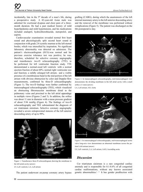
Figure 2. In transesophageal echocardiography, mid-transesophageal view<br />
demonstrates the dividing membrane in the left atrial cavity with a small<br />
orifice (11 mm) (arrow)<br />
LA, Left atrium; AO, Aorta<br />
Figure 3. In transesophageal echocardiography, mid-transesophageal aortic<br />
valve long-axis view demonstrates an abnormal membrane across the patient’s<br />
left atrium (arrow)<br />
LV, Left ventricle; LA, Left atrium; AAO, Ascending aorta<br />
Figure 1. Transthoracic three-D echocardiogram showing the membrane in<br />
the left atrial cavity (arrow)<br />
LV, Left ventricle; LA, Left atrium<br />
The patient underwent on-pump coronary artery bypass<br />
Discussion<br />
Cor triatriatum sinistrum is a rare congenital cardiac<br />
anomaly and is responsible for 0.1-0.4% <strong>of</strong> all congenital<br />
cardiac malformations, without any known associated<br />
genetic abnormalities.¹ , ² It has gender predilection with<br />
86