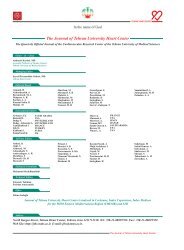
Journal of Tehran University Heart Center
Journal of Tehran University Heart Center
Journal of Tehran University Heart Center

You also want an ePaper? Increase the reach of your titles
YUMPU automatically turns print PDFs into web optimized ePapers that Google loves.
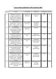
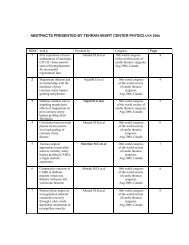
Demographics and Angiographic Findings in Patients under 35 Years <strong>of</strong> ...<br />
Introduction<br />
ST-segment elevation myocardial infarction (STEMI) is<br />
one <strong>of</strong> the most common causes <strong>of</strong> emergency department<br />
admissions and cardiovascular mortalities and thus currently<br />
accounts for a high burden on health care services in the<br />
world. Although STEMI is an uncommon entity in young<br />
patients, it has always attracted special attention because<br />
<strong>of</strong> its unusual features and devastating effect on their more<br />
active lifestyle. 1-5<br />
In recent years, whereas the mean age <strong>of</strong> coronary artery<br />
disease (CAD) has decreased, its prevalence seems to have<br />
been on the increase. 4 A number <strong>of</strong> studies, including our<br />
previous report, have shown significant differences in the risk<br />
factor pr<strong>of</strong>ile and coronary angiographic patterns between<br />
young and older patients with acute STEMI. 1-3, 6-8 Traditional<br />
risk factors <strong>of</strong> CAD are prevalent in young patients with<br />
acute STEMI but with a different pattern compared to their<br />
older counterparts; these differences may cause different<br />
treatment strategies and outcome among these patients. 3,<br />
5, 9<br />
There is evidence that a concentration <strong>of</strong> the novel risk<br />
factors <strong>of</strong> CAD such as LP (a) may be higher than normal in<br />
the <strong>of</strong>fspring <strong>of</strong> patients with a history <strong>of</strong> premature MI. 10<br />
There is a dearth <strong>of</strong> available data on very young adults with<br />
STEMI, as a life-threatening cardiac emergency condition.<br />
In an attempt to characterize patients 35 years <strong>of</strong> age or<br />
younger suffering STEMI, we reviewed the <strong>Tehran</strong> <strong>Heart</strong><br />
<strong>Center</strong> Angiography Registry (THCAR) and compared the<br />
demographics and clinical findings <strong>of</strong> these patients to those<br />
older than 35 years <strong>of</strong> age.<br />
Methods<br />
From all the patients admitted by cardiologists between<br />
January 2000 and March 2008 to the Angiography<br />
Department affiliated with the Academic <strong>Tehran</strong> <strong>Heart</strong> <strong>Center</strong>,<br />
we identified 5652 patients with a history <strong>of</strong> STEMI. The<br />
databank contains patients’ data collected by cardiologists and<br />
trained general practitioners, and the validity <strong>of</strong> all the data<br />
is checked by reabstracting 10% <strong>of</strong> the patients’ entries and<br />
by reentering 5% <strong>of</strong> the patients' records. The investigation<br />
was approved by the institutional Review Board, overseeing<br />
the participation <strong>of</strong> human subjects in research at <strong>Tehran</strong><br />
<strong>University</strong> <strong>of</strong> Medical Sciences. This study conforms to the<br />
principles outlined in the Declaration <strong>of</strong> Helsinki.<br />
The validation <strong>of</strong> acute myocardial infarction (AMI) events<br />
was based on information on medical history, symptoms,<br />
electrocardiogram, and cardiac enzymes. STEMI was<br />
diagnosed when new or presumed new ST-segment elevation<br />
≥ 1 mm ( ≥ 2 mm in V 1<br />
to V 3<br />
) was seen in any location in two<br />
or more contiguous leads or new left bundle branch block<br />
was found on the index or qualifying electrocardiogram<br />
with ≥ 1 positive cardiac biochemical marker <strong>of</strong> necrosis<br />
TEHRAN HEART CENTER<br />
(including CKMB-mass or quantitative cardiac troponin<br />
measurements). The cardiologist who performed the coronary<br />
angiography documented and recorded STEMI diagnosis<br />
in the datasheets. Coronary angiography was performed in<br />
almost all the patients as part <strong>of</strong> the pharmacoinvasive strategy<br />
or as the primary treatment option (primary percutaneous<br />
coronary intervention [PCI]) based on the American College<br />
<strong>of</strong> Cardiology/American <strong>Heart</strong> Association (ACC/AHA)<br />
Guidelines for the management <strong>of</strong> patients with STEMI. 11 To<br />
characterize the young patients, the patients were divided into<br />
2 subgroups <strong>of</strong> ≤ 35 years (Group I, n = 108) and those older<br />
than 35 years (Group II, n = 5544). The following data were<br />
included for analysis: demographic data (i.e. age and gender)<br />
and CAD risk factor pr<strong>of</strong>ile, comprised <strong>of</strong> current cigarette<br />
smoking history (patient regularly smokes a tobacco product/<br />
products one or more times per day or has smoked in the 30<br />
days prior to admission), hyperlipidemia (total cholesterol ≥<br />
5.0, HDL-cholesterol ≤ 1.0 in men or ≤ 1.1 in women, and<br />
triglycerides ≥ 2.0 mmol/l), family history <strong>of</strong> CAD (firstdegree<br />
relatives before the age <strong>of</strong> 55 in men and 65 years in<br />
women), hypertension (systolic blood pressure ≥ 140 and/or<br />
diastolic ≥ 90 mmHg and/or on anti-hypertensive treatment),<br />
diabetes mellitus (symptoms <strong>of</strong> diabetes and plasma glucose<br />
concentration ≥ 200 mg/dl (11.1 mmol/l), or fasting blood<br />
sugar (FBS) ≥ 126 mg/dl (7.0mmol/l) or 2-hp ≥ 200 mg/dl<br />
(11.1 mmol/l)), and opium consumption. 12<br />
Clinical manifestations, left ventricular ejection fraction<br />
(LVEF), hematologic indices, coronary angiographic findings,<br />
and treatment strategy were reported. Selective coronary<br />
arteriography was performed using standard technique in<br />
all the patients. Significant CAD was defined as a diameter<br />
stenosis > 50% in each major epicardial artery. A narrowing<br />
<strong>of</strong> < 50% was considered mild CAD. Normal vessels were<br />
defined as the complete absence <strong>of</strong> any disease in the left<br />
main coronary artery (LMCA), left anterior descending<br />
(LAD), right coronary artery (RCA), and left circumflex<br />
(LCx) as well as in their main branches (diagonal, obtuse<br />
marginal, ramus intermedius, posterior descending artery,<br />
and posterolateral branch). Even mild luminal irregularities<br />
were regarded as evidence <strong>of</strong> atherosclerosis.<br />
The results were reported as mean ± standard deviation<br />
(SD) for the quantitative variables and percentages for the<br />
categorical variables. The groups were compared using the<br />
Student t-test for the continuous variables and the Chi-square<br />
test for the dichotomous variables. This study was done with<br />
the power <strong>of</strong> 90%. P values <strong>of</strong> 0.05 or less were considered<br />
statistically significant. All the statistical analyses were<br />
carried out via Statistical Package for Social Sciences version<br />
16 (SPSS, IL, Chicago Inc., USA).<br />
Results<br />
The demographic and historical characteristics <strong>of</strong> the study<br />
The <strong>Journal</strong> <strong>of</strong> <strong>Tehran</strong> <strong>University</strong> <strong>Heart</strong> <strong>Center</strong>63