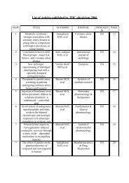
Journal of Tehran University Heart Center
Journal of Tehran University Heart Center
Journal of Tehran University Heart Center

Create successful ePaper yourself
Turn your PDF publications into a flip-book with our unique Google optimized e-Paper software.
Carotid Artery Wall Motion Estimation from Consecutive Ultrasonic Images:<br />
image analysis procedures. The two methods <strong>of</strong> blockmatching<br />
algorithm and a combined algorithm <strong>of</strong> dynamic<br />
programming and maximum-gradient are implemented for<br />
measuring the arterial wall changes <strong>of</strong> the common carotid<br />
in healthy subjects.<br />
Two-dimensional block-matching time domain approaches<br />
to speckle tracking have found widespread application<br />
because <strong>of</strong> their inherent simplicity and relative immunity to<br />
noise. 7 Golemati et al. demonstrated that the two-dimensional<br />
motion <strong>of</strong> the normal and diseased arterial wall as well as<br />
the surrounding tissue can be accurately quantified through<br />
cross-correlation. 5, 19 They compared block matching and<br />
optical flow and the effect <strong>of</strong> the block size on the arterial<br />
wall motion estimation <strong>of</strong> 10 healthy subjects in both radial<br />
and axial directions. 3 They also attempted to combine a<br />
FIELD-Π s<strong>of</strong>tware based ultrasound simulation approach<br />
with mathematical modeling <strong>of</strong> the arterial wall motion. The<br />
simulated sequential image data may be used to evaluate the<br />
performance <strong>of</strong> motion analysis algorithms. 7 Persson et al.<br />
designed and demonstrated a new non-invasive method for<br />
longitudinal and circumferential movement estimation. 20<br />
Cinthio et al. evaluated this echo-tracking system based<br />
on block matching for measuring radial and longitudinal<br />
movements <strong>of</strong> small regions <strong>of</strong> the intima-media complex<br />
<strong>of</strong> the arterial wall. They subsequently used this to estimate<br />
the longitudinal movement and resulting shear strain <strong>of</strong> the<br />
arterial wall. 21<br />
Other different image analysis algorithms have been<br />
investigated for automated ultrasonic boundary detection.<br />
These are the dynamic programming, maximum gradient,<br />
model-based, and matched filter algorithms. These methods<br />
were implemented to measure the intima-media thickness<br />
and internal diameter <strong>of</strong> arteries; and among them, dynamic<br />
programming and maximum gradient have the highest<br />
1, 4, 22<br />
accuracy.<br />
Wendelhag et al. used the dynamic programming algorithm<br />
for developing a computerized analyzing system to evaluate<br />
the boundaries <strong>of</strong> the intima-media. 23 Chang et al. proposed<br />
an automatic system for detecting the intima-media thickness<br />
<strong>of</strong> the common carotid artery using the snake techniques.<br />
They showed that the computerized system had the potential<br />
in automatically detecting the intimal and adventitial layers<br />
without any manual correction. 24 Jegelevicius et al. employed<br />
the dynamic programming and maximum-gradient algorithms<br />
for measuring the intima-media thickness in a single frame,<br />
separately.<br />
They suggested the maximum gradient for intima-media<br />
thickness measurement. 15 But all <strong>of</strong> the above-mentioned<br />
studies, was used the computerized analyzing method for<br />
detecting the lumen diameter or intima-media thickness in a<br />
single frame, and method for making the actual measurements<br />
over the entire cardiac cycle to capture the dynamic nature <strong>of</strong><br />
the vessel are spars.<br />
In the present study, we detected the absolute carotid<br />
TEHRAN HEART CENTER<br />
artery wall displacement in the radial direction by tracking<br />
the location <strong>of</strong> a block and using a maximum-gradient-based<br />
algorithm. The echo-tracking algorithm has been used most<br />
frequently in the studies on the arterial wall motion due<br />
to its computational simplicity and accuracy. On the other<br />
hand, ultrasonic boundary detection algorithms based on the<br />
maximum gradient and dynamic programming have been<br />
rarely used to extract arterial wall motion. The results <strong>of</strong> the<br />
present study showed that in conjunction with the arterial<br />
intima-media thickness and internal diameter measurements,<br />
boundary detection algorithms can be used for arterial wall<br />
motion detection. From the computation complexity view,<br />
maximum gradient was higher than block matching, and<br />
it required manually placing the approximate boundary<br />
points and was more time-consuming. The most important<br />
advantage <strong>of</strong> maximum gradient over block matching is its<br />
ability to detect the coincidental waveforms <strong>of</strong> the anterior<br />
and posterior wall motion and thus the arterial diameter<br />
changes.<br />
The results <strong>of</strong> the presents study showed that the maximum<br />
and mean radial displacements acquired from block matching<br />
were respectively 0.06 and 0.04 mm greater than the values<br />
gained from maximum gradient. Regarding the pixel size,<br />
the differences had the order <strong>of</strong> one pixel size. The greater<br />
values <strong>of</strong> the block-matching measurements can be related to<br />
its more accurate searching method. In the block-matching<br />
techniques, the intensity <strong>of</strong> a block <strong>of</strong> pixels is compared<br />
with the reference frame and the best match is found by<br />
maximizing the similarity. Consequently, more accurate<br />
two-dimensional tracking is possible. But in maximumgradient-based<br />
algorithms, searching is performed in just<br />
one direction, perpendicular to the boundary, and the point<br />
<strong>of</strong> maximum intensity gradient is picked up. Thus, with the<br />
presence <strong>of</strong> echo-dropout, it fails to track the boundary and<br />
the average <strong>of</strong> neighboring points is replaced.<br />
Our regression analysis demonstrated a good correlation,<br />
with a correlation coefficient <strong>of</strong> 0.89, between the two<br />
methods in measuring the radial displacement <strong>of</strong> the carotid<br />
artery wall. The Bland-Altman statistical analysis, with the<br />
mean difference and limits <strong>of</strong> agreement <strong>of</strong> -0.044, -0.12<br />
and 0.032 respectively, confirmed this correlation. Low<br />
dispersion coefficients showed high reproducibility <strong>of</strong> both<br />
methods, higher for the maximum-gradient method.<br />
Despite the differences in the radial displacements obtained<br />
from the two algorithms, it is notable that in seventeen out<br />
<strong>of</strong> twenty cardiac cycles, maximum displacements occurred<br />
in the same (± 1) frame, showing the coincidence <strong>of</strong> cardiac<br />
events for both algorithms.<br />
Conclusion<br />
The results <strong>of</strong> the present study showed that both blockmatching<br />
and maximum-gradient algorithms can be used<br />
The <strong>Journal</strong> <strong>of</strong> <strong>Tehran</strong> <strong>University</strong> <strong>Heart</strong> <strong>Center</strong>77