Journal of Tehran University Heart Center
Journal of Tehran University Heart Center
Journal of Tehran University Heart Center

Create successful ePaper yourself
Turn your PDF publications into a flip-book with our unique Google optimized e-Paper software.
The <strong>Journal</strong> <strong>of</strong> <strong>Tehran</strong> <strong>University</strong> <strong>Heart</strong> <strong>Center</strong><br />
Effat Soleimani et al.<br />
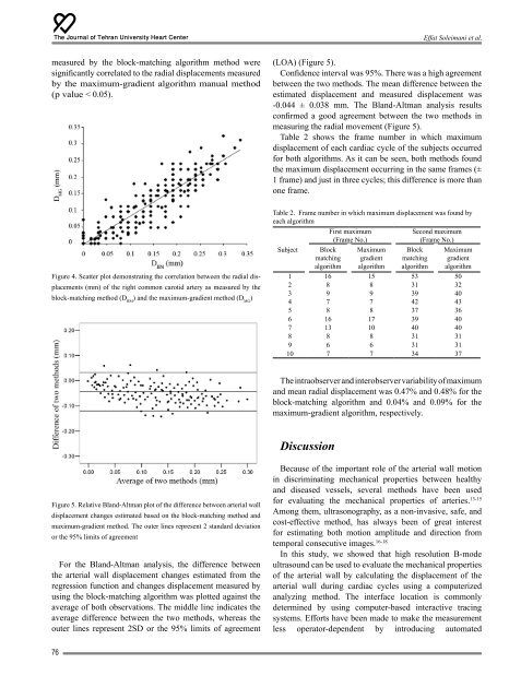
measured by the block-matching algorithm method were<br />
significantly correlated to the radial displacements measured<br />
by the maximum-gradient algorithm manual method<br />
(p value < 0.05).<br />
(LOA) (Figure 5).<br />
Confidence interval was 95%. There was a high agreement<br />
between the two methods. The mean difference between the<br />
estimated displacement and measured displacement was<br />
-0.044 ± 0.038 mm. The Bland-Altman analysis results<br />
confirmed a good agreement between the two methods in<br />
measuring the radial movement (Figure 5).<br />
Table 2 shows the frame number in which maximum<br />
displacement <strong>of</strong> each cardiac cycle <strong>of</strong> the subjects occurred<br />
for both algorithms. As it can be seen, both methods found<br />
the maximum displacement occurring in the same frames (±<br />
1 frame) and just in three cycles; this difference is more than<br />
one frame.<br />
Figure 4. Scatter plot demonstrating the correlation between the radial displacements<br />
(mm) <strong>of</strong> the right common carotid artery as measured by the<br />
block-matching method (D BM<br />
) and the maximum-gradient method (D MG<br />
)<br />
Table 2. Frame number in which maximum displacement was found by<br />
each algorithm<br />
Subject<br />
Block<br />
matching<br />
algorithm<br />
First maximum<br />
(Frame No.)<br />
Maximum<br />
gradient<br />
algorithm<br />
Second maximum<br />
(Frame No.)<br />
Block<br />
matching<br />
algorithm<br />
Maximum<br />
gradient<br />
algorithm<br />
1 16 15 53 50<br />
2 8 8 31 32<br />
3 9 9 39 40<br />
4 7 7 42 43<br />
5 8 8 37 36<br />
6 16 17 39 40<br />
7 13 10 40 40<br />
8 8 8 31 31<br />
9 6 6 31 31<br />
10 7 7 34 37<br />
The intraobserver and interobserver variability <strong>of</strong> maximum<br />
and mean radial displacement was 0.47% and 0.48% for the<br />
block-matching algorithm and 0.04% and 0.09% for the<br />
maximum-gradient algorithm, respectively.<br />
Discussion<br />
Figure 5. Relative Bland-Altman plot <strong>of</strong> the difference between arterial wall<br />
displacement changes estimated based on the block-matching method and<br />
maximum-gradient method. The outer lines represent 2 standard deviation<br />
or the 95% limits <strong>of</strong> agreement<br />
For the Bland-Altman analysis, the difference between<br />
the arterial wall displacement changes estimated from the<br />
regression function and changes displacement measured by<br />
using the block-matching algorithm was plotted against the<br />
average <strong>of</strong> both observations. The middle line indicates the<br />
average difference between the two methods, whereas the<br />
outer lines represent 2SD or the 95% limits <strong>of</strong> agreement<br />
Because <strong>of</strong> the important role <strong>of</strong> the arterial wall motion<br />
in discriminating mechanical properties between healthy<br />
and diseased vessels, several methods have been used<br />
for evaluating the mechanical properties <strong>of</strong> arteries. 13-15<br />
Among them, ultrasonography, as a non-invasive, safe, and<br />
cost-effective method, has always been <strong>of</strong> great interest<br />
for estimating both motion amplitude and direction from<br />
temporal consecutive images. 16-18<br />
In this study, we showed that high resolution B-mode<br />
ultrasound can be used to evaluate the mechanical properties<br />
<strong>of</strong> the arterial wall by calculating the displacement <strong>of</strong> the<br />
arterial wall during cardiac cycles using a computerized<br />
analyzing method. The interface location is commonly<br />
determined by using computer-based interactive tracing<br />
systems. Efforts have been made to make the measurement<br />
less operator-dependent by introducing automated<br />
76