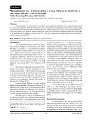
Impaction of the Maxillary Central Incisor Associated - People's ...
Impaction of the Maxillary Central Incisor Associated - People's ...
Impaction of the Maxillary Central Incisor Associated - People's ...

Create successful ePaper yourself
Turn your PDF publications into a flip-book with our unique Google optimized e-Paper software.
Case Report<br />
<strong>Impaction</strong> <strong>of</strong> <strong>the</strong> <strong>Maxillary</strong> <strong>Central</strong> <strong>Incisor</strong> <strong>Associated</strong> with Supernumerary Tooth:<br />
Surgical and Orthodontic Treatment<br />
(a)<br />
Raghavendra M. Shetty, (b) Uma Dixit, (c) Hanumanth Reddy, (d) Shivaprakash P. K., (e) Bhavneet<br />
Kaur<br />
(a)<br />
Department <strong>of</strong> Pediatric Preventive Dentistry, Chhattisgarh Dental College and Research Institute, Rajnandgaon,<br />
Chhattisgarh, (b) Dr. D. Y. Patil Dental College and Hospital, Navi Mumbai, (c) Department <strong>of</strong> Orthodontics, Marata Mandal<br />
Dental College and Hospital, Belgaum, (d) Department <strong>of</strong> Pediatric and Preventive Dentistry, P.M.N.M. Dental College and<br />
Hospital, Bagalkot, (e) Department <strong>of</strong> Pediatric and Preventive Dentistry, Institute <strong>of</strong> Dental Sciences, Jammu.<br />
Abstract:<br />
<strong>Impaction</strong> <strong>of</strong> maxillary permanent incisor is not a frequently case in dental practice, but its treatment is challenging<br />
because <strong>of</strong> its importance to facial es<strong>the</strong>tics. Supernumerary teeth are <strong>the</strong> main cause <strong>of</strong> impaction <strong>of</strong> upper incisor.<br />
Supernumerary teeth when present can cause both es<strong>the</strong>tic and pathologic problems. Supernumerary teeth in <strong>the</strong> maxillary<br />
midline are common. Early detection <strong>of</strong> such teeth is most important if complications are to be avoided.<br />
We report a case <strong>of</strong> 12 year old male with an impacted supernumerary tooth in <strong>the</strong> maxillary anterior region, which was<br />
interfering with <strong>the</strong> eruption <strong>of</strong> <strong>the</strong> permanent, left central incisor. The impacted supernumerary tooth was surgically<br />
removed. With <strong>the</strong> application <strong>of</strong> an orthodontic traction, impacted left maxillary central incisor was brought down to its<br />
proper position in <strong>the</strong> dental arch.<br />
Key Words: Impacted incisor, orthodontic traction, supernumerary.<br />
Introduction:<br />
Although <strong>the</strong> impaction <strong>of</strong> a permanent tooth<br />
is rarely diagnosed during <strong>the</strong> mixed dentition period,<br />
an impacted central incisor is usually diagnosed, when<br />
<strong>the</strong>re is a delay in <strong>the</strong> eruption <strong>of</strong> <strong>the</strong> tooth.<br />
Supernumerary teeth are <strong>the</strong> main cause <strong>of</strong> impaction<br />
<strong>of</strong> upper incisor (Smailene, 2006). Supernumerary teeth<br />
are <strong>the</strong> extra teeth formed due to <strong>the</strong> disturbances<br />
during <strong>the</strong> initiation and proliferation stages <strong>of</strong> tooth<br />
development (Bergstrom, 1977; Humerfelt, 1985). The<br />
supernumerary tooth present in <strong>the</strong> midline or just lateral<br />
to <strong>the</strong> midline is referred to as mesiodens.<br />
Supernumerary teeth are most frequently<br />
located in <strong>the</strong> maxillary incisor region (64.3%) with<br />
mesiodens accounting for 32.4% <strong>of</strong> such presentation.<br />
56-60% <strong>of</strong> premaxillary supernumerary teeth cause<br />
impaction <strong>of</strong> permanent incisor (Gregg and Kinirons<br />
1991; Becker, 1998) due to a direct obstruction for <strong>the</strong><br />
eruption tipping <strong>of</strong> adjacent teeth towards <strong>the</strong> place <strong>of</strong><br />
<strong>the</strong> impacted tooth, narrowing <strong>of</strong> <strong>the</strong> dental arch,<br />
displacement <strong>of</strong> <strong>the</strong> permanent teeth bud, or<br />
malformations <strong>of</strong> <strong>the</strong> unerupted tooth root (Rajab and<br />
Hamdan 2002; Roberts-Hary and Sandy 2004).<br />
An interesting case <strong>of</strong> an impacted<br />
supernumerary tooth in <strong>the</strong> maxillary anterior region,<br />
interfering with <strong>the</strong> eruption <strong>of</strong> <strong>the</strong> permanent left<br />
-----------------------------------------------------------------------------<br />
Corresponding Author: Dr.Raghavendra M. Shetty, Associate<br />
Pr<strong>of</strong>essor, Department <strong>of</strong> Pediatric and Preventive Dentistry,<br />
Chhattisgarh Dental College & Research Institute, Rajnandgaon - 491<br />
441 (Chhatisgarh)<br />
Phone No.: 9993987421<br />
E mail : raghavendra77@yahoo.com<br />
central incisor is presented. Combined surgical and<br />
orthodontic treatment employed, to bring <strong>the</strong> impacted<br />
left maxillary central incisor to its proper position in<br />
<strong>the</strong> dental arch is discussed.<br />
Case Report:<br />
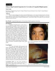
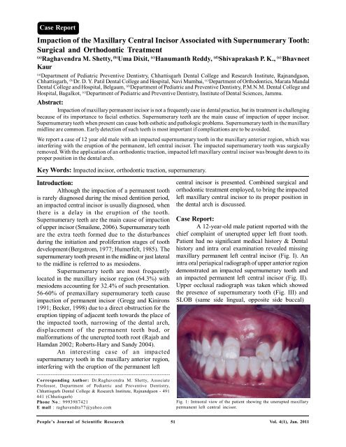
A 12-year-old male patient reported with <strong>the</strong><br />
chief complaint <strong>of</strong> unerupted upper left front tooth.<br />
Patient had no significant medical history & Dental<br />
history and intra oral examination revealed missing<br />
maxillary permanent left central incisor (Fig. I). An<br />
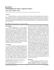
intra oral periapical radiograph <strong>of</strong> upper anterior region<br />
demonstrated an impacted supernumerary tooth and<br />
an impacted permanent left central incisor (Fig. II).<br />
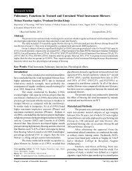
Upper occlusal radiograph was taken which showed<br />
<strong>the</strong> presence <strong>of</strong> supernumerary tooth (Fig. III) and<br />
SLOB (same side lingual, opposite side buccal)<br />
Fig. 1: Intraoral view <strong>of</strong> <strong>the</strong> patient showing <strong>the</strong> unerupted maxillary<br />
permanent left central incisor.<br />
People’s Journal <strong>of</strong> Scientific Research 51<br />
Vol. 4(1), Jan. 2011
<strong>Impaction</strong> <strong>of</strong> <strong>the</strong> <strong>Maxillary</strong> <strong>Central</strong> <strong>Incisor</strong> <strong>Associated</strong> ------- R.M. Shetty, UDixit, H. Reddy, Shivaprakash P. K., B. Kaur<br />
Fig. II: Intraoral periapical radiograph showing supernumerary tooth<br />
and an impacted maxillary left central incisor.<br />
Fig. IV: Operative view showing <strong>the</strong> supernumerary tooth on <strong>the</strong><br />
palatal side.<br />
Fig. III: Anterior maxillary occlusal radiograph showing<br />
supernumerary tooth.<br />
technique with two intra-oral periapical radiographs<br />
confirmed <strong>the</strong> presence <strong>of</strong> supernumerary tooth on <strong>the</strong><br />
palatal side and an impacted tooth in <strong>the</strong> buccal side<br />
The treatment plan comprised <strong>of</strong> surgical<br />
removal <strong>of</strong> <strong>the</strong> supernumerary tooth and orthodontic<br />
traction <strong>of</strong> <strong>the</strong> impacted incisor with closed eruption<br />
technician to bring it into proper position in <strong>the</strong> dental<br />
arch. With <strong>the</strong> patient under local anes<strong>the</strong>sia, full<br />
thickness mucoperiosteal flap on <strong>the</strong> palatal side was<br />
reflected. After careful elevation <strong>of</strong> <strong>the</strong> flap, adequate<br />
amount <strong>of</strong> bone was removed using <strong>the</strong> rotary cutting<br />
instruments and <strong>the</strong> impacted supernumerary tooth was<br />
exposed (Fig. IV). The supernumerary tooth was<br />
removed surgically and extraction socket was inspected<br />
for any pathology. The extracted supernumerary tooth<br />
was conical in shape. The palatal mucoperiosteal flap<br />
was repositioned but not sutured at this time. A full<br />
thickness mucoperiosteal flap was reflected labially,<br />
<strong>the</strong> bone and <strong>the</strong> follicular connective tissue covering<br />
Fig. V: Operative view showing <strong>the</strong> impacted left central incisor<br />
on <strong>the</strong> labial side.<br />
<strong>the</strong> impacted incisor was removed and adequate<br />
amount <strong>of</strong> crown was exposed for bonding <strong>of</strong> <strong>the</strong><br />
orthodontic bracket (Fig. V). Ligature was twisted to<br />
<strong>the</strong> flat Begg’s incisor bracket and made into a hook<br />
form and was bonded on <strong>the</strong> labial surface <strong>of</strong> <strong>the</strong><br />
impacted incisor. The labial and palatal flap was<br />
repositioned and sutured, keeping <strong>the</strong> ligature wire hook<br />
suspended in <strong>the</strong> oral cavity making sure <strong>the</strong> occlusion<br />
was not interfered (Fig. VI). After a week, <strong>the</strong> healing<br />
was normal and <strong>the</strong> sutures were removed. Begg’s<br />
bracket was bonded on lower permanent incisors and<br />
canines and 0.020 A. J. Wilcock arch wire (sectional)<br />
was used for anchorage. Yellow elastic was tied to <strong>the</strong><br />
ligature wire hook and was engaged to <strong>the</strong> lower<br />
brackets for <strong>the</strong> traction (Fig. VII). Elastic was<br />
engaged more towards <strong>the</strong> left side <strong>of</strong> <strong>the</strong> mandibular<br />
teeth so as to de-rotate <strong>the</strong> impacted incisor. The patient<br />
was demonstrated about how to engage <strong>the</strong> elastics<br />
and was told to disengage <strong>the</strong> elastics during eating<br />
and long speech. Elastics were changed every fifth<br />
day. After two weeks <strong>of</strong> traction with <strong>the</strong> yellow<br />
People’s Journal <strong>of</strong> Scientific Research 52<br />
Vol. 4(1), Jan. 2011
<strong>Impaction</strong> <strong>of</strong> <strong>the</strong> <strong>Maxillary</strong> <strong>Central</strong> <strong>Incisor</strong> <strong>Associated</strong> ------- R.M. Shetty, UDixit, H. Reddy, Shivaprakash P. K., B. Kaur<br />
Fig.VI: Post operative view showing sutured site and suspended ligature<br />
wire hook.<br />
Fig.IX: Placement <strong>of</strong> 0.016 NiTi round arch wire to align <strong>the</strong> left<br />
central incisor.<br />
Fig.VII: Yellow elastic tied to <strong>the</strong> ligature wire hook and engaged to<br />
<strong>the</strong> lower brackets for <strong>the</strong> traction<br />
Fig. X: OPG showing well aligned left central incisor without any<br />
bone resorption<br />
Fig. 8:<br />
Fig.VIII: Elastic thread tied from <strong>the</strong> slot <strong>of</strong> <strong>the</strong> bracket to <strong>the</strong><br />
sectional arch wire for fur<strong>the</strong>r traction.<br />
elastics, <strong>the</strong> incisor with <strong>the</strong> bracket was seen in <strong>the</strong><br />
oral cavity. Begg’s bracket was bonded on permanent<br />
maxillary left central incisor, lateral incisor, and canine<br />
and right lateral incisor and canine. 0.020 A. J. Wilcock<br />
arch wire was used for anchorage. The ligature wire<br />
hook was cut till <strong>the</strong> arch wire and <strong>the</strong> remaining part<br />
was passively tied to <strong>the</strong> arch wire. Elastic thread was<br />
Fig. XI: Six-month post treatment intraoral view <strong>of</strong> <strong>the</strong> patient<br />
showing well aligned left central incisor.<br />
tied from <strong>the</strong> slot <strong>of</strong> <strong>the</strong> bracket to <strong>the</strong> sectional arch<br />
wire for fur<strong>the</strong>r traction <strong>of</strong> left central incisor (Fig.<br />
VIII). After <strong>the</strong> crown <strong>of</strong> <strong>the</strong> impacted incisor was<br />
sufficiently erupted, 0.016 NiTi round arch wire<br />
(sectional) was used to align <strong>the</strong> incisor. Once <strong>the</strong><br />
incisor was well aligned <strong>the</strong> mammelons were trimmed<br />
and lingual fixed retention was given. The patient<br />
showed normal clinical crown length with acceptable<br />
People’s Journal <strong>of</strong> Scientific Research 53<br />
Vol. 4(1), Jan. 2011
<strong>Impaction</strong> <strong>of</strong> <strong>the</strong> <strong>Maxillary</strong> <strong>Central</strong> <strong>Incisor</strong> <strong>Associated</strong> ------- R.M. Shetty, UDixit, H. Reddy, Shivaprakash P. K., B. Kaur<br />
gingival contour (Fig. XI) and <strong>the</strong> tooth maintained its<br />
vitality with no evidence <strong>of</strong> root resorption (Fig. X).<br />
At six-month follow up (Fig. XI), <strong>the</strong> left maxillary<br />
incisor remained vital and responded normally to<br />
percussion and mobility and sensitivity testing with good<br />
width <strong>of</strong> attached gingiva.<br />
Discussion:<br />
Supernumerary teeth can affect <strong>the</strong> normal<br />
position and eruption <strong>of</strong> adjacent teeth and <strong>of</strong>ten require<br />
clinical intervention (Harris and Clark, 2008). The most<br />
common complication due to presence <strong>of</strong><br />
supernumerary teeth is <strong>the</strong> failure <strong>of</strong> eruption <strong>of</strong><br />
maxillary incisors (Rajab and Hamdan 2002).<br />
Supernumerary teeth in <strong>the</strong> premaxillary region are<br />
broadly <strong>of</strong> two types: one containing teeth <strong>of</strong> normal<br />
morphology known as supplemental teeth and <strong>the</strong> o<strong>the</strong>r<br />
<strong>of</strong> abnormal shape. The latter class has been fur<strong>the</strong>r<br />
categorized into <strong>the</strong> conical type (peg-shaped) and <strong>the</strong><br />
tuberculate type. The tuberculate supernumerary tooth<br />
seems to occur most frequently palatal to <strong>the</strong> upper<br />
central incisor and to be later in its development than<br />
<strong>the</strong> conical tooth. It also tends to delay or prevent <strong>the</strong><br />
eruption <strong>of</strong> <strong>the</strong> corresponding permanent central incisor,<br />
and is rarely seen erupted in childhood. It has also been<br />
documented that <strong>the</strong> conical-shaped supernumerary<br />
tooth does not usually affect <strong>the</strong> eruption <strong>of</strong> <strong>the</strong> adjacent<br />
permanent incisors but may cause <strong>the</strong>ir displacement,<br />
which may involve <strong>the</strong> crown, <strong>the</strong> root or <strong>the</strong> whole<br />
tooth. The conical supernumerary may be non-inverted<br />
or inverted. When non-inverted, it may remain<br />
unerupted palatal to <strong>the</strong> permanent incisors. When<br />
inverted, it may point posteriorly towards <strong>the</strong> nose or<br />
may even erupt into <strong>the</strong> nose (Mills, 1987; Pr<strong>of</strong>itt, 1992).<br />
In <strong>the</strong> present case <strong>the</strong> associated supernumerary tooth<br />
was conical, non -inverted and impacted on <strong>the</strong> palatal<br />
side and interfered with <strong>the</strong> eruption <strong>of</strong> <strong>the</strong> permanent<br />
tooth.<br />
The treatment protocol available for<br />
management <strong>of</strong> impacted permanent teeth due to<br />
supernumerary teeth are diverse. Methods <strong>of</strong><br />
management <strong>of</strong> crowding or impaction due to<br />
supernumerary tooth are; removal <strong>of</strong> supernumerary<br />
teeth or tooth only, removal <strong>of</strong> supernumerary teeth<br />
and bone overlying impacted teeth, incision <strong>of</strong> fibrous<br />
tissue over <strong>the</strong> alveolar ridge to promote <strong>the</strong> eruption<br />
with or without orthodontic traction (Regezi et al., 2003;<br />
Bhat, 2006).<br />
Spontaneous eruption <strong>of</strong> impacted maxillary<br />
incisors occurs in 54-76% <strong>of</strong> cases when supernumerary<br />
tooth is removed and it <strong>the</strong>re is enough space in <strong>the</strong><br />
dental arch (Crawford, 1997; Garvey et al., 1999).<br />
However, research data indicate that <strong>the</strong> spontaneous<br />
eruption <strong>of</strong> impacted maxillary incisor may take up to<br />
3 years and sometimes orthodontic treatment is<br />
necessary to achieve adequate alignment <strong>of</strong> <strong>the</strong><br />
erupted tooth in <strong>the</strong> dental arch (Witsenburg et al.,<br />
1981; Mason et al., 2000). If <strong>the</strong> root <strong>of</strong> <strong>the</strong> impacted<br />
tooth is still developing, <strong>the</strong> tooth may erupt normally;<br />
but, once <strong>the</strong> root apex has closed, <strong>the</strong> tooth has lost<br />
its potential to erupt (Kokich and Ma<strong>the</strong>ws, 1993). In<br />
<strong>the</strong> present case <strong>the</strong> root formation was almost<br />
complete and because <strong>of</strong> its rotation and labial<br />
placement, it was not desirable to wait for spontaneous<br />
eruption.<br />
After thorough clinical and radiographic<br />
examination, it was decided that <strong>the</strong> present case<br />
required a combination approach comprising <strong>of</strong> both<br />
surgical and orthodontic treatment to bring an unerupted<br />
maxillary central incisor into position as done by various<br />
authors (Cangialosi, 1982; Kamakura et al., 2002;<br />
Kocadereli and Turgut, 2005). Surgical exposures <strong>of</strong><br />
impacted incisors or surgical repositioning have also<br />
been used to bring impacted teeth into occlusion<br />
(Kamakura et al., 2002; Kocadereli and Turgut, 2005).<br />
Combination approach using conservative surgical<br />
treatment and careful orthodontic management to bring<br />
tooth into good position in <strong>the</strong> dental arch has also been<br />
reported with success (Cangialosi, 1982). Hence,<br />
extraction <strong>of</strong> <strong>the</strong> impacted supernumerary tooth was<br />
done and was followed by an innovative orthodontic<br />
traction <strong>of</strong> <strong>the</strong> unerupted permanent central incisor to<br />
bring <strong>the</strong> tooth into proper position in <strong>the</strong> arch.<br />
Three accepted ways <strong>of</strong> surgical exposure<br />
have been suggested by Becker (1998) as:<br />
a. Circular excision <strong>of</strong> <strong>the</strong> oral mucosa immediately<br />
overlying <strong>the</strong> impacted tooth.<br />
b. Apically repositioning <strong>of</strong> <strong>the</strong> raised flap that<br />
incorporates <strong>the</strong> attached gingiva overlying <strong>the</strong><br />
impacted tooth.<br />
c. Closed eruption technique in which <strong>the</strong> raised flap<br />
that incorporates attached gingiva is fully<br />
replaced back in its former position after an<br />
attachment has been bonded to <strong>the</strong> impacted<br />
tooth.<br />
The closed eruption technique has been<br />
favoured by many clinicians who claim that <strong>the</strong><br />
aes<strong>the</strong>tic and periodontal outcome is far more superior<br />
when compared with <strong>the</strong> circular excision and apically<br />
positioned flap technique (Lin, 1999; Uematsu et al.,<br />
2004; Paola et al., 2005). In <strong>the</strong> presented case <strong>the</strong><br />
People’s Journal <strong>of</strong> Scientific Research 54<br />
Vol. 4(1), Jan. 2011
<strong>Impaction</strong> <strong>of</strong> <strong>the</strong> <strong>Maxillary</strong> <strong>Central</strong> <strong>Incisor</strong> <strong>Associated</strong> ------- R.M. Shetty, UDixit, H. Reddy, Shivaprakash P. K., B. Kaur<br />
closed eruption technique was used for better and<br />
es<strong>the</strong>tic gingival margin. At <strong>the</strong> end <strong>of</strong> <strong>the</strong> treatment,<br />
patient showed normal clinical crown length with<br />
acceptable gingival contour.<br />
The extrusion force applied on <strong>the</strong> impacted<br />
central incisor in <strong>the</strong> present case was very light and<br />
measured in <strong>the</strong> range <strong>of</strong> 40-50 grams. This may have<br />
accounted for <strong>the</strong> little difference in <strong>the</strong> clinical crown<br />
length and maintenance <strong>of</strong> vitality <strong>of</strong> <strong>the</strong> impacted tooth<br />
post-alignment. The chances <strong>of</strong> nonvitality are naturally<br />
much lower when <strong>the</strong> treatment is initiated at a younger<br />
age due to <strong>the</strong> presence <strong>of</strong> a wide apical foramen<br />
(Chawla and Kapur , 2009), but in <strong>the</strong> present case <strong>the</strong><br />
patient was 12 years old at <strong>the</strong> time <strong>of</strong> initiation <strong>of</strong> <strong>the</strong><br />
treatment. In our view, forces for traction greater than<br />
50 grams should not be applied as it may be <strong>the</strong> cause<br />
<strong>of</strong> nonvitality as reported by Uematsu et al. (2004). In<br />
<strong>the</strong> present case <strong>the</strong> duration <strong>of</strong> treatment was around<br />
7-8 months and <strong>the</strong> aligned left maxillary incisor<br />
remained vital and responded normally to percussion<br />
and mobility and sensitivity testing.<br />
Early diagnosis <strong>of</strong> <strong>the</strong> maxillary central incisor<br />
impactions with surgical removal <strong>of</strong> supernumerary<br />
tooth coupled with adequate space spontaneous eruption<br />
<strong>of</strong> <strong>the</strong> impacted maxillary central incisors (Smailene,<br />
2006). If <strong>the</strong> impacted tooth is diagnosed at a later<br />
stage with its root completely formed or if present in<br />
<strong>the</strong> unfavorable position, combination <strong>of</strong> surgical and<br />
orthodontic treatment has to be carried out.<br />
Conclusion:<br />
Supernumerary teeth may result in <strong>the</strong> noneruption<br />
<strong>of</strong> adjacent permanent incisors. Early diagnosis<br />
<strong>of</strong> <strong>the</strong> presence and removal <strong>of</strong> supernumerary teeth<br />
is essential. <strong>Maxillary</strong> permanent left incisor was<br />
successfully positioned in <strong>the</strong> maxillary arch by surgical<br />
exposure and orthodontic traction, which showed good<br />
stability.<br />
Bibliography:<br />
1. Acikgoz G, Acikgoz A, Keskiner I, Turk T, Otan F.<br />
Aggressive periodontitis with supernumerary teeth: a<br />
retrospective study. J. Periodontol. 2004; 75: 1458- 1460.<br />
2. Bergstrom K. An orthopantomographic study <strong>of</strong><br />
hypodontia, supernumeraries and o<strong>the</strong>r anomalies in<br />
school children between <strong>the</strong> ages <strong>of</strong> 8-9 years. An<br />
epidemiological study. Swedish Dental Journal, 1977;<br />
1:145-57.<br />
3. Bodin I, Julin P, Thomsson M: Hyperodontia; Frequency<br />
and distribution <strong>of</strong> supernumerary teeth among 21,609<br />
patients. Dentomaxill<strong>of</strong>acial Radiology, 1978; 7:15-17.<br />
4. Bhat M. Supplemental mandibular incisor. J Indian Soc<br />
Pedod Prev Dent. 2006 ;( special issue):20–23.<br />
5. Becker A. The orthodontic treatment <strong>of</strong> impacted teeth.<br />
Mosby; 1998. p. 53-85.<br />
6. Cangialosi TJ. Management <strong>of</strong> a maxillary central incisor<br />
impacted by a supernumerary tooth. J Am Dent Assoc,<br />
1982; 105(5): 812-814.<br />
7. Chawla HS, Kapur A. Orthodontic management <strong>of</strong><br />
faciolingual horizontally impacted maxillary central<br />
incisor. J Indian Soc Pedod Prev Dent. 2009; 27(1): 65-<br />
69.<br />
8. Foster TD, Taylor GS. Characteristics <strong>of</strong> supernumerary<br />
teeth in <strong>the</strong> upper central incisor region. Dent Pract Dent<br />
Rec 1969; 20:8-12.<br />
9. Gregg TA, Kinirons MJ. The effect <strong>of</strong> <strong>the</strong> position and<br />
orientation <strong>of</strong> unerupted premaxillary supernumerary<br />
teeth on eruption and displacement <strong>of</strong> permanent incisor.<br />
Int. J. Paediatr. Dent. 1991; 1: 3- 7.<br />
10. Humerfelt D, Hurlen B, Humerfelt S. Hyperdontia in<br />
children below four years <strong>of</strong> age: A radiographic study.<br />
ASDC J Dent Child 1985;52:121-24.<br />
11. Huang WH, Tsai TP, Su HL. Mesiodens in <strong>the</strong> primary<br />
dentition stage: A radiographic study. J Dent Child 1992;<br />
59:186-89.<br />
12. Harris EF, Clark LL. An epidemiological study <strong>of</strong><br />
hyperdontia in American blacks and whites. Angle<br />
Orthod. 2008; 78:460–465.<br />
13. Jarvinen S. Supernumerary and congenitally missing<br />
permanent upper anterior teeth in 7-year-old Finnish<br />
children. A radiological study. Proc Finn Dent Soc<br />
1976;72:99-102<br />
14. Kamakura S, Matsui K, Katau F, Shirai N, Kochi S,<br />
Motegi K. Surgical and orthodontic management <strong>of</strong><br />
compound odontoma without removal <strong>of</strong> <strong>the</strong> impacted<br />
permanent tooth. Oral Surg Oral Med Oral Pathol Oral<br />
Radiol Endod. 2002; 4:540–542.<br />
15. Kocadereli Ý, Turgut MD. Surgical and orthodontic<br />
treatment <strong>of</strong> an impacted permanent incisor: case report.<br />
Dent Trauma. 2005; 21:234–239.<br />
16. Lin YT. Treatment <strong>of</strong> an impacted dilacerated maxillary<br />
central incisor. Am. J. Orthod. Dent<strong>of</strong>ac. Orthop. 1999;<br />
115: 406- 409.<br />
17. Luten JR. The prevalence <strong>of</strong> supernumerary teeth in<br />
primary and mixed dentition. Journal for Dentistry for<br />
Children, 1967; 34:346-353.<br />
18. Mills JRE. Principles and practice <strong>of</strong> orthodontics. 2nd<br />
ed. Edinburgh:Churchill Livingstone, 1987:49-52.<br />
19. Nazif MM, Ruffalo RC, Zullo T. Impacted supernumerary<br />
teeth: a survey <strong>of</strong> 50 cases. J Am Dent Assoc 1983;<br />
106:201-04.<br />
20. Oliver RG, Moxham BJ. Malformations <strong>of</strong> teeth. Curr.<br />
Pediatr. 1999; 9:257-261.<br />
21. Oliver R, Hodges ChGL. Delayed eruption <strong>of</strong> a maxillary<br />
central incisor associated with an odontome: report <strong>of</strong><br />
case. J. Dent. Child 1988; 55 (5): 368- 371.<br />
22. Pr<strong>of</strong>itt WR. Contemporary orthodontics. 2nd ed. St.<br />
Louis: CV Mosby Co, 1992:405.<br />
23. Paola C, Alessandra M, Roberta C. Orthodontic treatment<br />
<strong>of</strong> impacted dilacerated maxillary incisor. J. Clin. Pediatr.<br />
Dent. 2005; 30: 93-97.<br />
People’s Journal <strong>of</strong> Scientific Research 55<br />
Vol. 4(1), Jan. 2011
<strong>Impaction</strong> <strong>of</strong> <strong>the</strong> <strong>Maxillary</strong> <strong>Central</strong> <strong>Incisor</strong> <strong>Associated</strong> ------- R.M. Shetty, UDixit, H. Reddy, Shivaprakash P. K., B. Kaur<br />
24. Rajab LD, Hamdan M. Supernumerary teeth: review <strong>of</strong><br />
<strong>the</strong> literature and a survey <strong>of</strong> 152 cases. . Int. J. Paediatr.<br />
Dent. 2002; 12(4): 244-254.<br />
25. Roberts-Hary D, Sandy J. Orthodontics. Part 10:<br />
Impacted teeth. Br. Dent. J. 2004; 196 (6): 319- 327.<br />
26. Regezi JA, Sciubba JJ, Jordan RCK. Oral Pathology:<br />
Clinical Pathologic Correlations. 4th ed. Saunders:<br />
Elsevier Science; 2003. pp. 367–384.<br />
27. Smailene D, Sidlauskas A, Bucinskiene J: <strong>Impaction</strong><br />
<strong>of</strong> <strong>the</strong> central maxillary incisor associated with<br />
supernumerary teeth: Initial position and spontaneous<br />
eruption timing. Somatologiga, Baltic Dental and<br />
Maxill<strong>of</strong>acial Journal. 2006; 8(4): 103- 107.<br />
28. Scheiner MA, Sampson WJ. Supernumerary teeth. A<br />
review <strong>of</strong> <strong>the</strong> literature and four case reports. Aust Dent<br />
J. 1997; 42:160–165.<br />
29. Uematsu S, Uematsu T, Furusawa K, Deguchi T, Kurihara<br />
S. Orthodontic treatment <strong>of</strong> an impacted dilacerated<br />
maxillary central incisor combined with surgical exposure<br />
and apicoectomy. Angle Orthod. 2004; 74: 132-136.<br />
30. Vermette ME, Kokich VG, Kennedy DB. Uncovering<br />
labially impacted teeth: Apically positioned flap and<br />
closed eruption techniques. Angle Orhod. 1995; 65: 23-<br />
34.<br />
People’s Journal <strong>of</strong> Scientific Research 56<br />
Vol. 4(1), Jan. 2011