Giant Cell Lesion of the Jaw - People's Journal Of Scientific Research
Giant Cell Lesion of the Jaw - People's Journal Of Scientific Research
Giant Cell Lesion of the Jaw - People's Journal Of Scientific Research

Create successful ePaper yourself
Turn your PDF publications into a flip-book with our unique Google optimized e-Paper software.
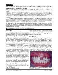
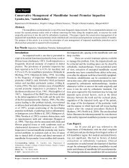
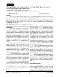
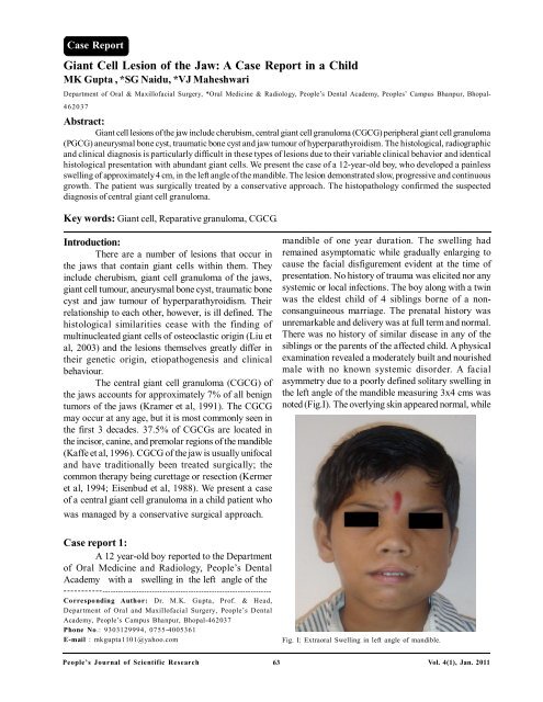
Case Report<strong>Giant</strong> <strong>Cell</strong> <strong>Lesion</strong> <strong>of</strong> <strong>the</strong> <strong>Jaw</strong>: A Case Report in a ChildMK Gupta , *SG Naidu, *VJ MaheshwariDepartment <strong>of</strong> Oral & Maxill<strong>of</strong>acial Surgery, *Oral Medicine & Radiology, People’s Dental Academy, Peoples’ Campus Bhanpur, Bhopal-462037Abstract:<strong>Giant</strong> cell lesions <strong>of</strong> <strong>the</strong> jaw include cherubism, central giant cell granuloma (CGCG) peripheral giant cell granuloma(PGCG) aneurysmal bone cyst, traumatic bone cyst and jaw tumour <strong>of</strong> hyperparathyroidism. The histological, radiographicand clinical diagnosis is particularly difficult in <strong>the</strong>se types <strong>of</strong> lesions due to <strong>the</strong>ir variable clinical behavior and identicalhistological presentation with abundant giant cells. We present <strong>the</strong> case <strong>of</strong> a 12-year-old boy, who developed a painlessswelling <strong>of</strong> approximately 4 cm, in <strong>the</strong> left angle <strong>of</strong> <strong>the</strong> mandible. The lesion demonstrated slow, progressive and continuousgrowth. The patient was surgically treated by a conservative approach. The histopathology confirmed <strong>the</strong> suspecteddiagnosis <strong>of</strong> central giant cell granuloma.Key words: <strong>Giant</strong> cell, Reparative granuloma, CGCG.Introduction:There are a number <strong>of</strong> lesions that occur in<strong>the</strong> jaws that contain giant cells within <strong>the</strong>m. Theyinclude cherubism, giant cell granuloma <strong>of</strong> <strong>the</strong> jaws,giant cell tumour, aneurysmal bone cyst, traumatic bonecyst and jaw tumour <strong>of</strong> hyperparathyroidism. Theirrelationship to each o<strong>the</strong>r, however, is ill defined. Thehistological similarities cease with <strong>the</strong> finding <strong>of</strong>multinucleated giant cells <strong>of</strong> osteoclastic origin (Liu etal, 2003) and <strong>the</strong> lesions <strong>the</strong>mselves greatly differ in<strong>the</strong>ir genetic origin, etiopathogenesis and clinicalbehaviour.The central giant cell granuloma (CGCG) <strong>of</strong><strong>the</strong> jaws accounts for approximately 7% <strong>of</strong> all benigntumors <strong>of</strong> <strong>the</strong> jaws (Kramer et al, 1991). The CGCGmay occur at any age, but it is most commonly seen in<strong>the</strong> first 3 decades. 37.5% <strong>of</strong> CGCGs are located in<strong>the</strong> incisor, canine, and premolar regions <strong>of</strong> <strong>the</strong> mandible(Kaffe et al, 1996). CGCG <strong>of</strong> <strong>the</strong> jaw is usually unifocaland have traditionally been treated surgically; <strong>the</strong>common <strong>the</strong>rapy being curettage or resection (Kermeret al, 1994; Eisenbud et al, 1988). We present a case<strong>of</strong> a central giant cell granuloma in a child patient whowas managed by a conservative surgical approach.mandible <strong>of</strong> one year duration. The swelling hadremained asymptomatic while gradually enlarging tocause <strong>the</strong> facial disfigurement evident at <strong>the</strong> time <strong>of</strong>presentation. No history <strong>of</strong> trauma was elicited nor anysystemic or local infections. The boy along with a twinwas <strong>the</strong> eldest child <strong>of</strong> 4 siblings borne <strong>of</strong> a nonconsanguineousmarriage. The prenatal history wasunremarkable and delivery was at full term and normal.There was no history <strong>of</strong> similar disease in any <strong>of</strong> <strong>the</strong>siblings or <strong>the</strong> parents <strong>of</strong> <strong>the</strong> affected child. A physicalexamination revealed a moderately built and nourishedmale with no known systemic disorder. A facialasymmetry due to a poorly defined solitary swelling in<strong>the</strong> left angle <strong>of</strong> <strong>the</strong> mandible measuring 3x4 cms wasnoted (Fig.I). The overlying skin appeared normal, whileCase report 1:A 12 year-old boy reported to <strong>the</strong> Department<strong>of</strong> Oral Medicine and Radiology, People’s DentalAcademy with a swelling in <strong>the</strong> left angle <strong>of</strong> <strong>the</strong>----------------------------------------------------------------------------Corresponding Author: Dr. M.K. Gupta, Pr<strong>of</strong>. & Head,Department <strong>of</strong> Oral and Maxill<strong>of</strong>acial Surgery, People’s DentalAcademy, People’s Campus Bhanpur, Bhopal-462037Phone No.: 9303129994, 0755-4005361E-mail : mkgupta1101@yahoo.comFig. I: Extraoral Swelling in left angle <strong>of</strong> mandible.People’s <strong>Journal</strong> <strong>of</strong> <strong>Scientific</strong> <strong>Research</strong> 63Vol. 4(1), Jan. 2011
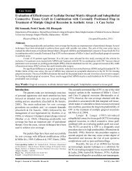
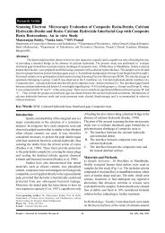
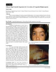
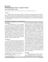
<strong>Giant</strong> <strong>Cell</strong> <strong>Lesion</strong> <strong>of</strong> <strong>the</strong> <strong>Jaw</strong>: A Case Report in a Child -------------------------- Gupta M.K., Naidu S.G. & Maheshwari V.J<strong>the</strong> swelling itself was bony hard and non tender topalpation. Intra oral examination showed a diffuseenlargement in <strong>the</strong> alveolar portion <strong>of</strong> <strong>the</strong> entire leftmandibular teeth # 36, 37, 38 region with obliteration<strong>of</strong> <strong>the</strong> buccal vestibule in <strong>the</strong> same region. Teeth # 37and 38 were clinically missing. Tooth displacement andmobility were not evident in <strong>the</strong> same quadrant. Theoverlying mucosa appeared normal.The swelling was non tender and bony hardon palpation with some areas <strong>of</strong> fluctuance in <strong>the</strong>buccal aspect. The adjacent dentition and <strong>the</strong> oralmucosa did not reveal any abnormality. A tooth vitalitytest revealed normal pulpal response <strong>of</strong> <strong>the</strong> teeth in <strong>the</strong>same quadrant. A provisional diagnosis <strong>of</strong> dentigerouscyst was made with <strong>the</strong> differential diagnosis <strong>of</strong>ameloblastoma, central giant cell granuloma,odontogenic myxoma and fibrous dysplasia.The radiographic examination with anorthopantomogram (Fig. III) revealed a solitary welldefined multilocular radiolucent lesion in <strong>the</strong> region <strong>of</strong>left mandibular angle and ramus. The lesion measured2 X4 cm extending to involve <strong>the</strong> left coronoid withdistinct and sclerotic borders except in <strong>the</strong> region <strong>of</strong><strong>the</strong> superior aspect. Multiple overlapping loculesappearing as ‘soap bubbles’ were noted within <strong>the</strong>lesion along with an unerupted tooth follicle <strong>of</strong>permanent left mandibular second molar. Themandibular canal was inferiorly displaced while areas<strong>of</strong> root resorption were not present. The maxilla didnot reveal any abnormalities. The radiographic featuresled us to consider a diagnosis <strong>of</strong> ameloblastoma or aFig. II: Intraoral photograph showing <strong>the</strong> site <strong>of</strong> surgery.central giant cell granuloma. Ameloblastoma andcentral giant cell granuloma may appear unilocular ormultilocular with a honeycomb or soap bubbleappearance. Odontogenic myxoma may appear as apoorly defined or well-circumscribed radiolucent defect,which may be unilocular or multilocular with a tennisracquet appearance. Serum calcium, phosphorous andalkaline phosphatase levels were found to be normal<strong>the</strong>reby excluding brown tumour <strong>of</strong> hyperparathyroidism.In view <strong>of</strong> <strong>the</strong> young age <strong>of</strong> <strong>the</strong> boy, aconservative surgery was performed under generalFig. III: Panoramic radiograph showing multilocular radiolucent lesion with faint curved septae in left ramus-body <strong>of</strong> mandible.People’s <strong>Journal</strong> <strong>of</strong> <strong>Scientific</strong> <strong>Research</strong> 64Vol. 4(1), Jan. 2011
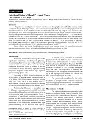
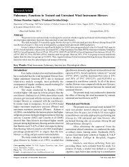
<strong>Giant</strong> <strong>Cell</strong> <strong>Lesion</strong> <strong>of</strong> <strong>the</strong> <strong>Jaw</strong>: A Case Report in a Child -------------------------- Gupta M.K., Naidu S.G. & Maheshwari V.Janes<strong>the</strong>sia to spare <strong>the</strong> mandible with an aggressivecurettage and marginal resection/peripheral osteotomy<strong>of</strong> <strong>the</strong> lesion. The unerupted permanent left mandibularsecond molar and <strong>the</strong> first molar were extracted during<strong>the</strong> procedure. The histopathological examination (Fig.IV) showed a highly vascularized fibrous stroma withseveral multinucleated giant cells with 20-30 nuclei.Randomly dispersed spindle shaped fibroblasts werealso noted and a conclusive diagnosis <strong>of</strong> a central giantcell granuloma was made. The post surgical follow up(Fig. II & V) has remained uneventful till date.Fig. IV: Photomicrograph (H and E stain, magnification 40 x) showingmultinucleated giant cells (~20 nuclei) and spindle shaped fibroblastsin a highly vascularized fibrous stroma.Discussion:<strong>Giant</strong> cell lesions <strong>of</strong> <strong>the</strong> jaws generally includecherubism, giant cell granuloma <strong>of</strong> <strong>the</strong> jaws, giant celltumour (GCT), aneurysmal bone cyst, traumatic bonecyst and jaw tumour <strong>of</strong> hyperparathyroidism. In 1953,Jaffe first described <strong>the</strong> “giant cell reparativegranulomas” and distinguished <strong>the</strong>m from <strong>the</strong> giant celltumor that usually is found in <strong>the</strong> epiphyseal regions <strong>of</strong>long bones. He established two pathological entities in<strong>the</strong> jaws, <strong>the</strong> central giant cell granuloma (CGCG),arising within bone and <strong>the</strong> peripheral giant cellgranuloma (PGCG) arising in s<strong>of</strong>t tissue mass (Jaffe,1953). <strong>Giant</strong> cell granuloma was described as anidiopathic non-neoplastic proliferative lesion and termeda reparative granuloma (Jaffe, 1953). Currentconsensus, however, is that <strong>the</strong>se are not reparativelesions and that if <strong>the</strong>y are not treated, <strong>the</strong>y areprogressive. The true nature <strong>of</strong> <strong>the</strong> central giant cellgranuloma remains speculative. It has been suggestedthat it may be an inflammatory lesion, a reactive lesion,a true tumor, or an endocrine lesion (Pogrel, 2004).Expression <strong>of</strong> <strong>the</strong> c-Src gene has been implicated in<strong>the</strong> development <strong>of</strong> CGCG, GCT and cherubism (Wanget al, 2006). In addition, histologically identical lesionsoccur in patients with known genetic defects such ascherubism, Noonan syndrome, or neur<strong>of</strong>ibromatosistype 1 (deLange et al, 2007). Central giant cellgranuloma has been shown in a report to be fur<strong>the</strong>rassociated with a reciprocal translocation t (X; 4) (q22;q31.3) (Buresh et al, 1999).Central giant cell granuloma is an uncommonlesion, usually asymptomatic and accounting for lessthan 7 % <strong>of</strong> all benign jaw lesions (Kramer et al, 1991).The lesion is found predominantly in children and youngadults with more than 60% <strong>of</strong> all cases occurring before<strong>the</strong> age <strong>of</strong> 30 years (Kaffe et al, 1996). <strong>Lesion</strong>s occurFig. V: Post operative panoramic radiograph showing extracted tooth no 36 and 37 with area <strong>of</strong> resection.People’s <strong>Journal</strong> <strong>of</strong> <strong>Scientific</strong> <strong>Research</strong> 65Vol. 4(1), Jan. 2011
<strong>Giant</strong> <strong>Cell</strong> <strong>Lesion</strong> <strong>of</strong> <strong>the</strong> <strong>Jaw</strong>: A Case Report in a Child -------------------------- Gupta M.K., Naidu S.G. & Maheshwari V.Jmore frequently in <strong>the</strong> mandible than in <strong>the</strong> maxilla in<strong>the</strong> anterior region <strong>of</strong> <strong>the</strong> jaws (Regezzi & Scuibba,1989). Radiographically, CGCG presents as radiolucentdefect, which may be unilocular or multilocular. Thedefect usually is well-circumscribed and, in some cases,displacement <strong>of</strong> teeth can be found (Chuong et al,1986). Central giant cell granuloma is expansive in itsgrowth; it does not grow around or invade nerve trunks.It also does not invade perineural sheaths or spreadvia perineural spaces. Histologically, CGCG containfocal arrangements <strong>of</strong> giant cells within a vascularstroma with thin-walled capillaries adjacent to <strong>the</strong> giantcells. There is a spindle cell stroma which may well be<strong>the</strong> cell <strong>of</strong> origin. The absence <strong>of</strong> perivascular cuffing(as seen in our case) can help differentiate CGCG fromcherubism (Pogrel, 2004). Presence <strong>of</strong> foreign bodytype giant cell (as seen in our case and absence <strong>of</strong>stromal tumour cells differentiate CGCG from a GCT.‘Solid‘aneurysmal bone cysts (ABC) are true benignneoplasms containing giant cells while trauma causingintramedullary hemorrhage has been implicated in <strong>the</strong>past as <strong>the</strong> etiology. Normal serum calcium, parathyroidhormone, alkaline phosphatase and phosphorous levelsdistinguish CGCG from o<strong>the</strong>r conditions like Browntumour <strong>of</strong> hyperparathyroidism (Pogrel, 2004).Cherubism is a self limiting condition, but giantcell granulomas can be aggressive with a tendency torecur and hence require treatment (Chuong et al, 1986).These lesions should be defined as “aggressive giantcell granulomas” <strong>of</strong> <strong>the</strong> jaws, ra<strong>the</strong>r than giant celltumor (Ficarra et al, 1987). <strong>Giant</strong> cell tumour, on <strong>the</strong>o<strong>the</strong>r hand is aggressive with a high recurrence ratebut also has a potential for malignant transformation(Hutter et al 1962). Aggressive/ recurring CGCG havea higher number and relative size index <strong>of</strong> giant cellsand a greater fractional surface area occupied by giantcells (Chuong et al, 1986).The conservative surgical treatment <strong>of</strong> CGCGusually involves curettage alone or alongwith peripheralostectomy with no evidence <strong>of</strong> disease in a 2 yearfollow up perior (Eisenbud et al, 1988). The margins <strong>of</strong><strong>the</strong> CGCG may also be <strong>the</strong>rmally sterilized with a laseror cryoprobes (Kermer et al, 1994). Radical surgicaltechniques <strong>of</strong> resection without continuity defect andperipheral ostectomy (Bataineh et al, 2002) and enblocresection have sometimes been justified for aggressiveCGCG (Chuong et al, 1986). However, recurrence withserious facial mutilation, loss <strong>of</strong> teeth and tooth germsseem unavoidable.Paediatric patients necessitate conservativemanagement to prevent long term developmentaldefects. Steroids and calcitonin have been advocatedin <strong>the</strong> recent past and <strong>the</strong>y at by inhibition <strong>of</strong> osteoclasticactivity. Equal parts <strong>of</strong> triamcinolone acetonide (10mg/ml) and 0.5% bupivacaine injected into <strong>the</strong> lesion for aperiod <strong>of</strong> 11 weeks has been shown to be effective ina child patient (Wendt et al, 2009). Relativecontraindications do exist in certain medical conditions,such as diabetes mellitus, peptic ulcer,and generalizedimmunocompromised states. Calcitonin nasal spray 200U/spray once or twice daily was reported to be safeand effective for <strong>the</strong> treatment <strong>of</strong> CGCG (Allon et al,2009). But, <strong>the</strong>rapy may be complicated owing to <strong>the</strong>great amount <strong>of</strong> discomfort and <strong>the</strong> relatively longduration <strong>of</strong> treatment, with poor compliance by children.Where surgery has been conservative dailysubcutaneous interferon alpha (3 million units/m 2 <strong>of</strong>body surface area) has been tried as an adjuvant dueto its anti-angiogenic properties; but significant sideeffects may limit its utility (Kaban et al, 2007). Acombination <strong>of</strong> interferon alpha and imatinib given for9 months has been shown to initiate regression <strong>of</strong> <strong>the</strong>lesion that continued after treatment had ceased (deLange et al, 2009). Bisphosphonates have also beenattempted intravenously with promising results (Daviset al, 1991). Never<strong>the</strong>less, recurrences <strong>of</strong> CGCG arenot uncommon and can be seen in upto 46% <strong>of</strong> cases(Whitaker et al, 1993).Conclusion:<strong>Giant</strong> cell lesions occur frequently in children<strong>of</strong>ten showing aggressive behaviour. Our approach toconservatively manage <strong>the</strong> lesion has shown goodresults with regular follow up till date. In conclusion,giant cell lesions may present as a diverse group <strong>of</strong>conditions peculiar to <strong>the</strong> jaw bones but, <strong>the</strong>ir diagnosisand management in <strong>the</strong> pediatric patient still remains achallenge to <strong>the</strong> clinician.Bibliography:1. Allon DM, Anavi Y, Calderon S, Central giant cell lesion<strong>of</strong> <strong>the</strong> jaw: nonsurgical treatment with calcitonin nasalspray. Oral Surgery, Oral Medicine, Oral PathologyOral Radiology & Endodontics, 2009 ;107(6):811-818.2. Bataineh AB, Al-Khateeb T, Rawashdeh MA. Thesurgical treatment <strong>of</strong> central giant cell granuloma <strong>of</strong> <strong>the</strong>mandible. <strong>Journal</strong> <strong>of</strong> Oral Maxill<strong>of</strong>acial Surgery , 2002;60(7): 756-776.People’s <strong>Journal</strong> <strong>of</strong> <strong>Scientific</strong> <strong>Research</strong> 66Vol. 4(1), Jan. 2011
<strong>Giant</strong> <strong>Cell</strong> <strong>Lesion</strong> <strong>of</strong> <strong>the</strong> <strong>Jaw</strong>: A Case Report in a Child -------------------------- Gupta M.K., Naidu S.G. & Maheshwari V.J3. Buresh CJ,Seemayer TA, Nelson M, Neff JR, DorfmanHD, Bridge J. t (X;4) ( q22;q31.3) in ginat cell reparativegranuloma.Cancer Genetics & Cytogenet, 1999; 115:80-81.4. Chuong R, Kaban LB, Kozakewich H, Perez-Atayde A:Central giant cell lesions <strong>of</strong> <strong>the</strong> jaws: a clinicopathologicstudy. <strong>Journal</strong> <strong>of</strong> Oral & Maxill<strong>of</strong>acial Surgery,1986;44(9):708-713.5. Davis JP, Archer DJ, Fisher C, Wimalawansa Sj, BaldwinD, Wimalawausa SJ: Multiple recurrent giant cell lesionsassociated with a high circulating level <strong>of</strong> parathyroidhormone related peptide in a young adult. British<strong>Journal</strong> <strong>of</strong> Oral & Maxill<strong>of</strong>acial Surgery, 1991;29(2):102-105.6. de Lange J, van den Akker HP, van den Berg H: Centralgiant cell granuloma <strong>of</strong> <strong>the</strong> jaw: a review <strong>of</strong> <strong>the</strong> literaturewith emphasis on <strong>the</strong>rapy options. Oral Surgery, OralMedicine, Oral Pathology, Oral Radiology &Endodontics, 2007;104(5):603-615.7. de Lange J, van Rijn RR, van den Berg H, van den AkkerHP: Regression <strong>of</strong> central giant cell granuloma by acombination <strong>of</strong> imatinib and interferon: a casereport.British <strong>Journal</strong> <strong>of</strong> Oral & Maxill<strong>of</strong>acial Surgery,2009;47(1):59-6.8. Eisenbud L, Stern M, Rothberg M, Sach S: Central giantcell granuloma <strong>of</strong> <strong>the</strong> jaws: Experiences in <strong>the</strong>management <strong>of</strong> thirty-seven cases. <strong>Journal</strong> <strong>of</strong> Oral andMaxill<strong>of</strong>acial Surgery, 1988:46(5):376-384.9. Ficarra G, Kaban LB, Hansen LS: Central giant cell lesions<strong>of</strong> <strong>the</strong> mandible and maxilla: A clinicopathologic andcytometric study. Oral Surgery Oral Medicine and OralPathology, 1987;49(1):64:44.10. Hutter RVP, Worcester JB, Francis KC: Benign andmalignant giant cell tumors <strong>of</strong> bone: A clinicopathologicalanalysis <strong>of</strong> <strong>the</strong> natural history <strong>of</strong> <strong>the</strong> disease. Cancer,1962 ;15(4):653-690.11. Jaffe HL: <strong>Giant</strong> cell reparative granuloma, traumatic bonecyst, and fibrous (fibro-osseous) dysplasia <strong>of</strong> <strong>the</strong>jawbones. Oral Surgery, Oral Madicin and OralPathology, 1953:6(1):159-175.12. Kaban LB, Troulis MJ, Wilkinson MS, Ebb D, DodsonTB: Adjuvant antiangiogenic <strong>the</strong>rapy for giant celltumors <strong>of</strong> <strong>the</strong> jaws. <strong>Journal</strong> <strong>of</strong> Oral & Maxill<strong>of</strong>acialSurgery, 2007;65(10) :2018-2024.13. Kaffe I, Ardekian L, Taicher S, Littner MM, Buchne A:Radiologic features <strong>of</strong> central giant cell granuloma <strong>of</strong><strong>the</strong> jaws. Oral Surgery Oral Medicine Oral PathologyOral Radiology & Endodontics, 81:720, 199614. Kermer C, Millesi W, Watzke IM: Local injection <strong>of</strong>corticosteroids for central giant cell granuloma. A casereport. International <strong>Journal</strong> Oral Maxill<strong>of</strong>acialSurgery, 1994;23:366-368.15. Kramer IRH, Pindborg JJ, Shear M: Histological Typing<strong>of</strong> Odontogenic Tumors. 2 nd Edn.; Springer-Verlag, Berlin1991;pp 31.16. Liu B, Yu SF, Li TJ:Multinucleated giant cells in variousforms <strong>of</strong> giant cell containing lesions <strong>of</strong> <strong>the</strong> jaws expressfeatures <strong>of</strong> osteoclasts. <strong>Journal</strong> <strong>of</strong> Oral Pathology &Medicine,2003: 32( 6): 367–375.17. Pogrel MA: Benign Non odontogenic <strong>Lesion</strong>s <strong>of</strong> <strong>the</strong><strong>Jaw</strong>s. In: Peterson’s Principles <strong>of</strong> Oral and Maxill<strong>of</strong>acialSurgery, (Vol. 1). M Miloro, GE Ghali, PE Larsen, PD Waite(Eds.); 2 nd Edn.; BC Decker Inc. London, 2004;pp 597-616.18. Stewart JCB: Begin Non odontogenic tumors. In: OralPathology-Clinical Pathologic Correlations. Regezi JA,Sciubba JJ, Jordan RCK (Eds.); 4 th Edn.; Saunders-AnImprint <strong>of</strong> Elsevier, Philadelphia, 2003;pp289-308.19. Wang C, Song Y, Peng B, Fan M, Li J, Zhu S, Bian Z.Expression <strong>of</strong> c-Src and comparison <strong>of</strong> cytologic featuresin cherubism, central giant cell granuloma and giant celltumors. Oncology Reports, 2006;15(3):589-594.20. Wendt FP, Torriani MA, Gomes AP, de Araujo LM,Torriani DD. Intralesional corticosteroid injection forcentral giant cell granuloma: an alternative treatment forchildren. <strong>Journal</strong> <strong>of</strong> Dentistry for Children ,2009; 76(3):229-232.21. Whitaker SB, Waldron CA.Central giant cell lesions <strong>of</strong><strong>the</strong> jaws. A clinical, radiologic, and histopathologicstudy.Oral Surgery Oral Medicine and Oral Pathology.1993;75(2):199-208People’s <strong>Journal</strong> <strong>of</strong> <strong>Scientific</strong> <strong>Research</strong> 67Vol. 4(1), Jan. 2011