Esthetic Closure of Diastema by Porcelain Laminate Veneers: A ...
Esthetic Closure of Diastema by Porcelain Laminate Veneers: A ...
Esthetic Closure of Diastema by Porcelain Laminate Veneers: A ...

Create successful ePaper yourself
Turn your PDF publications into a flip-book with our unique Google optimized e-Paper software.
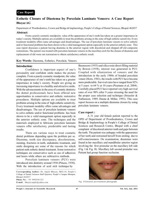
Case Report<strong>Esthetic</strong> <strong>Closure</strong> <strong>of</strong> <strong>Diastema</strong> <strong>by</strong> <strong>Porcelain</strong> <strong>Laminate</strong> <strong>Veneers</strong>: A Case ReportBhoyar AGDepartment <strong>of</strong> Prosthodontics, Crown and Bridge & Implantology, People’s College <strong>of</strong> Dental Sciences, Bhopal-462037Abstract:From a purely cosmetic standpoint, value <strong>of</strong> the appearance <strong>of</strong> one’s teeth has taken on a greater importance intoday’s society. Multiple options are available to treat the problems arising in the zone <strong>of</strong> high esthetic sensitivity. Everytreatment modality <strong>of</strong>fers some advantages and disadvantages. The use <strong>of</strong> porcelain laminate veneers to solve estheticand/or functional problems has been shown to be a valid management option especially in the anterior esthetic zone. Thiscase report discusses a patient having diastema in the anterior region with discolored and chipped <strong>of</strong>f old compositerestorations. The patient was treated with porcelain laminate veneers in the maxillary arch for the closure <strong>of</strong> diastema andreplacement <strong>of</strong> old worn out restoration to obtain satisfactory esthetic result.Key Words: <strong>Diastema</strong>, <strong>Esthetic</strong>s, <strong>Porcelain</strong>, <strong>Veneers</strong>.Introduction:Confidence is important aspect <strong>of</strong> one’spersonality and confident smile makes the picturecomplete. From a purely cosmetic standpoint, the value<strong>of</strong> the appearance <strong>of</strong> one’s teeth has taken on a greaterimportance in today’s society. People are giving anincreasing importance to a healthy and attractive smile.With the advancements in the area <strong>of</strong> cosmetic dentistrythe dental pr<strong>of</strong>essionals have been <strong>of</strong>fered newopportunities in conservative and esthetic restorativeprocedures. Multiple options are available to treatproblems arising in the zone <strong>of</strong> high esthetic sensitivity.Every treatment modality <strong>of</strong>fers some advantages anddisadvantages. The use <strong>of</strong> porcelain laminate veneersto solve esthetic and/or functional problems has beenshown to be a valid management option especially inthe anterior esthetic zone. The techniques and thematerials employed to fabricate porcelain laminateveneers <strong>of</strong>fer satisfactory, predictable and lastingresults.There are various ways to treat cosmeticdental problems depending upon the problem per se.<strong>Diastema</strong>, tooth size discrepancy, discolorations,staining, fractures in teeth, endodontic treatment, andsmile designing are some <strong>of</strong> the reasons for whichpatient seek esthetic dental treatment. Some restorativetechniques are conservative such as use <strong>of</strong> adhesivesand lasers while others are subtractive methods.<strong>Porcelain</strong> laminate veneers (PLV) wereintroduced into dentistry around 1938 (Pincus, 1938).With the introduction <strong>of</strong> acid etch technique <strong>by</strong>----------------------------------------------------------------------------Corresponding Author: Dr. Anjali Bhoyar, MIG-10, People’sCollege <strong>of</strong> Dental Sciences Staff Quarters, People’s Campus Bhanpur,Bhopal-462037Phone No.: +91 9993541123E-mail : bhoyar.anjali@yahoo.comBuonocore (1955) and silica resin direct filling material<strong>by</strong> Bowen (1958), interest was generated in PLV.Coupled with silanization <strong>of</strong> veneers and theintroduction in the early 1980s <strong>of</strong> bonded porcelainveneer (Horn, 1983), the results with PLV have becomemore predictable. Survival rates have ranged from 92%at 5 years to 64 % at 10 years (Peumans et al, 2004).Carefully placed PLV have reported very high survivalrates <strong>of</strong> over 90% after 9 years stressing the need forthe proper case selection and technique (Strassler &Nathanson, 1989; Dunne & Millar, 1993). This casereport focuses on a multiple diastema closure <strong>by</strong> usingporcelain laminate veneers.Case report :A 31 year old female patient reported to theOPD <strong>of</strong> Department <strong>of</strong> Prosthodontics, Crown andBridge & Implantology in People’s College <strong>of</strong> DentalSciences and Research Centre, Bhopal with a chiefcomplaint <strong>of</strong> discolored anterior teeth and gaps betweenthe teeth. The patient was unhappy with the appearance<strong>of</strong> her teeth and restrained herself from smiling due toself-consciousness. On examination, diastemas werefound in her maxillary and mandibular anterior regioninvolving the first premolar on the maxillary left side(Fig. I & Fig. II). Maxillary left second premolar wasPatient had group function occlusion.Fig. I:Extraoral pre-treatment photograph.People’s Journal <strong>of</strong> Scientific Research 47Vol. 4(1), Jan. 2011
<strong>Esthetic</strong> <strong>Closure</strong> <strong>of</strong> <strong>Diastema</strong> <strong>by</strong> <strong>Porcelain</strong> <strong>Laminate</strong> <strong>Veneers</strong>: A Case Report -------------------------- A.G. BhoyarFig. II: Intraoral pre-treatment photograph showing diastema anddiscolored composite restorations.missing. The first molar on same side <strong>of</strong> maxilla wasroot canal treated and had a full coverage gold crownwith occlusal perforation and short gingival margins.Patient had undergone composite veneering on hermaxillary incisors which got discolored and chipped <strong>of</strong>f.After thorough examination, impressions fordiagnostic models were made in irreversiblehydrocolloid (Heraplast, Heraeus Kulzer, USA). Themodels were studied to decide the shape and size <strong>of</strong>the restorations with help <strong>of</strong> a diagnostic wax up. Toprovide a long term solution , the patient was providedthe option <strong>of</strong> PLV. The patient agreed and opted formaxillary correction only as the mandibular anteriorswere less visible.At the onset <strong>of</strong> the treatment, thorough scalingand polishing was done. The gold crown on the maxillaryleft molar was replaced <strong>by</strong> a metal ceramic crown.Before proceeding for tooth preparation, shade wasselected using Vitapan Classical shade guide (VitaZahnfabrik, Germany). The maxillary teeth were thenprepared from right first premolar to the left firstpremolar to receive porcelain laminate veneers . Thetooth preparation was kept in enamel at a depth <strong>of</strong> 0.5mm using a depth cutting diamond and a tapereddiamond 1 mm in diameter. 0.25 mm chamfer wasmaintained in the cervical region (Fig. III). The chamferfinish lines were kept at the level <strong>of</strong> gingival margin.The length <strong>of</strong> the extruded left maxillary lateralincisor was adjusted corresponding to the incisal plane.The incisal chamfer was extended palatally as littleincrease in height was desirable. The centric stops werecarefully avoided during preparing the palatal finish line.The proximal preparation was extended beyond thecontact area to avoid visibility <strong>of</strong> the tooth restorationjunction.Fig. III: Photograph showing tooth preparation for porcelainveneersAfter finishing the sharp line angles and point angles,gingival retraction was performed. Impression <strong>of</strong> themaxillary arch was made in addition silicone (Affinis,Colte‘ne Whaledent) <strong>by</strong> single step double mixtechnique (Fig. IV). In this technique a prefabricatedperforated tray was coated with tray adhesive (Colteneadhesive, Coltene Whaledent) and putty consistencyaddition silicone was loaded on the tray. At the sametime light body material was syringed around theprepared teeth to record the fine details and thepreviously loaded tray was inserted in the mouth tomake the impression. Provisional restorations were notrequired as the tooth reduction was minimal andrestricted to enamel.The porcelain laminates werefabricated <strong>by</strong> refractory die technique (IPS d.SIGNIvoclar Vivadent, USA ). The laminates were tried infor shade, fit, marginal adaptation, shape, size,symmetry and contacts. First they were tried-inindividually using glycerin as a holding medium. Afterindividual evaluation, collective try-in was done toappreciate the esthetic enhancement. Patient’s approvalwas obtained at the time <strong>of</strong> try-in.The cementation appointment:<strong>Laminate</strong> Preparation: The laminates were arrangedon a wax sheet denoting the position <strong>of</strong> the tooth in thearch to avoid incorrect placement and inadvertentbreakage. The laminates were etched with 4 %Hydr<strong>of</strong>luoric acid (<strong>Porcelain</strong> Etchant , Bisco, USA)for 3 minutes carefully avoiding contact on the facialsurface (Fig.V). After etching, they were washedthoroughly using liberal amount <strong>of</strong> water. On drying, acoat <strong>of</strong> Silane coupling agent ( <strong>Porcelain</strong> Primer, Bisco,USA ) was applied (Fig.VI) .Tooth Preparation: The procedure for cementation wasPeople’s Journal <strong>of</strong> Scientific Research 48Vol. 4(1), Jan. 2011
<strong>Esthetic</strong> <strong>Closure</strong> <strong>of</strong> <strong>Diastema</strong> <strong>by</strong> <strong>Porcelain</strong> <strong>Laminate</strong> <strong>Veneers</strong>: A Case Report -------------------------- A.G. BhoyarFig. IV: Photograph showing impression made in addition siliconimpression material.performed on two teeth at a time starting at the midline.The prepared teeth were etched using 37% PhosphoricAcid (Meta Etchant- 37, Meta Biomed Co. Ltd, Korea)for 15 seconds. On air drying bonding agent (Meta P& Bond, Meta Biomed Co Ltd, Korea) was applied &light cured for 10 seconds. Dual cure composite crownand bridge luting agent (Duolink, Bisco, USA) was usedfor cementation. The laminates were spot cured for 5seconds initially. Excess cement was removed withexplorer and then complete curing was done for 20seconds. On completion <strong>of</strong> the cemen-tation procedure,the occlusion was checked in centric and eccentricpositions for interferences. The high points wereremoved and polished (Fig.VII & Fig. VIII).Fig.V: Photograph showing <strong>Laminate</strong>s after etching with hydr<strong>of</strong>luoricacid.Fig. VI: Photograph showing silane coupling agent applied on fittingsurface <strong>of</strong> veneers.Fig.VII: Intraoral post tre-atment photograph.Fig.VIII: Extraoral post tre-atment photograph.Discussion:The etiology <strong>of</strong> diastema may be attributed tothe following factors: (a) Hereditary- congenitallymissing teeth, tooth and jaw size discrepancy,supernumerary teeth & frenum attachments; (b)Developmental problems- habits, periodontal disease,tooth loss, posterior bite collapse (Oesterle & Shellhart,1999). Treatment planning for diastema correctioninclude orthodontic closure, restorative therapy, surgicalcorrection or multidisciplinary approach depending uponthe cause <strong>of</strong> diastema (Dlugokinski et al, 2002). Therestorative closure <strong>of</strong> diastema can be achieved <strong>by</strong>using any <strong>of</strong> the techniques mentioned ; directcomposite veneers, indirect composite veneers,porcelain laminate veneers, all ceramic crowns, metalceramic crowns and composite crowns ((Dlugokinskiet al, 2002; Rammelsberg et al, 2005).Composite resin and porcelain are the mostfrequently used veneering material for diastema closureconservatively. Smaller diastema can be closed withmicr<strong>of</strong>illed and hybrid resins if the diastema is about 1-1.5 mm in dimension. Composite resin is easy to use,requires less appointments, is economic but <strong>of</strong>fers lesswear resistance and surface staining, which makes itinferior to dental porcelain. Besides, failure <strong>of</strong> the sameprompted the patient to opt for porcelain laminates inthe current case (Cho et al, 1998).People’s Journal <strong>of</strong> Scientific Research 49Vol. 4(1), Jan. 2011
<strong>Esthetic</strong> <strong>Closure</strong> <strong>of</strong> <strong>Diastema</strong> <strong>by</strong> <strong>Porcelain</strong> <strong>Laminate</strong> <strong>Veneers</strong>: A Case Report -------------------------- A.G. BhoyarIt has become increasingly apparent thatconservation <strong>of</strong> tooth structure is a major factor indetermining the long term prognosis <strong>of</strong> any restorativeprocedure. One <strong>of</strong> the most important advantages <strong>of</strong>bonded porcelain veneers is that they are extremelyconservative in terms <strong>of</strong> tooth reduction. In the currentcase, only 0.5 mm reduction on the labial surface wasdone. This minimal reduction rarely, if ever, leads topulpal involvement which is a major advantage. Thehighly glazed surface <strong>of</strong> the porcelain laminates preventsplaque accumulation, considered important to attain ahealthy periodontal response. Excellent esthetics couldalso be achieved due to lifelike appearance <strong>of</strong> porcelainand scattering effect <strong>of</strong> the luting cement.However, porcelain laminates have their ownlimitations too. They should not be used when remainingenamel is inadequate to provide adequate retention.Large Class IV defects should probably not be restoredwith veneers because <strong>of</strong> the large amount <strong>of</strong>unsupported porcelain and the lack <strong>of</strong> tooth-coloredbacking. The amount <strong>of</strong> unsupported porcelain shouldbe carefully evaluated in cases with a large diastema .Darkly stained teeth are not optimally restored withveneers. The prognosis for veneers in bruxing isdoubtful. Certainly, such patients should be instructedto use a night guard after final restoration (Sheets &Taniguchi, 1990).Even, if the laminates fail in the long run, theconserved tooth can still be treated with a full crownrestoration. <strong>Porcelain</strong> laminate veneers <strong>of</strong>fer apredictable and successful treatment modality thatpreserves a maximum <strong>of</strong> sound tooth structure. Anincreased risk <strong>of</strong> failure is present only when veneersare partially bonded to dentin. The estimated survivalprobability <strong>of</strong> porcelain laminate veneers over a period<strong>of</strong> 10 years is 91% (Dumfahrt & Schäffer , 2000).Conclusion:Bonded porcelain veneers can providesuccessful esthetic and functional long-term servicefor patients. <strong>Porcelain</strong> laminate veneers <strong>of</strong>fers moreextensiveapplications when they are used cautiouslyand the results achieved have been gratifying for thecosmetic dentist and the patient alike. It has becomeincreasingly apparent that conservation <strong>of</strong> toothstructure is a major factor in determining the long-termprognosis <strong>of</strong> any restorative procedure.Bibliography:1. Bowen RL: Development <strong>of</strong> a silica-resin direct fillingmaterial. Report no. 6333. National Bureau <strong>of</strong> Standards,1958, Washington DC.2. Buonocore MG: A simple method <strong>of</strong> increasing theadhesion <strong>of</strong> acrylic filling materials to enamel surfaces.Journal <strong>of</strong> Dental Research,1955;34(6):849-853.3. Calamia JR: Clinical evaluation <strong>of</strong> etched porcelainveneers. American Journal <strong>of</strong> Dentistry,1989;2(1): 9-15.4. Cho GC, Donovan TE, Chee WW: Clinical experienceswith bonded porcelain laminate veneers. Journal <strong>of</strong>California Dental Association, 1998;26(2):121-127.5. Dlugokinski MD, Frazier KB, Goldstein RE: Restorativetreatment <strong>of</strong> <strong>Diastema</strong>. In: <strong>Esthetic</strong> in Dentistry (Vol.2).RE Goldstein, VB Hoywood (Eds.); 2 nd Edn.; BC DeckerInc. London, 2002;pp703-732.6. Dumfahrt H, Schäffer H: <strong>Porcelain</strong> laminate veneers. Aretrospective evaluation after 1 to 10 years <strong>of</strong> service:Part II-Clinical results. International Journal <strong>of</strong>Prosthodontics, 2000;13(1):9-18.7. Dunne SM, Millar BJ: A longitudinal study <strong>of</strong> the clinicalperformance <strong>of</strong> porcelain veneers. British DentalJournal,1993,175:317-321.8. Horn HR: <strong>Porcelain</strong> laminate veneers bonded to etchedenamel. Dental Clinics <strong>of</strong> North America, 1983;27(4):671-684.9. Jordan RE, Suzuki M, Senda A: Clinical evaluation <strong>of</strong>porcelain laminate veneers: a four-year recall report.Journal <strong>of</strong> <strong>Esthetic</strong> & Restorative Dentistry,1989;1(4):126-132.10. Karlsson S, Landahl I, Stegersjo G , Milleding P: A clinicalevaluation <strong>of</strong> ceramic laminate veneer. The InternationalJournal <strong>of</strong> Prosthodontics, 1992; 5(5):447-451.11. Oesterle LJ, Shellhart WE: Maxillary midline diastemas:a look at the causes. Journal <strong>of</strong> the American DentalAssociation, 1999;130(1):85-94.12. Peumans M, De-Munck J, Fieuws S, Lambrechts P,Vanherle G , Van Meerbeek B: A prospective ten-yearclinical trial <strong>of</strong> porcelain veneers. The Journal <strong>of</strong>Adhesive Dentistry, 2004;6(1):65-76.13. Pincus CR: Building mouth personality. Journal <strong>of</strong> SouthCalifornia Dental Association, 1938;14:125-129.14. Pippin DJ, Mixson JM, Soldan-Els AP: Clinical evaluation<strong>of</strong> restored maxillary incisors: veneers vs PFM crowns.Journal <strong>of</strong> the American Dental Association, 1995;126(11): 1523-1529.15. Rammelsberg P, Spiegl K, Eickemeyer G , Schimitter M:Clinical performance <strong>of</strong> metal free polymer crowns after3 years in service. Journal <strong>of</strong> Dentistry, 2005;33:517-523.16. Sheets CG, Taniguchi T: Advantages and limitations inthe use <strong>of</strong> porcelain veneer restorations. The Journal<strong>of</strong> Prosthetic Dentistry, 1990; 64(4):406-411.17. Strassler HE, Nathanson D: Clinical evaluation <strong>of</strong> etchedporcelain veneers over a period <strong>of</strong> 18 to 42 months.Journal <strong>of</strong> <strong>Esthetic</strong> Dentistry, 1989; 1(1):21-28.People’s Journal <strong>of</strong> Scientific Research 50Vol. 4(1), Jan. 2011