Efficacy of Temporalis Myofascial Flap as an Interpositional Graft ...
Efficacy of Temporalis Myofascial Flap as an Interpositional Graft ...
Efficacy of Temporalis Myofascial Flap as an Interpositional Graft ...

Create successful ePaper yourself
Turn your PDF publications into a flip-book with our unique Google optimized e-Paper software.
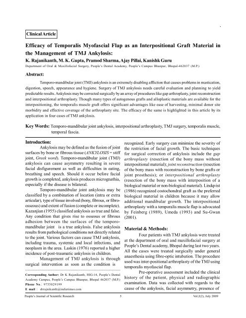
Clinical Article<strong>Efficacy</strong> <strong>of</strong> <strong>Temporalis</strong> My<strong>of</strong><strong>as</strong>cial <strong>Flap</strong> <strong>as</strong> <strong>an</strong> <strong>Interpositional</strong> <strong>Graft</strong> Material inthe M<strong>an</strong>agement <strong>of</strong> TMJ Ankylosis:K. Raj<strong>an</strong>ik<strong>an</strong>th, M. K. Gupta, Pramod Sharma, Ajay Pillai, K<strong>an</strong>ishk GuruDepartment <strong>of</strong> Oral & Maxill<strong>of</strong>acial Surgery, People’s Dental Academy, People’s Campus Bh<strong>an</strong>pur, Bhopal-462037 (M.P.)Abstract:Temporo-m<strong>an</strong>dibular joint (TMJ) <strong>an</strong>kylosis is <strong>an</strong> extremely disabling affliction that causes problems in m<strong>as</strong>tication,digestion, speech, appear<strong>an</strong>ce <strong>an</strong>d hygiene. Surgery <strong>of</strong> TMJ <strong>an</strong>kylosis needs careful evaluation <strong>an</strong>d pl<strong>an</strong>ning to yieldpredictable results. Ankylosis may be corrected surgically by <strong>an</strong> array <strong>of</strong> procedures like gap arthropl<strong>as</strong>ty, joint reconstruction<strong>an</strong>d interpositional arthropl<strong>as</strong>ty. Though m<strong>an</strong>y types <strong>of</strong> autogenous grafts <strong>an</strong>d allopl<strong>as</strong>tic materials are available for theinterpositioning, the temporalis muscle graft <strong>of</strong>fers signific<strong>an</strong>t adv<strong>an</strong>tages like e<strong>as</strong>e <strong>of</strong> harvesting, minimal donor sitemorbidity <strong>an</strong>d effective coverage <strong>of</strong> the arthropl<strong>as</strong>ty site. The efficacy <strong>of</strong> the same is highlighted in this article by itsapplication in four c<strong>as</strong>es <strong>of</strong> TMJ <strong>an</strong>kylosis.Key Words: Temporo-m<strong>an</strong>dibular joint <strong>an</strong>kylosis, interpositional arthropl<strong>as</strong>ty, TMJ surgery, temporalis muscle,temporal f<strong>as</strong>cia.Introduction:Ankylosis may be defined <strong>as</strong> the fusion <strong>of</strong> jointsurfaces by bone or fibrous tissue (ANKYLOSIS = stiffjoint, Greek word). Temporo-m<strong>an</strong>dibular joint (TMJ)<strong>an</strong>kylosis c<strong>an</strong> cause <strong>as</strong>ymmetry resulting in severefacial disfigurement <strong>as</strong> well <strong>as</strong> difficulties in eating,breathing <strong>an</strong>d speech. Should it occur before facialgrowth is completed, <strong>an</strong>kylosis produces micrognathia,especially if the dise<strong>as</strong>e is bilateral.Temporo-m<strong>an</strong>dibular joint <strong>an</strong>kylosis may becl<strong>as</strong>sified by a combination <strong>of</strong> location (intra or extraarticular), type <strong>of</strong> tissue involved (bony, fibrous, or fibroosseous)<strong>an</strong>d extent <strong>of</strong> fusion (complete or incomplete).Kaz<strong>an</strong>ji<strong>an</strong> (1955) cl<strong>as</strong>sified <strong>an</strong>kylosis <strong>as</strong> true <strong>an</strong>d false.Any condition that gives rise to osseous or fibrousadhesion between the surfaces <strong>of</strong> the temporom<strong>an</strong>dibularjoint is a true <strong>an</strong>kylosis. False <strong>an</strong>kylosisresults from pathological conditions not directly relatedto the joint. Various factors c<strong>an</strong> cause TMJ <strong>an</strong>kylosis,including trauma, systemic <strong>an</strong>d local infections, <strong>an</strong>dneopl<strong>as</strong>m in the area. L<strong>as</strong>kin (1976) reported a higherincidence <strong>of</strong> post-traumatic <strong>an</strong>kylosis in children.M<strong>an</strong>agement <strong>of</strong> TMJ <strong>an</strong>kylosis is throughsurgical intervention <strong>as</strong> soon <strong>as</strong> the condition is-----------------------------------------------------------------------------Corresponding Author: Dr K Raj<strong>an</strong>ik<strong>an</strong>th, HIG-14, People’s DentalAcademy Campus, People’s Campus Bh<strong>an</strong>pur, Bhopal 462037 (M.P.)Phone No.: 9755029199E mail : drrajnik<strong>an</strong>th@indiatimes.comrecognized. Early surgery c<strong>an</strong> minimize the severity <strong>of</strong>the restriction <strong>of</strong> facial growth. The b<strong>as</strong>ic techniquesfor surgical correction <strong>of</strong> <strong>an</strong>kylosis include the gaparthropl<strong>as</strong>ty (resection <strong>of</strong> the bony m<strong>as</strong>s withoutinterpositional material); joint reconstruction (resection<strong>of</strong> the bony m<strong>as</strong>s with reconstruction by bone grafts orjoint prosthesis); or interpositional arthropl<strong>as</strong>ty(resection <strong>of</strong> the bony m<strong>as</strong>s with interposition <strong>of</strong> abiological material or non-biological material). Lindqvist(1986) recognized costochondral graft <strong>as</strong> the preferredbiological material in children because it may allowadditional m<strong>an</strong>dibular growth. The interpositionalarthropl<strong>as</strong>ty with a temporalis muscle flap is advocatedby Feinberg (1989), Umeda (1993) <strong>an</strong>d Su-Gw<strong>an</strong>(2001).Material & Methods:Four patients with TMJ <strong>an</strong>kylosis were treatedat the department <strong>of</strong> oral <strong>an</strong>d maxill<strong>of</strong>acial surgery atPeople’s Dental academy, Bhopal during l<strong>as</strong>t two years.All the c<strong>as</strong>es were treated surgically under general<strong>an</strong>aesthesia using fibre-optic intubation. The procedureused w<strong>as</strong> inter-positional arthropl<strong>as</strong>ty <strong>of</strong> the TMJ usingtemporalis my<strong>of</strong><strong>as</strong>cial flap.Pre-operative <strong>as</strong>sessment included the clinicalhistory <strong>of</strong> the patient, physical <strong>an</strong>d radiographicexamination. Data w<strong>as</strong> collected with regards to thecause <strong>of</strong> the <strong>an</strong>kylosis, facial <strong>as</strong>ymmetry, presence <strong>of</strong>People’s Journal <strong>of</strong> Scientific Research 5 Vol.2(2), July 2009
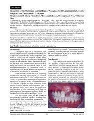
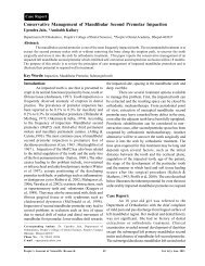
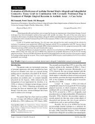
<strong>Efficacy</strong> <strong>of</strong> <strong>Temporalis</strong> My<strong>of</strong><strong>as</strong>cial <strong>Flap</strong> <strong>as</strong> <strong>an</strong> <strong>Interpositional</strong> <strong>Graft</strong> ------- K Raj<strong>an</strong>ik<strong>an</strong>th, M K Gupta, P Sharma, A Pillai, K Gurumicrognathia, the time <strong>of</strong> onset <strong>of</strong> the <strong>an</strong>kylosis, theside affected, <strong>an</strong>d the nature <strong>of</strong> the union (fibrous /bony). Me<strong>as</strong>urements <strong>of</strong> maximal inter-incisal opening(Fig.I), lateral movements <strong>an</strong>d protrusion were made aday before the surgical procedure. The radiographicexamination included p<strong>an</strong>oramic radiographs <strong>an</strong>dcomputerized axial tomograms (Fig. II) to determinethe <strong>an</strong>atomic boundaries <strong>of</strong> <strong>an</strong>kylosis <strong>an</strong>d the type <strong>of</strong><strong>an</strong>kylosis.retracted <strong>an</strong>teriorly, <strong>an</strong> incision w<strong>as</strong> made through thesuperficial layer <strong>of</strong> temporal f<strong>as</strong>cia beginning from theroot <strong>of</strong> the zygomatic arch just in front <strong>of</strong> the tragus<strong>an</strong>tero-posteriorly towards the upper corner <strong>of</strong> theretracted flap.After exposing the joint <strong>an</strong>d identification <strong>of</strong>the site <strong>of</strong> the <strong>an</strong>kylosis (Fig. III), aggressive excision<strong>of</strong> the fibrous <strong>an</strong>d/or bony m<strong>as</strong>s w<strong>as</strong> done initially withdrills <strong>an</strong>d completed with a chisel (Fig. IV).Fig. I: Photograph showing mouth opening pre-operatively in bonytype <strong>of</strong> <strong>an</strong>kylosis.Fig. II: Photograph showing three dimensional CT reconstruction <strong>of</strong>affected TMJ.Operative Procedure:A written, informed <strong>an</strong>d verbal consent w<strong>as</strong>taken for the procedure. The temporal region w<strong>as</strong>prepared. Approach to the TMJ region w<strong>as</strong> gained usingAl-kayat & Bramely’s (1979) modified pre-auricularincision. Dissection w<strong>as</strong> carried out through thesuperficial temporal f<strong>as</strong>cia, which w<strong>as</strong> retracted<strong>an</strong>teriorly to protect the facial nerve, <strong>an</strong>d the periosteumover the zygomatic arch w<strong>as</strong> incised. With the flapFig. III: Photograph showing <strong>an</strong>kylosed TMJ.Care w<strong>as</strong> taken to avoid injury to the internal maxillaryartery underneath the condyle. It w<strong>as</strong> followed by theexcision <strong>of</strong> the coronoid process <strong>an</strong>d burring <strong>of</strong> theglenoid fossa creating a gap <strong>of</strong> at le<strong>as</strong>t 15 mm betweenthe ro<strong>of</strong> <strong>of</strong> the fossa <strong>an</strong>d the m<strong>an</strong>dible. A p<strong>as</strong>sive interincisalopening <strong>of</strong> at le<strong>as</strong>t 30 mm w<strong>as</strong> achieved. Copiousirrigation w<strong>as</strong> done with saline. Contra lateralcoronoidectomy w<strong>as</strong> performed when necessary, inaccord<strong>an</strong>ce with Kab<strong>an</strong>’s protocol (1990).A finger-shaped flap <strong>of</strong> sufficient length w<strong>as</strong>marked on the temporal f<strong>as</strong>cia so that after rotation itwould reach the joint site e<strong>as</strong>ily without <strong>an</strong>y unduestretching (Fig.V). The composite flap <strong>of</strong> temporal f<strong>as</strong>cia<strong>an</strong>d muscle w<strong>as</strong> developed which would be pedicled onthe br<strong>an</strong>ches <strong>of</strong> superficial temporal artery. The flapw<strong>as</strong> rotated <strong>an</strong>d sutured to the medial, <strong>an</strong>terior <strong>an</strong>dposterior regions <strong>of</strong> the site <strong>of</strong> arthropl<strong>as</strong>ty (Fig.VI) sothat it covers the cut ends <strong>of</strong> the arthropl<strong>as</strong>ty completely<strong>an</strong>d effectively. Layer-wise closure w<strong>as</strong> done <strong>an</strong>d <strong>as</strong>urgical drain placed. The physiotherapy beg<strong>an</strong> aftertwo days post-operatively with jaw exercises undersupervision. It w<strong>as</strong> intensified gradually in the postoperative<strong>an</strong>d recall period (Fig.VII).People’s Journal <strong>of</strong> Scientific Research 6 Vol.2(2), July 2009
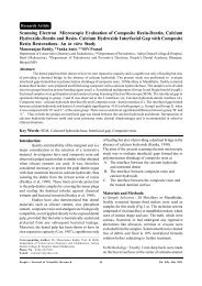
<strong>Efficacy</strong> <strong>of</strong> <strong>Temporalis</strong> My<strong>of</strong><strong>as</strong>cial <strong>Flap</strong> <strong>as</strong> <strong>an</strong> <strong>Interpositional</strong> <strong>Graft</strong> ------- K Raj<strong>an</strong>ik<strong>an</strong>th, M K Gupta, P Sharma, A Pillai, K GuruResults:Four patients were subjected to TMJ surgery:one male <strong>an</strong>d three females. All the females hadunilateral bony <strong>an</strong>kylosis (CT findings) with minimalmouth opening. The male w<strong>as</strong> having fibrous <strong>an</strong>kylosis<strong>of</strong> right TMJ with <strong>an</strong> opening <strong>of</strong> 11 mm pre-operatively.Fig.VII: Photograph showing inter-incisal post-operatively <strong>of</strong> thesame patient <strong>as</strong> showing the fig. I.Fig.IV: Photograph showing resected <strong>an</strong>kylosed m<strong>as</strong>s.The me<strong>an</strong> age w<strong>as</strong> 14.25 years <strong>an</strong>d aetiology includedtrauma in all the c<strong>as</strong>es. Only one c<strong>as</strong>e w<strong>as</strong> operatedpreviously elsewhere <strong>an</strong>d had recurrence within 1 year.Follow-up period r<strong>an</strong>ged from 3 months to 2 years. Thevarious me<strong>as</strong>urements are grouped <strong>an</strong>d depicted inTable 1.Table I: Pre & Post-Operative me<strong>as</strong>urements <strong>of</strong> inter incisalopening in the patientsS.NoAge/SexType <strong>of</strong><strong>an</strong>kylosisPre-operativemouth openingPost-operativemouth openingPost-operativefollow-up period1.14 / FBonyNil34 mm2 years2.11/ MFibrous11 mm39 mm1yr 8 months3.15/ FBonyNil36 mm7 monthsFig.V: Photograph showing elevation <strong>of</strong> <strong>Temporalis</strong> my<strong>of</strong><strong>as</strong>cial flap.4.17/FBonyNil38 mm3 monthsFig.VI: Photograph showing interpositioning <strong>of</strong> the temporalismy<strong>of</strong><strong>as</strong>cial flap.Discussion:Early <strong>an</strong>kylosis <strong>of</strong> TMJ in children c<strong>an</strong> be adeterrent to normal m<strong>an</strong>dibular growth. Therefore, earlydiagnosis <strong>of</strong> TMJ <strong>an</strong>kylosis <strong>an</strong>d early surgicalintervention is import<strong>an</strong>t. M<strong>an</strong>agement <strong>of</strong> TMJ <strong>an</strong>kylosisis mainly performed through surgical intervention.Various techniques for the m<strong>an</strong>agement <strong>of</strong> TMJ<strong>an</strong>kylosis have been described. However, no singletechnique h<strong>as</strong> proved entirely satisfactory. Thecharacteristic pathology <strong>of</strong> <strong>an</strong>kylosis is the formation<strong>of</strong> a bony m<strong>as</strong>s, which replaces the articulation, resultingin restriction <strong>of</strong> m<strong>an</strong>dibular movements. For this re<strong>as</strong>on,treatment <strong>of</strong> TMJ <strong>an</strong>kylosis requires removal <strong>of</strong> <strong>as</strong>ufficient amount <strong>of</strong> bone to allow for free movement<strong>of</strong> the m<strong>an</strong>dibular stump <strong>an</strong>d interposition <strong>of</strong> somePeople’s Journal <strong>of</strong> Scientific Research 7 Vol.2(2), July 2009
<strong>Efficacy</strong> <strong>of</strong> <strong>Temporalis</strong> My<strong>of</strong><strong>as</strong>cial <strong>Flap</strong> <strong>as</strong> <strong>an</strong> <strong>Interpositional</strong> <strong>Graft</strong> ------- K Raj<strong>an</strong>ik<strong>an</strong>th, M K Gupta, P Sharma, A Pillai, K Gurumaterial between the remaining ramus <strong>an</strong>d skull b<strong>as</strong>e.It is necessary to use <strong>an</strong> interpositional material toprevent TMJ re-<strong>an</strong>kylosis after arthropl<strong>as</strong>ty (orcondylectomy). This particular <strong>as</strong>pect <strong>of</strong> the treatmenth<strong>as</strong> been the subject <strong>of</strong> numerous discussions. The use<strong>of</strong> various allogenic interpositional materials h<strong>as</strong> led toserious complications, including foreign body reaction<strong>an</strong>d migration. Homografts, such <strong>as</strong> skin, temporalismuscle, or f<strong>as</strong>cia lata, are considered <strong>as</strong> the material<strong>of</strong> choice for interposition.In recent years, a pedicled temporalismy<strong>of</strong><strong>as</strong>cial or temporal f<strong>as</strong>cia flap h<strong>as</strong> been advocatedin TMJ surgery to treat the TMJ <strong>an</strong>kylosis (Feinberg& Larsen, 1989). Adv<strong>an</strong>tages <strong>of</strong> these flaps in TMJreconstruction include close proximity to the TMJwithout involving <strong>an</strong> additional surgical site, adequateblood supply, autogenous origin, <strong>an</strong>d mainten<strong>an</strong>ce <strong>of</strong>attachment to the coronoid process which providesmovement <strong>of</strong> the flap during function, simulatingphysiologic action <strong>of</strong> the disc. Its proximity to the jointallows for a pedicled tr<strong>an</strong>sfer <strong>of</strong> v<strong>as</strong>cularized tissueinto the joint area. In this c<strong>as</strong>e a composite (f<strong>as</strong>cia,muscle, <strong>an</strong>d periosteum) axial flap w<strong>as</strong> harvested, <strong>as</strong>described by Herbosa & Rotsk<strong>of</strong>f (1990). The axialflaps were e<strong>as</strong>ily rotated inferiorly into the joint space.Rotation under the zygomatic arch prevents bulkiness<strong>an</strong>d avoids the need for surgically reducing the thickness<strong>of</strong> the zygomatic arch, <strong>as</strong> suggested by Pogrel & Kab<strong>an</strong>(1990), when rotating the muscle over the arch.Regarding the fate <strong>of</strong> temporalis muscle graft,Umeda et al (1993) have demonstrated by magneticreson<strong>an</strong>ce imaging that the flaps appeared to be viable<strong>an</strong>d the tissue signal w<strong>as</strong> compatible with vital muscle<strong>an</strong>d / or fat <strong>as</strong> opposed to tissue scarring.for M<strong>an</strong>agement <strong>of</strong> Temporo-m<strong>an</strong>dibular Joint Ankylosis.Journal <strong>of</strong> Oral <strong>an</strong>d Maxill<strong>of</strong>acial Surgery, 1990;48(11):1145-1151.5. Kaz<strong>an</strong>ji<strong>an</strong> VH: Temporo-m<strong>an</strong>dibular <strong>an</strong>kylosis withm<strong>an</strong>dibular retrusion. Americ<strong>an</strong> Journal <strong>of</strong> Surgery,1955;90: 905-910.6. L<strong>as</strong>kin DM: Role <strong>of</strong> the meniscus in the etiology <strong>of</strong> posttraumatic temporom<strong>an</strong>dibular joint <strong>an</strong>kylosis.International Journal <strong>of</strong> Oral Surgery, 1978;7(4):340-345.7. Lindqvist C, Pihakari A, T<strong>as</strong><strong>an</strong>en A, Hampf G: Autogenouscostochondral grafts in TMJ arthropl<strong>as</strong>ty. A survey <strong>of</strong>66 arthropl<strong>as</strong>ties in 60 patients. Journal <strong>of</strong> Maxill<strong>of</strong>acialSurgery, 1986; 14(3):143-149.8. Pogrel MA, Kab<strong>an</strong> LB: The role <strong>of</strong> a temporalis f<strong>as</strong>cia<strong>an</strong>d muscle flap in temporom<strong>an</strong>dibular joint surgery.Journal <strong>of</strong> Oral <strong>an</strong>d Maxill<strong>of</strong>acial Surgery, 1990; 48(1):14-19.9. Smith JA, S<strong>an</strong>dler NA, Ozaki WH, Braun TW: Subjective<strong>an</strong>d objective <strong>as</strong>sessment <strong>of</strong> the temporal my<strong>of</strong><strong>as</strong>cialflap in previously operated temporom<strong>an</strong>dibular joints.Journal <strong>of</strong> Oral <strong>an</strong>d Maxill<strong>of</strong>acial Surgery, 1999;57(9):1058-1065.10. Su-Gw<strong>an</strong> K: Treatment <strong>of</strong> temporom<strong>an</strong>dibular joint<strong>an</strong>kylosis with temporalis muscle <strong>an</strong>d f<strong>as</strong>cia flap.International Journal <strong>of</strong> Oral <strong>an</strong>d Maxill<strong>of</strong>acialSurgery, 2001;30(3):189-193.11. Umeda H, Kab<strong>an</strong> LB, Pogrel MA,Stren M, Rotsk<strong>of</strong>f KS:Long-term viability <strong>of</strong> temporalis muscle/f<strong>as</strong>cia flap usedfor temporom<strong>an</strong>dibular reconstruction. Journal <strong>of</strong> Oral<strong>an</strong>d Maxill<strong>of</strong>acial Surgery, 1993;51(5):530-534.Bibliography:1. Al kayat A, Bramley P: A modified pre-auricular approachto the Temporo-m<strong>an</strong>dibular joint <strong>an</strong>d molar arch. BritishJournal <strong>of</strong> Oral Surgery, 1979; 17:91-103.2. Feinberg SE, Larsen PE: The use <strong>of</strong> a pedicled temporalismuscle-pericr<strong>an</strong>ial flap for replacement <strong>of</strong> the TMJ disc:preliminary report. Journal <strong>of</strong> Oral <strong>an</strong>d Maxill<strong>of</strong>acialSurgery, 1989; 47: 142-146.3. Herbosa EG, Rotsk<strong>of</strong>f KS: Composite temporalis pedicleflap <strong>as</strong> <strong>an</strong> interpositional graft in temporom<strong>an</strong>dibularjoint arthropl<strong>as</strong>ty: a preliminary report. Journal <strong>of</strong> Oral<strong>an</strong>d Maxill<strong>of</strong>acial Surgery, 1990; 48(11): 1049-1056.4. Kab<strong>an</strong> LB, Perrott DH, Fisher K, Topazi<strong>an</strong> RG: A ProtocolPeople’s Journal <strong>of</strong> Scientific Research 8 Vol.2(2), July 2009