Peripheral vision and pattern recognition: a review - strasburger - main
Peripheral vision and pattern recognition: a review - strasburger - main
Peripheral vision and pattern recognition: a review - strasburger - main

You also want an ePaper? Increase the reach of your titles
YUMPU automatically turns print PDFs into web optimized ePapers that Google loves.
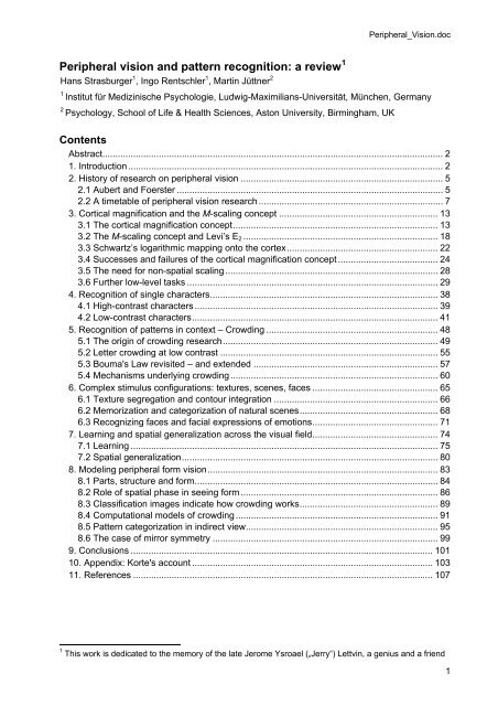
<strong>Peripheral</strong>_Vision.doc<br />
<strong>Peripheral</strong> <strong>vision</strong> <strong>and</strong> <strong>pattern</strong> <strong>recognition</strong>: a <strong>review</strong> 1<br />
Hans Strasburger 1 , Ingo Rentschler 1 , Martin Jüttner 2<br />
1<br />
Institut für Medizinische Psychologie, Ludwig-Maximilians-Universität, München, Germany<br />
2<br />
Psychology, School of Life & Health Sciences, Aston University, Birmingham, UK<br />
Contents<br />
Abstract..................................................................................................................................... 2<br />
1. Introduction........................................................................................................................... 2<br />
2. History of research on peripheral <strong>vision</strong> ............................................................................... 5<br />
2.1 Aubert <strong>and</strong> Foerster ........................................................................................................ 5<br />
2.2 A timetable of peripheral <strong>vision</strong> research ........................................................................ 7<br />
3. Cortical magnification <strong>and</strong> the M-scaling concept .............................................................. 13<br />
3.1 The cortical magnification concept................................................................................ 13<br />
3.2 The M-scaling concept <strong>and</strong> Levi’s E 2 ............................................................................ 18<br />
3.3 Schwartz’s logarithmic mapping onto the cortex........................................................... 22<br />
3.4 Successes <strong>and</strong> failures of the cortical magnification concept ....................................... 24<br />
3.5 The need for non-spatial scaling ................................................................................... 28<br />
3.6 Further low-level tasks .................................................................................................. 29<br />
4. Recognition of single characters......................................................................................... 38<br />
4.1 High-contrast characters ............................................................................................... 39<br />
4.2 Low-contrast characters................................................................................................ 41<br />
5. Recognition of <strong>pattern</strong>s in context – Crowding ................................................................... 48<br />
5.1 The origin of crowding research.................................................................................... 49<br />
5.2 Letter crowding at low contrast ..................................................................................... 55<br />
5.3 Bouma's Law revisited – <strong>and</strong> extended ........................................................................ 57<br />
5.4 Mechanisms underlying crowding ................................................................................. 60<br />
6. Complex stimulus configurations: textures, scenes, faces ................................................. 65<br />
6.1 Texture segregation <strong>and</strong> contour integration ................................................................ 66<br />
6.2 Memorization <strong>and</strong> categorization of natural scenes...................................................... 68<br />
6.3 Recognizing faces <strong>and</strong> facial expressions of emotions................................................. 71<br />
7. Learning <strong>and</strong> spatial generalization across the visual field................................................. 74<br />
7.1 Learning ........................................................................................................................ 75<br />
7.2 Spatial generalization.................................................................................................... 80<br />
8. Modeling peripheral form <strong>vision</strong>.......................................................................................... 83<br />
8.1 Parts, structure <strong>and</strong> form............................................................................................... 84<br />
8.2 Role of spatial phase in seeing form ............................................................................. 86<br />
8.3 Classification images indicate how crowding works...................................................... 89<br />
8.4 Computational models of crowding ............................................................................... 91<br />
8.5 Pattern categorization in indirect view........................................................................... 95<br />
8.6 The case of mirror symmetry ........................................................................................ 99<br />
9. Conclusions ...................................................................................................................... 101<br />
10. Appendix: Korte's account .............................................................................................. 103<br />
11. References ..................................................................................................................... 107<br />
1 This work is dedicated to the memory of the late Jerome Ysroael („Jerry“) Lettvin, a genius <strong>and</strong> a friend<br />
1
<strong>Peripheral</strong>_Vision.doc<br />
Abstract<br />
We summarize the various str<strong>and</strong>s of research on peripheral <strong>vision</strong> <strong>and</strong> relate them to theories<br />
of form perception. After a historical overview, we describe quantifications of the cortical<br />
magnification hypothesis, including an extension of Schwartz’s cortical mapping function. The<br />
merits of this concept are considered across a wide range of psychophysical tasks, followed by<br />
a discussion of its limitations <strong>and</strong> the need for non-spatial scaling. We also <strong>review</strong> the<br />
eccentricity dependence of other low-level functions including reaction time, temporal resolution<br />
<strong>and</strong> spatial summation, as well as perimetric methods. A central topic is then the <strong>recognition</strong> of<br />
characters in peripheral <strong>vision</strong>, both at low <strong>and</strong> high levels of contrast, <strong>and</strong> the impact of<br />
surrounding contours known as crowding. We demonstrate how Bouma’s law, specifying the<br />
critical distance for the onset of crowding, can be stated in terms of the retino-cortical mapping.<br />
The <strong>recognition</strong> of more complex stimuli, like textures, faces <strong>and</strong> scenes reveals a substantial<br />
impact of mid-level <strong>vision</strong> <strong>and</strong> cognitive factors. We further consider eccentricity-dependent<br />
limitations of learning, both at the level of perceptual learning <strong>and</strong> <strong>pattern</strong> category learning.<br />
Generic limitations of extrafoveal <strong>vision</strong> are observed for the latter in categorization tasks<br />
involving multiple stimulus classes. Finally, models of peripheral form <strong>vision</strong> are discussed. We<br />
report that peripheral <strong>vision</strong> is limited with regard to <strong>pattern</strong> categorization by a distinctly lower<br />
representational complexity <strong>and</strong> processing speed. Taken together, the limitations of cognitive<br />
processing in peripheral <strong>vision</strong> appear to be as significant as those imposed on low-level<br />
functions <strong>and</strong> by way of crowding.<br />
Keywords: <strong>Peripheral</strong> <strong>vision</strong>, visual field, acuity, contrast sensitivity, temporal resolution,<br />
crowding effect, perceptual learning, computational models, categorization, object <strong>recognition</strong>,<br />
faces, facial expression, natural scenes, scene gist, texture, contour, learning, perceptual<br />
learning, category learning, generalization, invariance, translation invariance, shift invariance,<br />
representational complexity.<br />
1. Introduction<br />
The driver of a car traveling at high speed, a shy person avoiding to directly look at the object of<br />
her or his interest, a patient suffering from age-related macular degeneration, they all face the<br />
problem of getting the most out of seeing sidelong. It is commonly thought that blurriness of<br />
<strong>vision</strong> is the <strong>main</strong> characteristic of that condition. Yet Lettvin (1976) picked up the thread where<br />
Aubert <strong>and</strong> Foerster (1857) had left it when he insisted that any theory of peripheral <strong>vision</strong><br />
exclusively based on the assumption of blurriness is bound to fail: ”When I look at something it<br />
is as if a pointer extends from my eye to an object. The ‘pointer’ is my gaze, <strong>and</strong> what it touches<br />
I see most clearly. Things are less distinct as they lie farther from my gaze. It is not as if these<br />
things go out of focus – but rather it’s as if somehow they lose the quality of form” (Lettvin,<br />
1976, p. 10, cf. Figure 1).<br />
2
<strong>Peripheral</strong>_Vision.doc<br />
Figure 1. One of Lettvin’s demonstrations. “Finally, there are two images that carry an amusing<br />
lesson. The first is illustrated by the O composed of small o's as below. It is a quite clearly circular<br />
array, not as vivid as the continuous O, but certainly definite. Compare this with the same large O<br />
surrounded by only two letters to make the word HOE. I note that the small o's are completely visible<br />
still, but that the large O cannot be told at all well. It simply looks like an aggregate of small o's.”<br />
(Lettvin, 1976, p.14)<br />
To account for a great number of meticulous observations on peripheral form <strong>vision</strong>, Lettvin<br />
(1976, p. 20) suggested “that texture somehow redefined is the primitive stuff out of which form<br />
is constructed”. His proposal can be taken further by noting that texture perception was<br />
redefined by Julesz <strong>and</strong> co-workers (Julesz, Gilbert, Shepp, & Frisch, 1973; Caelli & Julesz,<br />
1978; Caelli, Julesz, & Gilbert, 1978; Julesz, 1981). These authors succeeded to show that<br />
texture perception ignores relative spatial position, whereas form perception from local scrutiny<br />
does not. Julesz (1981, p. 97) concluded that cortical feature analyzers are “not connected<br />
directly to each other” in peripheral <strong>vision</strong> <strong>and</strong> interact “only in aggregate”. By contrast, research<br />
on form <strong>vision</strong> indicated the existence in visual cortex of co-operative mechanisms that locally<br />
connect feature analyzers (e.g., Grossberg & Mingolla, 1985; Phillips & Singer, 1997;<br />
Carpenter, Grossberg, & Mehanian, 1989; Shapley, Caelli, Grossberg, Morgan, & Rentschler,<br />
1990; Lee, Mumford, Romero, & Lamme, 1998).<br />
Our interest in peripheral <strong>vision</strong> was aroused by the work of Lettvin (1976). Our principal goal<br />
since was to better underst<strong>and</strong> form <strong>vision</strong> in the peripheral visual field. However, the specifics<br />
of form <strong>vision</strong> can only be appreciated in the light of what we know about lower-level functions.<br />
We therefore proceed from low-level functions to the <strong>recognition</strong> of characters <strong>and</strong> more<br />
complex <strong>pattern</strong>s. We then turn to the question of how the <strong>recognition</strong> of form is learned.<br />
Finally, we consider models of peripheral form <strong>vision</strong>. As all that constitutes a huge field of<br />
research, we had to exclude important areas of work. We omitted work on optical aspects, on<br />
motion (cf. the paper by Nishida in this issue), on color, <strong>and</strong> on reading. We also ignored most<br />
clinical aspects including the large field of perimetry. We just touch on applied aspects, in<br />
particular insights from aviation <strong>and</strong> road traffic.<br />
More specifically, we <strong>review</strong> in Chapter 2 research on peripheral <strong>vision</strong> in ophthalmology,<br />
optometry, psychology, <strong>and</strong> in the engineering sciences with a historical perspective. Chapter 3<br />
addresses the variation of spatial scale as a major contributor to differences in performance<br />
across the visual field. Here the concept of size-scaling inspired by cortical magnification is the<br />
<strong>main</strong> topic. Levi’s E 2 value is introduced <strong>and</strong> we summarize E 2 values over a wide range of<br />
tasks. However, non-spatial stimulus dimensions, in particular <strong>pattern</strong> contrast, are also<br />
important. Single-cell-recording <strong>and</strong> fMRI studies support the concept for which we present<br />
3
<strong>Peripheral</strong>_Vision.doc<br />
empirical values <strong>and</strong> a logarithmic retino-cortical mapping function which matches the inverselinear<br />
law Further low-level tasks <strong>review</strong>ed are the measurements of visual reaction time,<br />
apparent brightness, temporal resolution, flicker detection, <strong>and</strong> spatial summation. These tasks<br />
have found application as diagnostic tools for perimetry, both in clinical <strong>and</strong> non-clinical<br />
settings.<br />
<strong>Peripheral</strong> letter <strong>recognition</strong> is a central topic in our <strong>review</strong>. In Chapter 4, we first consider its<br />
dependence on stimulus contrast. We then proceed to crowding, the phenomenon traditionally<br />
defined as loss of <strong>recognition</strong> performance for letter targets appearing in the context of other,<br />
distracting letters (Chapter 5). Crowding occurs when the distracters are closer than a critical<br />
distance specified by Bouma’s law (1970). We demonstrate its relationship with size-scaling<br />
according to cortical magnification <strong>and</strong> derive the equivalent of Bouma’s law in retinotopic<br />
cortical visual areas. Furthermore, we discuss how crowding is related to low-level contour<br />
interactions, such as lateral masking <strong>and</strong> surround suppression, <strong>and</strong> how it is modulated by<br />
attentional factors.<br />
Regarding the <strong>recognition</strong> of scenes, objects, <strong>and</strong> faces in peripheral <strong>vision</strong>, a key question is<br />
whether observer performance follows predictions based on cortical magnification <strong>and</strong> acuity<br />
measures (Chapter 6). Alternatively, it might be that configural information plays a role in the<br />
peripheral <strong>recognition</strong> of complex stimuli. Such information could result from mid-level<br />
processes of perceptual organization integrating local features into contours <strong>and</strong> contours into<br />
parts of objects or scenes.<br />
Of particular relevance for basic <strong>and</strong> clinical research is the possibility of improving peripheral<br />
form <strong>vision</strong> by way of learning (Chapter 7). Perceptual learning may enhance elementary<br />
functions such as orientation discrimination, contrast sensitivity, <strong>and</strong> types of acuity. This entails<br />
the question of whether crowding can be ameliorated or even removed by perceptual learning.<br />
We shall then proceed to consider possibilities of acquiring <strong>pattern</strong> categories through learning<br />
in indirect view. Of special interest is the extent of shift invariance of learned <strong>recognition</strong><br />
performance, <strong>and</strong> whether this imposes similar limitations on low-level <strong>and</strong> cognitive functions in<br />
peripheral <strong>vision</strong>.<br />
In Chapter 8 we <strong>review</strong> modeling peripheral form <strong>vision</strong> by employing concepts from computer<br />
<strong>vision</strong>, artificial neural networks, <strong>and</strong> <strong>pattern</strong> <strong>recognition</strong>. The most successful of these<br />
approaches are rooted in the above-mentioned work of Lettvin <strong>and</strong> Julesz <strong>and</strong> co-workers. That<br />
is, they modeled peripheral form <strong>vision</strong> by deteriorating structure within image parts using some<br />
sort of summary statistics. An alternative approach, termed the method of classification images,<br />
uses techniques of system identification. Finally, cognitive limitations of peripheral form <strong>vision</strong><br />
are explored using the analysis of category learning by means of psychometric methodologies<br />
based on statistical <strong>pattern</strong> <strong>recognition</strong>.<br />
4
<strong>Peripheral</strong>_Vision.doc<br />
Some remarks on terminology: The transition between the fovea <strong>and</strong> the region outside the<br />
fovea is smooth <strong>and</strong> there is no well-defined boundary between them. The uncertainty is<br />
reflected in a somewhat vague terminology. Speaking of foveal <strong>vision</strong>, we typically refer to the<br />
performance of the foveola having a diameter of 1 deg of arc (W<strong>and</strong>ell, 1995). The fovea’s<br />
diameter according to W<strong>and</strong>ell (1995) is 5.2°. The parafovea (~ 5°– 9° Ø) <strong>and</strong> the perifovea (~<br />
9°– 17° Ø) extend around the fovea. Together they make up the macula with a diameter of ~<br />
17°. In perimetry, one might refer to the central visual field with 60° diameter. <strong>Peripheral</strong> <strong>vision</strong><br />
would then occur within the area from 60° up to nearly 180° horizontal diameter. However, as<br />
Korte (1923) noted, the functional differences for form <strong>recognition</strong> already occur at a few<br />
degrees eccentricity. He therefore used the term indirect <strong>vision</strong>. Here, we will refer to the central<br />
visual field as roughly that of the fovea <strong>and</strong> perifovea (< 8° radius), to foveal <strong>vision</strong> below 2°<br />
eccentricity, <strong>and</strong> to peripheral <strong>vision</strong> for anything outside 2° eccentricity.<br />
2. History of research on peripheral <strong>vision</strong><br />
2.1 Aubert <strong>and</strong> Foerster<br />
The first quantitative measurements of indirect <strong>vision</strong> were conducted by Hück (1840). As he<br />
measured only closely around the fovea, the first extensive study is the treatise by the<br />
physiologist Hermann Rudolph Aubert <strong>and</strong> the ophthalmologist Carl Friedrich Richard Foerster,<br />
in Breslau (1857). Their perimeter (Figure 2a) allowed presentation of many different stimuli up<br />
to 60°eccentricity <strong>and</strong> used an electric arc for brief presentation to avoid eye movements. Letter<br />
acuity measurements were performed in a dark room that just allowed accommodation after 15<br />
min. dark adaptation. Using another apparatus, they also measured two-point resolution, i.e. the<br />
minimum resolvable distance of two black points (Figure 2b), in analogy (as they explain) to<br />
Ernst Heinrich Weber’s resolution measurements with compass points on the skin in 1852.<br />
5
<strong>Peripheral</strong>_Vision.doc<br />
a<br />
b<br />
9° 14.5° 22°<br />
Figure 2. (a) The perimeter built by Hermann Aubert <strong>and</strong> Carl Foerster in Breslau in 1855 to<br />
measure letter acuity in dark adaptation. “We had digits <strong>and</strong> letters printed on 2 feet wide <strong>and</strong> 5<br />
feet long paper at equal distances. That paper sheet could be scrolled by two cylinders, such that<br />
new characters could always be brought into the visual field. The frame was adjustable between<br />
0.1 <strong>and</strong> 1m viewing distance ...” (Aubert & Foerster, 1857). The use of an electric arc (“Riesssche<br />
Flasche”) for brief presentation dates back to Volkmann <strong>and</strong> Ernst Heinrich Weber. (b) Aubert <strong>and</strong><br />
Foerster’s (1857) results for photopic two-point resolution (measured with a different apparatus).<br />
The inner circle corresponds to 9° visual angle; measurements go out to 22°. Note the linear<br />
increase up to 14.5° radius, <strong>and</strong> steeper increase further out.<br />
Aubert <strong>and</strong> Foerster’s measurements of letter acuity demonstrated that, up to the blind spot, the<br />
minimum discernible size is essentially proportional to the maximum eccentricity angle.<br />
Minimum size increases (i.e. acuity decreases) at a steeper rate farther out. They also<br />
described the isopters (lines of equal acuity) as being elliptic rather than circular in shape, with<br />
the <strong>main</strong> axis along the horizontal meridian. For a more detailed description of the isopters they<br />
performed a second experiment in which they measured with a different apparatus two-point<br />
separation under photopic conditions with unlimited viewing time. Here, the subjects were<br />
trained to fixate well. The <strong>pattern</strong> of results was more complex, showing a nasal/temporal<br />
anisotropy <strong>and</strong> considerable interindividual variation, but on the whole, the first experiment was<br />
confirmed.<br />
These results are well known. What is less well known is Aubert <strong>and</strong> Foerster’s insight that<br />
peripheral <strong>vision</strong> seems to be qualitatively different from foveal <strong>vision</strong> in some rather strange<br />
way:<br />
“When the two points cease to be distinguished as two, that is when they lie beyond the limiting<br />
point, they are not seen as a single point but quite peculiarly undetermined as something black, the<br />
form of which cannot be further stated. Also on the skin, in those bluntly sensing areas, two<br />
dividers’ points never make qualitatively quite the same impression like a single dividers’ point. …<br />
6
<strong>Peripheral</strong>_Vision.doc<br />
One either sees something black of indetermined form or one sees two points.” (Aubert & Foerster<br />
1857, p. 30) 2<br />
The nature of this qualitative difference later became an issue for the Gestalt psychologists <strong>and</strong><br />
is of particular interest for the present <strong>review</strong>.<br />
2.2 A timetable of peripheral <strong>vision</strong> research<br />
Table 1 provides an overview of important dates in peripheral <strong>vision</strong> research. A first l<strong>and</strong>mark<br />
was the publication of Fechner’s book “Elemente der Psychophysik” in Leipzig (1860). Among<br />
other things, it presents a systematization of threshold measurement where Fechner coins the<br />
term “Weber’s law”, <strong>and</strong> develops his well-known logarithmic psychophysical scale. Many<br />
consider this book to be the birth of psychophysics. However, we are not certain to what extent<br />
it directly influenced threshold measurements. Few of the psychophysical papers <strong>review</strong>ed here<br />
cite Fechner. Wertheim (1894), for example, whose isopters for square-wave grating acuity are<br />
shown in Figure 3, quotes Purkinje, Hück, Volkmann, Aubert <strong>and</strong> Foerster, Weber, L<strong>and</strong>olt,<br />
Helmholtz, but not Fechner. Possibly, Fechner had more influence on the area of psychometric<br />
scaling, <strong>and</strong> it seems that the traditions of psychophysics <strong>and</strong> psychometrics have stayed quite<br />
separate ever since – with a few notable exceptions (Macmillan, 2003, Klein & Macmillan,<br />
2003). The foundations for the psychometric function, for example, were laid in the<br />
psychometrics tradition by F. M. Urban in three papers between 1907 <strong>and</strong> 1910. Urban (1910),<br />
in particular, introduced the term psychometric function (in analogy to the then established<br />
“biometric function”; 1910, p. 230) which is nowadays commonly used in threshold<br />
measurement (cf. Klein & Macmillan, 2003).<br />
With regard to peripheral <strong>vision</strong>, the second half of the 19 th century saw a refinement of acuity<br />
measurement. We will <strong>review</strong> this briefly in Section 4.1.1 but mention a few milestones here.<br />
Wertheim (1894) explained that, while optotypes are important for the practicing<br />
ophthalmologist, simple <strong>and</strong> well-defined stimuli are required to obtain precise visual-field<br />
topography. He used gratings produced by high-precision wire frames where the thickness <strong>and</strong><br />
distance of the wires were measured in micrometers under a microscope ( Helmholtz, 1867,<br />
had used similar objects). With respect to interindividual differences, Wertheim highlighted the<br />
importance of perceptual learning (cf. Section 7.1). He further pointed out that acuity depends<br />
on stimulus size (cf. our <strong>review</strong> of spatial summation in Section 3.6.4).<br />
2 „Wenn die zwei Punkte aufhören, als zwei unterschieden zu werden, also jenseits des Gränzpunktes<br />
liegen, so sieht man sie nicht als einen Punkt, sondern ganz eigenthümlich unbestimmt als etwas<br />
Schwarzes, dessen Form weiter nicht anzugeben ist. Auch auf der Haut machen in den stumpfer<br />
fühlenden Gegenden zwei Zirkelspitzen nie qualitativ ganz denselben Eindruck, wie eine einzige<br />
Zirkelspitze. ... Man sieht entweder etwas Schwarzes von unbestimmter Form, oder man sieht zwei<br />
Punkte.“ (p. 30).<br />
7
<strong>Peripheral</strong>_Vision.doc<br />
Figure 3. Square-wave grating acuity<br />
results by Theodor Wertheim (1894) in<br />
Berlin. The markings on the lines of<br />
constant acuity (isopters) are, from the<br />
inside outwards: 1; 0.333; 0.2; 0.143;<br />
0.1; 0.074; 0.056; 0.045; 0.04; 0.033;<br />
0.026. These were relative readings<br />
where central acuity is set equal to 1.<br />
Stimuli were constructed from wire<br />
frames.<br />
8
<strong>Peripheral</strong>_Vision.doc<br />
A Timetable of <strong>Peripheral</strong> Vision Research<br />
1857<br />
Hermann Aubert & Carl Friedrich<br />
Richard Foerster (Breslau)<br />
First quantitative characterization of indirect <strong>vision</strong><br />
1860 Gustav Theodor Fechner (Leipzig)<br />
Birth of psychophysics, systematization of threshold<br />
measurement („Elemente der Psychophysik“)<br />
1871<br />
Hermann von Helmholtz (i.a.<br />
Berlin)<br />
Independence of attentional focus from fixation<br />
1894 Theodor Wertheim (Berlin) <strong>Peripheral</strong> grating acuity<br />
1906 Adolf Basler (Tübingen) <strong>Peripheral</strong> motion perception<br />
1909<br />
Tatsuji Inouye (Tokyo), (<strong>and</strong> 1916<br />
Gordon Holmes)<br />
Retinotopy in V1<br />
1910<br />
Friedrich Johann Viktor (F.M.)<br />
Urban (Pennsylvania)<br />
Concept of the psychometric function<br />
1935 Gustav Østerberg (Copenhagen) Retinal receptor density<br />
1958 Frank Weymouth (Los Angeles) Minimal angle of resolution (MAR)<br />
1961 P. M. Daniel & David Whitteridge Introduction of the cortical magnification factor<br />
1972/3 Ernst Pöppel & Lewis O. Harvey Jr. Performance plateau in perimetry<br />
1975 Stuart Anstis Popular demo of the crowding effect<br />
1976 Jerome Ysroael Lettvin “On Seeing Sidelong”<br />
1979 Jyrki Rovamo & Veijo Virsu Strong (untenable) cortical magnification hypothesis<br />
1985 Dennis Levi Introduction of E 2 parameter<br />
1985<br />
Ingo Rentschler & Bernhard<br />
Treutwein<br />
Loss of positional relationships in extrafoveal <strong>vision</strong><br />
1989<br />
Ken Nakayama & Manfred<br />
MacKeben<br />
Sustained <strong>and</strong> transient attention<br />
1991<br />
Hans Strasburger, Lewis O.<br />
Harvey Jr., Ingo Rentschler<br />
Low contrast character <strong>recognition</strong> <strong>and</strong> crowding<br />
1996 Martin Jüttner & Ingo Rentschler<br />
Pattern categorization along one perceptual dimension<br />
only<br />
1998 Roger B.H. Tootell, Rainer Goebel Retinotopy by functional MRI<br />
1999 Manfred MacKeben Sustained attention <strong>and</strong> letter <strong>recognition</strong><br />
2000 Thomas Kammer Retinotopy by functional transcranial magnetic stimulation<br />
2007<br />
Mark Schira, Alex R. Wade,<br />
Christopher W. Tyler<br />
Retinotopic map of the fovea/parafovea<br />
Table 1. L<strong>and</strong>marks of peripheral-<strong>vision</strong> research<br />
Two noteworthy papers were published by Basler (1906, 1908). They dealt with the minimum<br />
shift at which a movement is seen, in photopic <strong>vision</strong> <strong>and</strong> in the dark. For photopic <strong>vision</strong> the<br />
surprising finding was that the minimum shift is in the range of Vernier acuity, “such that a<br />
movement can be seen between two points that would not be resolved on the retina” (p. 587).<br />
That minimum distance is 1/3 rd of a degree of arc in the fovea <strong>and</strong> steeply increases towards<br />
the periphery. The increase is shallower horizontally than vertically. The threshold is lower at<br />
higher speed <strong>and</strong> at higher luminance. In the dark, when there are no comparisons, the<br />
threshold increased around four-fold (Basler, 1908). Despite the key role played by motion<br />
perception in peripheral <strong>vision</strong> we will not <strong>review</strong> motion-related work in this paper for reasons<br />
of space.<br />
9
<strong>Peripheral</strong>_Vision.doc<br />
Concerning the physiological substrate underlying the psychophysical measurements,<br />
Wertheim (1894) <strong>and</strong> Fick (1898) related them to the density of retinal receptor cells. Excellent<br />
data on retinal cone <strong>and</strong> rod receptor densities were provided by Østerberg (1935) (Figure 4;<br />
note the detail with which these measurements were taken) <strong>and</strong> still underlie many current<br />
textbook figures. Polyak (1932) went one step further <strong>and</strong> concluded from his anatomical<br />
studies that there must be a mathematical function which describes the retino-cortical mapping.<br />
Talbot <strong>and</strong> Marshall (1941) studied this in the central part of the visual field <strong>and</strong> derived a<br />
projection factor that could be expressed by a single number. Yet acuity data <strong>and</strong> receptor<br />
densities re<strong>main</strong>ed in the center of interest (e.g. Pirenne, 1962). Weymouth (1958) concluded<br />
that receptor densities cannot underlie many of the decline functions from his extensive<br />
overview of acuity <strong>and</strong> other spatial visual performance measures (Figure 5), as well as of the<br />
neurophysiological literature. Instead he proposed retinal ganglion cells as the possible<br />
neurophysiological substrate (cf. Curcio & Allen, 1990).<br />
Figure 4. Cone <strong>and</strong> rod receptor density results by<br />
Østerberg (1935). These data underlie many of the<br />
current textbook figures.<br />
Figure 5. MAR functions <strong>review</strong>ed by<br />
Weymouth (1958). “Comparison of vernier<br />
threshold, minimal angle of resolution,<br />
motion threshold, <strong>and</strong> mean variation of the<br />
settings of horopter rods” (1958, Fig. 13)<br />
For decades acuities had been plotted on the ordinate of a typical graph – i.e., the inverse of a<br />
spatial threshold – but Weymouth advocated going back to showing the spatial thresholds<br />
directly. He called the latter “minimum angle of resolution” (MAR), a term still used today.<br />
Daniel <strong>and</strong> Whitteridge (1961) <strong>and</strong> Cowey <strong>and</strong> Rolls (1974) were next to study the relationship<br />
between the retinal <strong>and</strong> the primary cortical mapping, a str<strong>and</strong> of research that had started with<br />
10
<strong>Peripheral</strong>_Vision.doc<br />
the cortical maps provided by Inouye (1909) <strong>and</strong> Holmes (1916, 1945) (Figure 6). We will come<br />
back to the cortical magnification concept in Section 3.1.<br />
a<br />
b<br />
Figure 6. (a) Retinotopic organization of area V1 by Daniel <strong>and</strong> Whitteridge (1961). Vertical lines show<br />
eccentricity boundaries, horizontal curved lines show radians as in the visual half-field in (b). “This<br />
surface is folded along the heavy dotted lines so that F touches E, that D <strong>and</strong> C touch B, <strong>and</strong> A folds<br />
round so that it touches <strong>and</strong> overlaps the deep surface of B.” (1961, p. 213)<br />
The history of peripheral <strong>vision</strong> research is also that of a peculiar neglect of the role of visual<br />
spatial attention. In the 19 th <strong>and</strong> beginning of the 20 th centuries, perceptual scientists were well<br />
aware of spatial attention. Johannes Müller in 1825 explained that fixation <strong>and</strong> attention can be<br />
decoupled. Hermann von Helmholtz (1871) showed this experimentally <strong>and</strong> pointed out that<br />
spatial attention is more important than fixation for perceptual performance. The Gestalt<br />
psychologists also discussed the role of attention (Wagner, 1918; Korte, 1923). However, at<br />
some point, awareness was lost in the study of “low-level” functions, like acuity or light<br />
sensitivity, <strong>and</strong> the study of spatial attention became confined to the predecessor of cognitive<br />
psychology (Eriksen & Rohrbaugh, 1970; Trevarthen, 1968; Posner, Snyder, & Davidson, 1980;<br />
Jonides, 1981; Yantis & Jonides, 1984). Nakayama <strong>and</strong> MacKeben (1989) at last brought the<br />
concept of attention back to perception research. They pointed out differences in time constants<br />
between slow, consciously controlled “sustained”, <strong>and</strong> fast, reflex-like “transient” attention.<br />
Pertinent to peripheral <strong>vision</strong>, MacKeben (1999) showed that sustained attention is anisotropic<br />
with a dominance of the horizontal meridian. Since most, if not all, visual acuity measurements<br />
outside the fovea were conducted using paradigms where the location of the next target was<br />
known to the subject, the anisotropy will have an impact on the results. The modulating<br />
influence of spatial attention on perceptual performance, including tasks considered low-level,<br />
has since been shown in numerous studies (e.g. Carrasco, Penpeci-Talgar, & Eckstein, 2000,<br />
2002; Talgar, Pelli, & Carrasco, 2004; Poggel, Strasburger, & MacKeben, 2007). We return to<br />
the role of spatial attention in peripheral <strong>vision</strong> in Chapter 5.<br />
11
<strong>Peripheral</strong>_Vision.doc<br />
We finish this brief historical overview with three psychophysical papers. Anstis (1974) helped<br />
to popularize phenomena of indirect <strong>vision</strong> by providing demonstration charts that nicely capture<br />
some essentials. Figure 7 shows peripheral letter acuity. Compare this chart with his<br />
demonstration of crowding from the same paper which is shown in Figure 19 in Chapter 5. The<br />
complementary approach for characterizing the visual field is by measuring luminance<br />
increment (or contrast) thresholds. Harvey <strong>and</strong> Pöppel (1972) presented detailed perimetry data<br />
(Figure 8a) <strong>and</strong> derived a schematic characterization of the visual field with respect to sensitivity<br />
(Pöppel & Harvey, 1973). The interesting point is that isopters are isotropic in the center part of<br />
the field but elongated horizontally further out. At the transition, there is a performance plateau<br />
on the horizontal, but not on the vertical meridian (Figure 8b). We will come back to this in<br />
Section 4.2.<br />
Figure 7. Demonstration of<br />
peripheral letter acuity by Anstis<br />
(1974) (cut-out). Letter sizes are<br />
chosen such that they are at the<br />
size threshold (2 sj’s, 216 cd/m²)<br />
during central fixation.<br />
Surprisingly, this is true almost<br />
regardless of viewing distance, as<br />
eccentricity angle <strong>and</strong> viewing<br />
angle vary proportionally with<br />
viewing distance. (To obtain the<br />
chart in original size, enlarge it<br />
such that the center of the lower<br />
“R” is 66 mm from the fixation<br />
point).<br />
12
<strong>Peripheral</strong>_Vision.doc<br />
Figure 8. Characterization of the visual field by Pöppel <strong>and</strong> Harvey. (a) Perimetry data by Harvey<br />
<strong>and</strong> Pöppel (1972), i.e. light increment thresholds. (b) Schematic representation of the visual field<br />
by Pöppel <strong>and</strong> Harvey (1973) based on the data in a. They distinguish five regions: (A) the fovea<br />
which shows highest photopic sensitivity; (B) the perifovea with a radius of around 10° where<br />
photopic thresholds increase with eccentricity; (C) a performance plateau extending to around 20°<br />
vertically <strong>and</strong> 35° horizontally where the dashed circle shows the nasal border; (D) peripheral field<br />
where thresholds increase up to the border of binocular <strong>vision</strong>; (E) monocular temporal border<br />
region. The two black dots are the blind spots.<br />
3. Cortical magnification <strong>and</strong> the M-scaling concept<br />
3.1 The cortical magnification concept<br />
Most visual functions 3 including form <strong>vision</strong> in the primate are mediated by the primary<br />
retinocortical pathway (receptors – ganglion cells – LGN – area V1), <strong>and</strong> the pathway’s<br />
retinotopic organization is reflected in the psychophysical results. If in a given neural layer the<br />
circuitry is assumed to be similar across the visual field, it makes sense to consider for the<br />
processing power just the neural volume or even just the area dedicated to processing of any<br />
small region of the visual field. This idea underlies the concept of cortical magnification. The<br />
linear cortical magnification factor M was defined by Daniel <strong>and</strong> Whitteridge (1961) as “the<br />
diameter in the primary visual cortex onto which 1 deg of the visual field project”. It can be used<br />
as linear or as areal factor, where the latter is the square of the former. M can be considered for<br />
every structure that is retinotopically organized <strong>and</strong> indeed there are now good estimates for<br />
many areas, obtained by single cell studies or fMRI (cf. Section 3.3) (for <strong>review</strong>s of cortical<br />
magnification <strong>and</strong> M-scaling see, e.g. Pointer, 1986, Virsu, Näsänen, & Osmoviita, 1987,<br />
Wässle, Grünert, Röhrenbeck, & Boycott, 1990, Van Essen & Anderson, 1995; Slotnick, Klein,<br />
Carney, & Sutter, 2001, Drasdo, 1991, Strasburger, Rentschler, & Harvey, 1994).<br />
Even though M describes neuroanatomical properties, it can be well approximated by<br />
psychophysical methods involving low-level tasks (Daniel & Whitteridge, 1961, Cowey & Rolls,<br />
1974, Rovamo, Virsu, & Näsänen, 1978, Rovamo & Virsu, 1979, Koenderink, Bouman, Bueno<br />
de Mesquita, & Slappendel, 1978). Two estimation approaches can be distinguished, direct <strong>and</strong><br />
3 Most but not all because there are alternative visual pathways mediated by collaterals to the tectum,<br />
pretectum, tegmentum, <strong>and</strong> hypothalamus which do not pass through the LGN.<br />
13
<strong>Peripheral</strong>_Vision.doc<br />
indirect estimation. Direct estimation determines the variation of a size threshold across the<br />
visual field. Examples are optotype acuity, grating acuity, <strong>and</strong> vernier acuity, i.e., tasks where a<br />
size threshold can be meaningfully determined (Weymouth, 1958). In the indirect approach, the<br />
targets are size-scaled such that performance on some non-spatial measure like contrast<br />
sensitivity equals the foveal performance. It is applicable whenever target size <strong>and</strong> the criterion<br />
measure are in some inverse relationship. Particularly popular has been the application to<br />
grating contrast sensitivity by Rovamo, Virsu, <strong>and</strong> Näsänen (1978). Both in the direct <strong>and</strong><br />
indirect approach, the foveal value M 0 re<strong>main</strong>s a free parameter <strong>and</strong> needs to be obtained by<br />
some other way.<br />
Measurements should be taken in polar coordinates, i.e., along iso-eccentric or iso-polar lines in<br />
the visual field. M can be determined from anatomical <strong>and</strong> physiological data (Van Essen,<br />
Newsome, & Maunsell, 1984; Horton & Hoyt, 1991, Slotnick et al., 2001, Duncan & Boynton,<br />
2003, Larsson & Heeger, 2006) or psychophysically by the minimal angle of resolution (MAR)<br />
or the size threshold in low-level psychophysical tasks (Rovamo & Virsu, 1979; Virsu &<br />
Rovamo, 1979; Virsu et al., 1987). Figure 9 shows several examples. Weymouth (19581958)<br />
had proposed plotting MAR on the ordinate instead of its inverse (as was customary before),<br />
since the MAR varies as an approximately linear function with eccentricity. In line with that<br />
suggestion, Figure 9 shows the inverse of M, which corresponds to visual angle per tissue size.<br />
14
<strong>Peripheral</strong>_Vision.doc<br />
3.0<br />
1/M [deg/mm]<br />
2.5<br />
2.0<br />
1.5<br />
1.0<br />
(c) Van Essen et al. (1984) for the macaque<br />
(d) Tolhurst & Ling (1988)<br />
(e) Horton & Hoyt (1991)<br />
(b) (a+bE) 1.1 /M<br />
o<br />
(h) Duncan&Boynton (2003)<br />
(g) own new fit<br />
(f) Schira et al. (2007)<br />
0.5<br />
0.0<br />
(a) Rovamo & Virsu (1979)<br />
homogeneity<br />
0 10 20 30 40<br />
Eccentricity [deg]<br />
Figure 9. Examples of M scaling functions. By definition, only size is considered in the scaling<br />
(modified from Strasburger, 2003b). For easy comparison these functions disregard the horizontal/<br />
vertical anisotropy.<br />
−1<br />
3 −1<br />
Curve (a): The function used by Rovamo <strong>and</strong> Virsu (1979), M = (1 + aE + bE ) ⋅ M , with the<br />
0<br />
values a=0.33; b=0.00007; M o = 7.99 mm/° (for the nasal horizontal meridian).<br />
Curve (b) (dashed line): Power function with exponent 1.1 used by van Essen et al. (1984) for their<br />
anatomical results,<br />
−1<br />
1.1 −1<br />
M = (1 + aE)<br />
⋅ M , but with parameters a <strong>and</strong> M<br />
0<br />
o like in (a) for a comparison of<br />
the curves’ shapes.<br />
Curve (c): Same function as in (b) but with values given by van Essen et al. (1984) for the macaque,<br />
a=1.282 <strong>and</strong> M o =15.55 mm/°.<br />
Curve (d): Same function as in (b) but with values estimated by Tolhurst <strong>and</strong> Ling (1988) for the<br />
human, M o estimated by 1.6-fold larger: M o =24.88mm/°.<br />
Curve (e) (green, dashed): Inverse linear function with values from Horton <strong>and</strong> Hoyt (1991): E 2 =0.75<br />
<strong>and</strong> M 0 =23.07 mm/°.<br />
Curve (f) (red, long dashes): Inverse linear function with values from Schira et al. (2007): E 2 =0.77<br />
<strong>and</strong> M 0 =24.9 mm/° (root of areal factor).<br />
Curve (g) (blue, long dashes): Inverse linear function with own fit to Larsson <strong>and</strong> Heeger's (2006)<br />
area-V1 location data: M 0 =22.5; E 2 =0.785<br />
Curve (h) (purple, dash-dotted): Inverse linear function with values from Duncan <strong>and</strong> Boynton (2003):<br />
M 0 =18.5; E 2 =0. 0.831.<br />
15
<strong>Peripheral</strong>_Vision.doc<br />
Equation Source Comment eq.<br />
−1<br />
−1<br />
M = M ⋅(1+<br />
aE)<br />
0<br />
−1<br />
−1<br />
M = M0<br />
⋅( 1+<br />
E E2)<br />
−1<br />
−1<br />
M = M ⋅(1+<br />
aE + bE<br />
0<br />
3<br />
)<br />
e.g. Cowey & Rolls (1974) 4 simple <strong>and</strong> useful (1)<br />
Levi et al. (1985)<br />
Rovamo & Virsu (1979)<br />
Same as above using E 2 .<br />
Caution: E 2 alone does not<br />
predict slope (a foveal value is<br />
needed)<br />
3 rd -order term adds little<br />
precision<br />
(2)<br />
(3)<br />
−1<br />
−1<br />
M = M ⋅(1+<br />
aE)<br />
0<br />
1<br />
M = a + b sin( E)<br />
α<br />
Van Essen et al. (1984), α=1.1<br />
Tolhurst & Ling (1988) , α=1.1<br />
Sereno et al (1995), α=1.26<br />
− Virsu & Hari (1996),<br />
Näsänen & O'Leary (2001)<br />
another way to introduce a slight<br />
non-linearity; α is close to 1<br />
only 1/8 of the sine period is<br />
used<br />
(4)<br />
(5)<br />
Table 2. Scaling equations proposed by various authors (modified from Strasburger, 2003b).<br />
Various analytic functions have been used to describe the relationship shown in Figure 9; they<br />
are summarized in Table 2. However, as already apparent from Wertheim’s (1894) data (also<br />
used by Cowey & Rolls, 1974), an inverse linear function fits those data nicely:<br />
−1<br />
−1<br />
−1<br />
M = M ⋅ 1+<br />
aE)<br />
= M ⋅(1+<br />
E E ) = bE + c,<br />
0<br />
(<br />
0<br />
2<br />
with b = a / M = 1/ M E <strong>and</strong> c = M<br />
0<br />
0<br />
2<br />
−1<br />
0<br />
(6)<br />
Rovamo <strong>and</strong> Virsu added a third-order term to capture the slight nonlinearity which they<br />
observed in their data (Equation 3 above) (Rovamo & Virsu, 1979; Virsu & Rovamo, 1979;<br />
Rovamo et al., 1978). They based their estimate on retinal ganglion cell densities, on the<br />
assumption that the subsequent mapping in the lateral geniculate is 1:1, such that the scale<br />
would be the same in the retina <strong>and</strong> cortex. This assumption has been shown to be incorrect<br />
(see below). The third-order term is small <strong>and</strong> is not needed in central <strong>vision</strong>. Note, however,<br />
that when it is used (i.e., when b≠0) it will affect both the linear coefficient <strong>and</strong> the foveal value<br />
M –1 0 considerably so that they are not directly comparable to the corresponding values in<br />
Equation 1.<br />
Van Essen et al. (1984) used an exponent different from 1 to achieve a slight nonlinearity<br />
(Equation 4). Tolhurst <strong>and</strong> Ling (1988) extrapolated data from the macaque (reported by Van<br />
Essen et al.) to the human using the same function. Virsu <strong>and</strong> Hari (1996) derived, from<br />
geometric considerations, a sine function of which only one eighth of a period is used for<br />
describing that relationship (Equation 5).<br />
Whether M –1 (E) is indeed linear at small eccentricities seems still an unresolved question.<br />
Drasdo (1989) explicates this (Figure 10). Drasdo’s figure refers to retinal ganglion cell density<br />
(the ordinate showing the square root of areal ganglion cell density), but the same argument<br />
applies to the cortical cell density. The problem arises from the fact that the density of ganglion<br />
4 Using the data of Wertheim (1894)<br />
16
<strong>Peripheral</strong>_Vision.doc<br />
cells onto which the receptors in the foveola project, cannot be determined directly but needs to<br />
be inferred from more peripheral measurements. The anatomical reason is that central ganglion<br />
cells are displaced laterally in the retina to not obscure the imaging onto the central receptors.<br />
Then again, the length of the connecting fibers of Henle is difficult to measure (e.g. Wässle et<br />
al., 1990). For the estimation, in the figure, the hatched area under the curve is set equal to the<br />
area under the dashed line. Even if the steep increase of the curve towards smallest<br />
eccentricity (corresponding to a decreasing ganglion cell density towards the very center) might<br />
overstate the issue, there is no guarantee that ganglion cell density keeps increasing towards<br />
the center. More recently, Drasdo, Millican, Katholi, <strong>and</strong> Curcio (2007) have provided a more<br />
precise estimate of the length of the Henle fibers (406–675 μm) <strong>and</strong>, based on that, estimated<br />
the ganglion-cell-to-cone ratio in the fovea’s center as 2.24:1 – not too different from the value<br />
of 3–4:1 previously reported by Wässle et al. (Wässle et al., 1990; Wässle & Boycott, 1991).<br />
Figure 10. Estimation of ganglion cell density by Drasdo (1989). The continuous line shows the<br />
inverse of the linear ganglion cell density as a function of eccentricity. According to the model, the<br />
hatched area under the curve is equal to the area under the dashed-line (from Strasburger, 2003b,<br />
modified from Drasdo, 1989, Fig. 1).<br />
Estimates of cortical magnification that rest on estimates of retinal ganglion cell density are<br />
based on the assumption that the mapping scale is more or less preserved in the LGN.<br />
However, already work from the 90s suggests that this assumption is highly inaccurate (e.g.<br />
Azzopardi & Cowey, 1993, Azzopardi & Cowey, 1996a, Azzopardi & Cowey, 1996b).<br />
Furthermore, the mapping scale within the LGN varies with eccentricity <strong>and</strong> differently for parvo<br />
(P) <strong>and</strong> magno (M) cells: For example, Azzopardi et al. (1999) reported that the P/M ratio<br />
decreases from 35:1 in the fovea (
<strong>Peripheral</strong>_Vision.doc<br />
estimates of 10:1 to 16:1 (Grünert, Greferath, Boycott, & Wässle, 1993), the fact re<strong>main</strong>s that<br />
the P/M ratio changes with eccentricity. Many perceptual tasks are mediated by both the parvo<strong>and</strong><br />
magno-cellular pathways where the relative contribution of the two is governed by stimulus<br />
characteristics. Thus, even for elementary perceptual tasks that are believed to rely on precortical<br />
processing, different scaling functions would be required, depending upon whether – for<br />
that task – pre- or post-geniculate processing dominates <strong>and</strong> whether the parvo or the magno<br />
stream contributes more. Drasdo (1991) thus advocates a multi-channel <strong>and</strong> multi-level<br />
modeling for the pre-cortical stream. In this context it should be noted that current views of the<br />
roles of M <strong>and</strong> P pathways differ from earlier textbook accounts. For example, contrary to<br />
previous assumptions, the spatial resolution of P <strong>and</strong> M pathways seems to be comparable,<br />
with parasol (P <strong>and</strong> M) retinal ganglion cells showing a similar size of their receptive field<br />
centers <strong>and</strong> a similar dependency on retinal eccentricity (see <strong>review</strong> by Lee, Martin, & Grünert,<br />
2010, Fig. 5). Lee et al. (2010) further contend that the parvo-cellular pathway does not support<br />
an achromatic spatial channel. Also, Vernier acuity tasks appear to rely on the magno rather<br />
than the parvo cellular pathway (Lee, Wehrhahn, Westheimer, & Kremers, 1995; see the <strong>review</strong><br />
by Lee, 2011). The conceptual link between afferent peripheral pathways <strong>and</strong> psychophysical<br />
tasks considered here is further complicated by the fact that those pathways can show higher<br />
sensitivity than the central mechanisms. For example, parvo cells respond to chromatic<br />
modulation at high temporal frequencies (30–40 Hz), whereas chromatic psychophysical<br />
sensitivity decreases steeply above 4 Hz. Thus, signals of the parvo pathway do not, in this<br />
case, reach conscious perception (Lee, 2011, Fig. 2).<br />
3.2 The M-scaling concept <strong>and</strong> Levi’s E 2<br />
It is now well established that for many visual functions the variation of performance across the<br />
visual field is based – partly or fully – on the projection properties of the afferent visual pathway.<br />
Performance variations with eccentricity can therefore be minimized by using appropriately<br />
scaled stimuli, i.e. stimuli which are larger in the periphery. However, just which anatomical<br />
factor or factors to choose for the scaling for any given task is a matter of debate. Many authors<br />
have opted to use size scaling as a predominantly psychophysical rather than a<br />
neuroanatomical concept (e.g. Levi, Klein, & Aitsebaomo, 1984; Levi & Klein, 1985; Virsu et al.,<br />
1987; Watson, 1987b). Watson (1987b) coined the term local spatial scale effective at a given<br />
visual field location, to emphasize that an assumption as to which substrate underlies<br />
performance for any particular visual task is not required. As Watson (1987b showed, a valid<br />
empirical estimate of local spatial scale can be obtained by equalizing the high-spatialfrequency<br />
limb of the contrast sensitivity function.<br />
To compensate for the influence of M, the inverse of any of the functions given in Table 2 can<br />
be used, e.g.,<br />
18
<strong>Peripheral</strong>_Vision.doc<br />
S = S ⋅ 1+<br />
E ), (7)<br />
0<br />
( E2<br />
where S is the stimulus size at eccentricity E, S 0 is the threshold size at E=0, i.e., in the center<br />
of the fovea, <strong>and</strong> E 2 is a constant related to the slope b of the function:<br />
b = S E 0 2<br />
(8)<br />
Stimuli according to Equation 7 are called M-scaled, or simply scaled. With E 2 properly chosen<br />
they project onto equal cortical areas independent of eccentricity. For a stimulus of arbitrary<br />
size S, its projection size S c (in mm cortical diameter) is predicted by Equation (9):<br />
S c<br />
= S ⋅M<br />
1+<br />
E )<br />
(9)<br />
0<br />
( E2<br />
The parameter E 2 in these equations was introduced by Levi <strong>and</strong> Klein (Levi et al., 1984, Levi,<br />
Klein, & Aitsebaomo, 1985) as a single summary descriptor providing a quick way of comparing<br />
the eccentricity dependencies across visual tasks. From Equation 7 it can be seen that it<br />
corresponds to the eccentricity at which S is twice the foveal value. Another, graphical<br />
interpretation is that E 2 is the function’s intercept with the abscissa as shown in Figure 11. Note<br />
that the function’s slope is not determined by E 2 alone <strong>and</strong> can be inferred from E 2 only if the<br />
function’s foveal value is fixed <strong>and</strong> known. The intended comparison of slopes on the basis of<br />
E 2 is thus meaningful, e.g., for fovea-normalized functions. Furthermore, since the empirical<br />
functions deviate somewhat from linearity <strong>and</strong> these deviations are more apparent at larger<br />
eccentricities, E 2 comparisons are best restricted to central <strong>vision</strong>. These limitations of using E 2<br />
are illustrated in Figure 11 <strong>and</strong> listed in Table 3. Finally, since E 2 can get very small, a ratio of<br />
E 2 values is not necessarily well defined. Levi et al.’s (1985, Table 1) values vary in a range of<br />
1:40. Mäkelä et al. (1992) point out that the ratio can get as large as 1:200.<br />
In summary, caution in interpreting E 2 should be used (a) if the foveal value is not measured but<br />
is inferred only (e.g. for ganglion cell densitiy) or is unreliable, (b) if the foveal value is not<br />
representative for the function, e.g., because the deviation from linearity is substantial, or (c) if a<br />
normalization is not meaningful, for example when the same visual task is compared across<br />
subjects (Table 3).<br />
19
<strong>Peripheral</strong>_Vision.doc<br />
Size = 1 / Sensitivity<br />
L<strong>and</strong>olt<br />
acuity<br />
E 2 Eccentricity<br />
60°<br />
Figure 11. Schematic illustration of the E 2 value. Four functions<br />
with same E 2 are shown, two linear functions with different<br />
foveal values, <strong>and</strong> two non-linear functions with same foveal<br />
value (from Strasburger, 2003b, Chpt. 4).<br />
Comparisons of slope on the basis of the E2 value are not meaningful if ...<br />
a) the foveal value is inferred rather than measured or is unreliable;<br />
b) the foveal value is not representative, e.g. because of deviations from linearity;<br />
c) normalization is not meaningful.<br />
d) Do not interpret ratios of E 2 values.<br />
Table 3. Caveats for using E 2<br />
With these caveats in mind, Tables 4, 5, <strong>and</strong> 6 show a collection of E 2 values taken or inferred<br />
from the literature.<br />
20
<strong>Peripheral</strong>_Vision.doc<br />
E 2 Values of Assorted Acuity Measures<br />
Visual Function E 2 Value Literature<br />
Source<br />
Slope *<br />
1/E 2<br />
Beard et al. (1997) ______________________________________________________<br />
Vernier acuity 0.8 ± 0.2 Beard et al. (1997) 1.25<br />
Drasdo (1991) __________________________________________________________<br />
Grating acuity 2.6 Klein & Levi (2001) 0.38<br />
Grating acuity 2.7 Virsu et al. (1987) 0.37<br />
L<strong>and</strong>olt-C acuity 1.14 Virsu et al. (1987) 0.88<br />
L<strong>and</strong>olt-C acuity 1.0 Weymouth (19581958) 1.0<br />
Vernier acuity 0.7 Levi et al. (1985) 1.43<br />
Vernier acuity 0.64 Bourdon (1902) 1.56<br />
Levi et al. (1985) ________________________________________________________<br />
M –1 0.77 Dow et al. (1981) 1.30<br />
M –1 0.82 Van Essen et al. (1984) 1.22<br />
Grating acuity (diff. subjects) 2.6 – 3.0 Levi et al. (1985) 0.38 – 0.33<br />
Vernier acuity (diff. subjects) 0.62 – 0.77 Levi et al. (1985) 1.61 – 1.30<br />
Weymouth (1958) _______________________________________________________<br />
Grating acuity ≈ 2.5 Wertheim (1894) 0.4<br />
L<strong>and</strong>olt-C acuity (students) 1.0 Weymouth (19581958) 1.0<br />
L<strong>and</strong>olt-C acuity (diff. subjects) 1.8 – 2.6 Weymouth (1958) 0.55 – 0.38<br />
Virsu et al. (1987) _______________________________________________________<br />
Two-point hyperacuity ≈ Grating acuity Virsu et al. (1987)<br />
Two-point resolution ≈ Grating acuity Virsu et al. (1987)<br />
Snellen E acuity ≈ Grating acuity Virsu et al. (1987)<br />
L<strong>and</strong>olt-C acuity<br />
factor of 2 difference Virsu et al. (1987)<br />
to grating acuity<br />
bisection hyperacuity<br />
factor of 2 difference Virsu et al. (1987)<br />
to grating acuity<br />
Anstis (1974) __________________________________________________________<br />
Letter acuity 2.3* Anstis (1974) 0.43<br />
(*Anstis reports y = 0.031 + 0.046 E. The E 2 results with assuming a foveal value of 0.1°)<br />
Further _______________________________________________________________<br />
Letter identification 3.3 Higgins (2002) 0.3<br />
B<strong>and</strong>-pass filtered h<strong>and</strong>-written<br />
numerals<br />
0.93* Näsänen & O’Leary<br />
(2001)<br />
1.08<br />
Phosphenes from cortical<br />
stimulation<br />
4.9* Drasdo, 1977 data from<br />
Brindley & Lewin, 1968<br />
Migraine scotoma size 4.41* Grüsser, 1995, Fig. 3 0.23<br />
Differential motion, upper & 1.77* McKee & Nakayama 0.57<br />
lower field<br />
(1984)<br />
smallest print size for<br />
maximum<br />
reading speed (CPS)<br />
0.91° Chung & Tjan (2009) 1.10<br />
Table 4. E 2 values for various visual tasks <strong>and</strong> anatomical estimates (first three columns). The last<br />
column shows the resulting slope b in Equation (6) <strong>and</strong> (8), with the foveal value M 0 or S 0 set to 1. (Table<br />
extended from Strasburger, 2003b, p. 78; *Asterisks denote values added by Strasburger).<br />
0.5<br />
21
<strong>Peripheral</strong>_Vision.doc<br />
E 2 <strong>and</strong> M values estimated from psychophysics, fMRI, <strong>and</strong> EEG<br />
Study Task / Stimuli E 2 (deg) M 0<br />
Methodology<br />
ΨΦ Cowey & Rolls (1974) Phosphenes (Brindley &<br />
Lewin, 1968) + MAR<br />
(Wertheim 1894)<br />
ΨΦ<br />
ΨΦ<br />
Rovamo & Virsu<br />
(1979)<br />
Vakrou, Whitaker,<br />
McGraw, McKeefry<br />
(2005)<br />
MRI /<br />
lesions<br />
mfVEP Slotnick, Klein,<br />
Carney, Sutter (2001)<br />
fMRI<br />
fMRI<br />
fMRI<br />
1.746 M 0 =8.55<br />
mm/°<br />
Scaled gratings 3.0 M 0 =7.99<br />
mm/°<br />
temporal 2-afc<br />
L/M: 0.91 or 0.75<br />
L/M: 0.1°<br />
color grating CSF<br />
S/(L+M): 8.1 or 8.5<br />
S/(L+M): 0.15°<br />
Achrom.: 2.4 or 1.6<br />
Horton & Hoyt (1991) Perimetry, 3 patients 0.75 M 0 =23.1<br />
mm/°<br />
M-scaled checkerboard 0.20±0.26 • 0.92±0.28 (sj TC) M 0 =43,4 ±<br />
segments, 37.5 Hz. 0.10±0.39 • 0.48±0.18 (sj HB) 9,6 mm/° (*)<br />
Dipole source distance <strong>and</strong> 0.68±0.49 • 0.52±0.11(sj SD) (goes up to<br />
200!)<br />
size<br />
Weighted mean 0.50±0.08<br />
Duncan & Boynton<br />
(2003)<br />
Larsson & Heeger<br />
(2006)<br />
Henriksson, Nurminen,<br />
Hyvärinen, Vanni<br />
(2008)<br />
Achrom: 0.8°<br />
Checkerboard rings 8Hz 0.831 M 0 =18.5<br />
mm/°<br />
Checkerboard exp<strong>and</strong>ing 0.785 M 0 =22.5<br />
ring 0.375°/TR) +<br />
mm/°(*)<br />
rotating wedge 15°/TR<br />
b/w sinewave-modulated<br />
rings<br />
1,007 (ν=1/optimum_SF for<br />
V1, derived from text to Fig. 6,<br />
p. 7 top, r 2 =99%)<br />
ν=0.55°(*)<br />
Table 5. E 2 <strong>and</strong> M 0 values obtained with non-invasive objective techniques, with psychophysical studies (ΨΦ) added<br />
for comparison. Asterisks (*) denote values added by Strasburger.<br />
Threshold task<br />
(K) Foveal S E 2 (S –1 ) Source on which estimate is based<br />
value<br />
(arc min)<br />
Unreferenced motion 0.56 0.18 5.6 Levi et al., 1984<br />
Panum’s areas 6.5 0.18 5.6 Ogle & Schwartz, 1959<br />
Grating acuity 0.625<br />
0.6<br />
0.38<br />
0.37<br />
2.6<br />
2.7<br />
Slotnick et al., 2001<br />
Virsu et al., 1987<br />
L<strong>and</strong>olt C acuity 0.57 0.88 1.14 Virsu et al., 1987<br />
1.5 1.0 1.0 Weymouth, 1958Weymouth, 1958 (low luminance <strong>and</strong><br />
short exposure)<br />
Referenced or relative motion 0.19 0.95 1.05 Levi et al., 1984<br />
Stereoscopic acuity 0.1 1.23 0.81 Fendick & Westheimer, 1983<br />
Vernier acuity 0.16<br />
0.44<br />
1.43<br />
1.57<br />
0.7<br />
0.64<br />
Levi et al., 1985<br />
Weymouth, 1958 (Bourdon’s data)<br />
Table 6. E 2 values from Drasdo (1991, Table 19.2 on p. 258) for the horizontal meridian.<br />
3.3 Schwartz’s logarithmic mapping onto the cortex<br />
The cortical magnification factor M relates cortical sizes to retinal sizes. It is a local mapping in<br />
that a small circular patch in the visual field is mapped onto an elliptical area in one of the early<br />
visual areas. From the relationship M(E), one can, under the assumption of retinotopy, derive<br />
22
<strong>Peripheral</strong>_Vision.doc<br />
the global mapping function for that cortical area by integrating the function along a meridian<br />
starting from the fovea:<br />
E<br />
∫<br />
δ = M(<br />
E)<br />
dE , (10)<br />
0<br />
where δ is the distance, in mm, on the cortical surface from the cortical representation of the<br />
fovea’s center along the meridian’s projection. Schwartz (1980) has exposed this in his<br />
cybernetic treatise on cortical architecture <strong>and</strong> has noted that, if M –1 is proportional to<br />
eccentricity, the cortical distance is proportional to the logarithm of eccentricity, i.e.,<br />
δ ∝ lnE<br />
(11)<br />
with scaling factors that can be chosen differently between meridians. Empirical mapping<br />
functions obtained by fMRI are provided in Sereno (1995), Engel (1997), Popovic (2001),<br />
Duncan <strong>and</strong> Boynton (2003), Larsson <strong>and</strong> Heeger (2006), <strong>and</strong> Schira et al. (2007, 2009 ).<br />
Schwartz’s proportionality assumption corresponds to c=0 <strong>and</strong> E 2 =0 in Equation 6. It is useful<br />
for sufficiently large eccentricities which are of primary interest in anatomical <strong>and</strong> physiological<br />
studies. However, the assumption becomes highly inaccurate below about 3°, <strong>and</strong> in the center<br />
of the fovea (i.e., E=0) Equations 6–11 are undefined or diverge. To solve this problem, we can<br />
use the st<strong>and</strong>ard inverse linear cortical magnification rule as stated in Equation 6 above <strong>and</strong><br />
plotted in Figure 9 <strong>and</strong> Figure 11. Using Equation 6 <strong>and</strong> 10, we arrive at<br />
δ<br />
E<br />
E<br />
M0<br />
M ( E)<br />
dE = dE = M0E2<br />
ln(1+<br />
E E ) , i.e.,<br />
2<br />
1+<br />
E E<br />
= ∫ ∫<br />
0 0 2<br />
δ = M<br />
0E2<br />
ln( 1+<br />
E<br />
E )<br />
(12)<br />
2<br />
with notations as before (Strasburger & Malania, 2011). This equation uses the notation<br />
established in psychophysics, holds over a large range of eccentricities, <strong>and</strong> is well-defined in<br />
the fovea.<br />
In the neuroscience literature, often the inverse function E=E(δ) is used. Engel et al. (1997), for<br />
example, use E=exp(aδ+b), i.e., the inverse function to Equation 11. It corresponds to Equation<br />
13, with the constant term “–1” being dismissed, <strong>and</strong> is undefined in the fovea. With the<br />
notations used here, the inverse function to Equation 12 is given by<br />
E<br />
= E<br />
δ<br />
M0E2<br />
2( e −<br />
1)<br />
. (13)<br />
Again, this equation uses well-established notation, holds over a large eccentricity range, <strong>and</strong> is<br />
well-defined in the fovea.<br />
23
<strong>Peripheral</strong>_Vision.doc<br />
3.4 Successes <strong>and</strong> failures of the cortical magnification concept<br />
The cortical magnification hypothesis has been a story of successes <strong>and</strong> failures. That in many<br />
visual tasks thresholds vary linearly with eccentricity had been long known since Aubert <strong>and</strong><br />
Foerster’s report. It was summarized concisely by Weymouth (19581958), who had conjectured<br />
that retinal properties are at the basis of this property. The cortical magnification hypothesis,<br />
then, brought forward by Daniel <strong>and</strong> Whitteridge (1989) <strong>and</strong> Cowey <strong>and</strong> Rolls (1974), again<br />
gave rise to a large number of studies. It culminated in a pointed statement by Rovamo et al.<br />
(1978, p. 56) that “a picture can be made equally visible at any eccentricity by scaling its size<br />
by the magnification factor, because the contrast sensitivity function represents the spatial<br />
modulation transfer function of the visual system for near-threshold contrasts.” By invoking the<br />
systems-theoretical concept of the modulation transfer function (MTF, see e.g. Caelli, 1981) this<br />
seemed to provide a causal explanation as to why the first stage of visual processing could be<br />
modelled by a signal-processing module, the characteristics of which are captured by a mere<br />
change of spatial scale. It was considered a breath of fresh air by visual physiologists since it<br />
refuted the prevailing view of separate systems in cognitive psychology (e.g.,Trevarthen, 1968)<br />
<strong>and</strong> allowed for a uniform treatment of fovea <strong>and</strong> periphery. A great many studies were<br />
subsequently published in support of the cortical magnification concept. However, not only was<br />
the invoking of the MTF inappropriate in this context, but in the prevailing enthusiasm also a<br />
great number of incompatible empirical findings were hushed up, as Westheimer pointedly<br />
criticized (Westheimer, 1982, p. 161 5 ). Even today, Westheimer’s critique appears valid <strong>and</strong> upto-date.<br />
Exactly what constitutes a success or a failure is less clear cut as it seems. It will depend on<br />
how narrow the criteria of fulfillment are set by the researcher, <strong>and</strong> conflicting conclusions may<br />
result. The strong, all-embracing hypothesis put forward by Rovamo <strong>and</strong> Virsu (1979) (see<br />
above) is hardly, if ever, satisfied. Even in the specific case of the grating contrast sensitivity<br />
function (CSF), where it had originally been offered, an unexplained factor of two in the change<br />
of this function re<strong>main</strong>s. A more cautious explanation with respect to the generality of the claim<br />
was given by Koenderink et al. (1978, p. 854) who propose that “if the just resolvable distance<br />
at any eccentricity is taken as a yardstick <strong>and</strong> (stimuli) are scaled accordingly, then the spatiotemporal<br />
contrast detection thresholds become identical over the whole visual field. (…) The<br />
just resolvable distance correlates well (…) with the cortical magnification factor”. A third, still<br />
5 “There is a rather insistent opinion abroad that spatial visual processing has identical properties right<br />
across the visual field save for a multiplicative factor which is a function of eccentricity. Evidence is<br />
sought in the concordance of values of minimum angle of resolution <strong>and</strong> the reciprocal of the<br />
magnification factor in various eccentricities. The modulation sensitivity function has also been included<br />
under this rubric” (Westheimer, 1982, p. 161).<br />
Westheimer bases his critique on his extensive studies on hyperacuities, concluding that these increase<br />
much steeper with eccentricity than do st<strong>and</strong>ard acuities. Levi et al. (1985, 1987) <strong>and</strong> Virsu et al. (1987)<br />
<strong>main</strong>tain that hyperacuities do not form a homogeneous group such that some fit in with cortical<br />
magnification <strong>and</strong> others do not. Wilson (1991) tries to explain the steeper rate by incorporating further<br />
(non-spatial) properties of the retino-cortical pathway.<br />
24
<strong>Peripheral</strong>_Vision.doc<br />
weaker claim would be to give up constraints with respect to just what the “correct” M factor is,<br />
<strong>and</strong> use size scaling such that it optimally equalizes performance (e.g. Watson, 1987b). In the<br />
light of the difficulties pointed out in Section 3.2, this pragmatic approach appears highly useful<br />
<strong>and</strong> the M <strong>and</strong> E 2 values summarized above can still be used as a yardstick. Even though the<br />
M(E) function that is then used might differ considerably from the anatomical functions, the term<br />
“M scaling” is still often used as a shortcut. A fourth, again more general concept is that spatial<br />
scaling is used together with scaling of further, non-spatial variables (e.g. Virsu et al., 1987). We<br />
will return to that case in the following Section 3.5.<br />
A bewildering variety of visual functions have been studied with respect to whether or not they<br />
are scalable. They are summarized in Table 7 <strong>and</strong> organized in terms of direct <strong>and</strong> indirect<br />
estimation (cf. Section 3.1), with a further subdi<strong>vision</strong> into two cases, where size measurement<br />
itself is the criterion: D1, where the size threshold is compared to M, <strong>and</strong> D2, where a suprathreshold<br />
size is compared to M. A typical example for D1 is acuity; an example for D2 would be<br />
migraine scotoma size as studied by Grüsser (1995).<br />
Perceptual functions which have been reported as successfully scalable are a variety of acuity<br />
<strong>and</strong> low-level discrimination tasks, as well as various low-level biopsychological measures like<br />
the diameter of Panum's fusion area, migraine scotoma size, <strong>and</strong> phosphenes from cortical<br />
stimulation. An often cited success is grating contrast sensitivity as a function of both spatial<br />
<strong>and</strong> temporal frequency. However, for grating contrast sensitivity García-Pérez <strong>and</strong> Sierra-<br />
Vásquez (1996) vehemently contradict scalability, listing as many as 46 empirical reports that<br />
show a steeper than tolerable, if only moderate, decline with eccentricity.<br />
Then there are perceptual functions with conflicting evidence. Best known are hyperacuity<br />
tasks, where pro-scaling reports include a crowding Vernier acuity task <strong>and</strong> contra-scaling<br />
reports include bisection hyperacuity. The consensus is that these tasks (like acuities) do not<br />
form a homogeneous group. However, there is also disagreement about tasks that have<br />
traditionally been considered scaling successes (e.g., orientation sensitivity, two-dot<br />
separation). For example, two-dot separation-discrimination, which seemed to be size-scalable<br />
from the graph in Aubert <strong>and</strong> Foerster's classical paper (1857), was shown to be a scaling<br />
failure in the near periphery by Foster et al. (1989). Finally, there are the clear failures of M-<br />
scaling which include a wide variety of tasks, as listed in the table. Tyler (1999) even reports<br />
reverse eccentricity scaling for symmetry detection. In our own work we have concentrated on<br />
low-contrast character <strong>recognition</strong>.<br />
It is difficult to discern a common <strong>pattern</strong> as to which visual tasks are scalable. Also, over the<br />
years, tasks that were assumed to be prime examples of scalability were dismissed as beset<br />
with problems. Perhaps, a common characteristic of the scalable tasks would be that they are<br />
mostly considered depending upon low-level processing (up to V1). From the failure of scaling<br />
25
<strong>Peripheral</strong>_Vision.doc<br />
for results on low-contrast character <strong>recognition</strong>, Strasburger et al. (1994, 1996) concluded that<br />
higher-level tasks require additional scaling along non-spatial variables. This topic is taken up in<br />
the next section.<br />
26
<strong>Peripheral</strong>_Vision.doc<br />
Approach<br />
Visual tasks<br />
Literature source<br />
Perceptual functions which were reported to be successfully scalable<br />
Acuity tasks<br />
D1 grating acuity Wertheim, 1894, from his graph; Weymouth, 1958;<br />
Daniel & Whitteridge, 1961; Cowey & Rolls, 1974;<br />
Drasdo, 1977; Rovamo & Virsu, 1979; Virsu et al.,<br />
1987<br />
D1 Snellen acuity Ludvigh, 1941; Virsu et al., 1987<br />
D1 spatial-frequency <strong>and</strong> orientation Thomas, 1987; Levi, Klein, & Sharma, 1999<br />
discrimination<br />
Further tasks<br />
D2 diameter of Panum's fusion area Ogle & Schwartz, 1959<br />
D2 migraine scotoma size Drasdo, 1977, based on the data from Lashley,<br />
1941<br />
D2 phosphenes from cortical stimulation Drasdo, 1977 using data from Brindley & Lewin,<br />
1968<br />
Ind grating contrast sensitivity as a<br />
function of temporal frequency<br />
Virsu, Rovamo, Laurinen, & Näsänen, 1982; Kelly,<br />
1984<br />
Perceptual functions with conflicting reports<br />
grating contrast sensitivity as a<br />
function of spatial frequency:<br />
Ind pro scaling Hilz & Cavonius, 1974; Koenderink et al., 1978;<br />
Rovamo et al., 1978; Virsu & Rovamo, 1979;<br />
Rovamo & Virsu, 1979<br />
Ind contra scaling García-Pérez & Sierra-Vásquez (1996), listing as<br />
many as 46 empirical reports that show steeper<br />
than tolerable, if moderate, decline with eccentricity.<br />
hyperacuity tasks:<br />
D1 pro scaling Levi et al., 1985, including a crowding vernier acuity<br />
task; Virsu et al., 1987<br />
D1 contra scaling Hering, 1899; Bourdon, 1902; Weymouth,<br />
1958Weymouth, 1958; Westheimer, 1982; Virsu et<br />
al., 1987 for bisection hyperacuity<br />
non-scalar model Beard et al., 1997<br />
orientation sensitivity:<br />
Ind pro scaling Virsu et al., 1987<br />
Ind contra scaling Di Russo et al., 2005<br />
Two-dot separation-discrimination<br />
threshold in the near periphery:<br />
D1 pro scaling Aubert & Foerster, 1857, derived from the data<br />
plots<br />
D1 contra scaling Foster et al., 1989<br />
Clear failures of M-scaling<br />
D1<br />
two-point separation in the far<br />
periphery<br />
Aubert & Foerster, 1857, derived from the data<br />
plots<br />
D1 stereo acuity Fendick & Westheimer, 1983<br />
Ind scotopic contrast sensitivity Koenderink et al., 1978<br />
Ind blur detection in colored borders Blatherwick & Hallett, 1989<br />
D1 line bisection Levi & Klein, 1986; Virsu et al., 1987<br />
27
<strong>Peripheral</strong>_Vision.doc<br />
Ind numerosity judgment Parth & Rentschler, 1984<br />
~D1 positional relation of image<br />
components<br />
Rentschler & Treutwein, 1985; Harvey, Rentschler,<br />
& Weiss, 1985; Bennett & Banks, 1987; Saarinen,<br />
1987, 1988<br />
Ind symmetry detection Tyler, 1999; note that Tyler even reports reverse<br />
eccentricity scaling<br />
~D1 spatial phase resolution Harvey, Rentschler, & Weiss, 1985<br />
Ind face masking by spatially correlated Hübner, Rentschler & Encke, 1985<br />
<strong>pattern</strong>s<br />
D1, D2 low-contrast character <strong>recognition</strong> Strasburger et al., 1991, Strasburger et al., 1994<br />
Motion<br />
Ind apparent grating movement Hilz, Rentschler, & Brettel, 1981<br />
Ind unreferenced grating motion Levi et al., 1984<br />
D1 acuity for fine-grain motion Foster et al., 1989<br />
D1 first- <strong>and</strong> second-order motion Solomon & Sperling, 1995<br />
Table 7. Summary of literature reports on successes <strong>and</strong> failures of cortical magnification <strong>and</strong> M-scaling.<br />
In the first column, three approaches are distinguished: direct estimation of the first kind (D1) where a<br />
size threshold is compared with M, direct estimation of the second kind (D2), where a (supra-threshold)<br />
size is compared with M, <strong>and</strong> indirect estimation (Ind), where some other measure is equalized by<br />
scaling.<br />
3.5 The need for non-spatial scaling<br />
For many visual tasks, M-scaling removes perhaps not all but still a large portion of<br />
performance variation across the visual field. Virsu et al. (Virsu et al., 1987) show in their<br />
analysis of seven spatial threshold tasks (including two hyperacuity tasks) that between 85%<br />
<strong>and</strong> 97% of the variance were accounted for. In the cases were unexplained variance re<strong>main</strong>s,<br />
additional scaling along some other, non-spatial variable may equalize performance. We can<br />
therefore distinguish errors of the first kind, which relate to the specific scaling factor chosen,<br />
from errors of a second kind that indicate a fundamental inadequacy of spatial scaling per se. In<br />
discussions on the cortical magnification concept, the latter errors have often been played down<br />
as being exceptions rather than the rule. Rovamo und Raninen (1984), for example, introduced<br />
scaling of retinal illumination, which they call “F-scaling”, as part of their concept. The neglect of<br />
of non-spatial scaling variables led us to call for attendance to contrast as a key variable in<br />
peripheral <strong>pattern</strong> <strong>recognition</strong> (Strasburger, Harvey, & Rentschler, 1991, Strasburger et al.,<br />
1994, Strasburger & Rentschler, 1996, Strasburger, 1997a, Strasburger & Pöppel, 1997,<br />
Strasburger, 2001b, Strasburger, 2003a; cf. Chapter 4).<br />
The need for scaling non-spatial variables <strong>and</strong> the crucial role played by contrast are now well<br />
accepted. Mäkelä et al. (2001) contend that, for the identification of facial images in peripheral<br />
<strong>vision</strong>, spatial scaling alone is not sufficient, but that additional contrast scaling does equalize<br />
performance. Melmoth <strong>and</strong> Rovamo (2003) confirm that scaling of letter size <strong>and</strong> contrast<br />
equalizes perception across eccentricities <strong>and</strong> set size, where set size is the number of<br />
alternatives for the letters.<br />
28
<strong>Peripheral</strong>_Vision.doc<br />
3.6 Further low-level tasks<br />
We wish to finish the chapter with a brief <strong>review</strong> of visual functions that had not been<br />
considered in the above.<br />
3.6.1 Reaction time<br />
Reaction time shows large intra- <strong>and</strong> inter-subject variability. Nevertheless, there are some<br />
factors that have small but systematic effects, like age, eccentricity, luminance, size, duration,<br />
monocular/binocular viewing <strong>and</strong> (temporal vs. nasal) side (for <strong>review</strong>s see Teichner & Krebs,<br />
1972, Schiefer et al., 2001). While reaction time is, on the whole, probably the best studied<br />
human performance indicator, information on its dependency on retinal eccentricity is relatively<br />
scarce. Poffenberger (1912) found an increase of 0.53 ms/° in the temporal <strong>and</strong> 0.33 ms/° in the<br />
nasal visual field. Rains (1963) observed an increase of 5 ms/° in the nasal perifovea <strong>and</strong> a<br />
further shallow increase of 0.4 ms/deg up to 30°nasally, but no RT increase in the temporal<br />
visual field. Osaka (1976, 1978) studied visual reaction time on the nasal <strong>and</strong> temporal<br />
horizontal meridian from the fovea up to 50° eccentricity in six steps, using four target sizes<br />
between 0.3° <strong>and</strong> 1.9° (luminance 8.5 cd/m²). The studies confirmed the superiority of nasal<br />
over temporal RT at any retinal eccentricity <strong>and</strong> found a steady increase with eccentricity, at a<br />
rate between 1.08 ms/° <strong>and</strong> 1.56ms/° temporally, <strong>and</strong> 0.84 ms/° <strong>and</strong> 1.42 ms/° nasally.<br />
More recently, Schiefer, Strasburger et al. (2001) observed for an age-homogeneous group of<br />
twelve young adults a slope of 1.8 ms/° in the mean up to 30° eccentricity (ecc. 0°–15°: 0.5<br />
ms/°, ecc. 15°–20°: 3.6 ms/°, ecc. 20°–30°: 1.6 ms/°). Interestingly, eccentricity accounted for<br />
6% of the total variance, ranking second after the factor subject (accounting for 13% of the<br />
variance). In another study, Poggel, Calmanti, Treutwein <strong>and</strong> Strasburger (2011) tested 95<br />
subjects in the age range of 10 to 90 years (mean age: 47.8 years) at 474 locations in the<br />
central visual field up to ±27° horizontally <strong>and</strong> ±22.5° vertically. Again, simple visual reaction<br />
times (RT) showed a steady increase with increasing eccentricity in the visual field of 1.66 ms/°<br />
on average, which concurs with the earlier findings.<br />
It seems likely that part of the RT increase with eccentricity is linked to retinal properties <strong>and</strong><br />
stems from reduced spatial summation. An indirect indicator is that RT both in the fovea <strong>and</strong> the<br />
periphery depends systematically on target luminance but is largely independent of target<br />
brightness (Osaka, 1982). More direct evidence comes from spatial summation, which is closely<br />
linked to retinal receptive field sizes (cf. Chapter 3.6.4). Receptive field center sizes of broad<br />
b<strong>and</strong> cells increase about 13-fold from the fovea (0.1°) to 30 deg eccentricity (1.2°) (Equation<br />
18 below; data: De Monasterio & Gouras, 1975). Stimuli in Schiefer et al. (2001) had a diameter<br />
of 0.43° <strong>and</strong> were thus much larger than foveal receptive fields but only about a quarter of the<br />
average receptive field size at 30° eccentricity. Targets in Osaka (1978) had 1° diameter,<br />
leading to the same effect. Osaka (1976) reported summation up to 1.15° in the fovea but more<br />
than their maximum target size of 1.9° at 50°. Indeed, Carrasco (1997) showed that a reaction-<br />
29
<strong>Peripheral</strong>_Vision.doc<br />
time increase of 0.15 ms/° between 1.5° <strong>and</strong> 7° eccentricity was fully neutralized when using<br />
stimuli that are scaled according to Rovamo <strong>and</strong> Virsu’s (1979) equation (cf. Equation 3 above).<br />
3.6.2 Apparent brightness<br />
In the sixties up to the early eighties, a line of research sprung up in the following of S. Stevens<br />
(e.g. Stevens & Galanter, 1957, Stevens, 1966) to study the perceptual counterpart of<br />
luminance: brightness. The newly established method of magnitude estimation was used to<br />
assess supra-threshold perceptual properties of the most basic of the visual senses, that of light<br />
<strong>and</strong> dark. In the present context we are only interested in studies on brightness in the visual<br />
periphery (Marks, 1966, Pöppel & Harvey, 1973, Osaka, 1977, 1980, 1981, Zihl, Lissy, &<br />
Pöppel, 1980).<br />
Brightness of a patch of light in the visual field is not to be confused with lightness, the<br />
perceived reflectance of an object (Gilchrist, 2006), even though under restricted conditions the<br />
two are indistinguishable. Another separate concept is that of the intensity of the illumination of<br />
an object or a scene. Illumination <strong>and</strong> reflectance together determine the luminance of a<br />
surface, which is the proximal (i.e. retinal) stimulus for both the surface’s lightness <strong>and</strong> the<br />
corresponding visual area’s brightness. To emphasize that brightness is a perceptual rather<br />
than a physical measure, the older literature speaks of apparent or subjective brightness.<br />
For the peripheral visual field, the amazing overall finding is that the brightness vs. luminance<br />
function for small patches of light in scotopic, mesopic <strong>and</strong> photopic <strong>vision</strong> at all retinal loci<br />
closely follows a power function as described in Stevens’s law (Marks, 1966, Pöppel & Harvey,<br />
1973, Osaka, 1977, 1980, 1981, Zihl et al., 1980). However, the exponent of the power function<br />
varies substantially. Osaka (1977) studied scotopic brightness summation over time in the<br />
range of 1 – 1000 ms for target sizes of 0.27° – 1.9°, at 0°–60° eccentricity with target<br />
luminances of 0.86– 8.6 cd/m². With increasing stimulus duration, brightness increased up to<br />
100 ms (concurrent with Bloch’s law) <strong>and</strong> then stayed mostly constant at all retinal loci (with a<br />
slight overshoot at certain durations, dependent on locus <strong>and</strong> duration, known as the Broca-<br />
Sulzer effect). Brightness increased a little less than twofold with a 7-fold increase of stimulus<br />
size. Osaka (1980) followed up on these findings <strong>and</strong> looked more closely at the brightness<br />
exponent (Stevens constant) as a function of retinal eccentricity (10°, 20°, 30°, 40°, <strong>and</strong> 60°)<br />
under dark- <strong>and</strong> light-adapted conditions. Stimulus duration was kept fixed at 1 s to be in the<br />
constant range observed in Osaka (1977). The exponent was 0.33 foveally, in both adaptation<br />
conditions, <strong>and</strong> increased slightly with eccentricity, to about 0.35 in light-adapted <strong>and</strong> to 0.38 in<br />
dark adapted conditions. Finally, Osaka (1981) extended the range of stimulus durations tested.<br />
The brightness exponent was found to be constant at 0.33 between 100 ms <strong>and</strong> 3 s (cubic-root<br />
power function), but increased to much higher values, up to 0.9, for small <strong>and</strong> large durations.<br />
30
<strong>Peripheral</strong>_Vision.doc<br />
As an effect of the described relationships, brightness varies across the visual field in a manner<br />
different from that of the luminance threshold, i.e. of st<strong>and</strong>ard perimetric measurements. Marks<br />
(1966) stated that with dark adaptation a stimulus of fixed luminance appears brighter in the<br />
periphery than in the fovea <strong>and</strong> found it to be maximal at 20° eccentricity. Pöppel <strong>and</strong> Harvey<br />
(1973, p. 145), by contrast, reported subjective brightness of a supra-threshold target to be<br />
independent from its position in the visual field, for both photopic <strong>and</strong> scotopic conditions: “A<br />
target with a given luminance will elicit the same brightness sensation at all retinal positions. As<br />
a consequence of this brightness constancy throughout the visual field, peripheral targets at<br />
threshold appear brighter than foveal targets at threshold because a peripheral target at<br />
threshold has more luminance than a foveal target at threshold.” Zihl, Lissy, <strong>and</strong> Pöppel (1980)<br />
confirmed this finding in case of photopic <strong>and</strong> mesopic adaptation; yet for scotopic adaptation,<br />
brightness of constant luminance stimuli decreased beyond 20° eccentricity.<br />
Astonishingly, this research on peripheral apparent brightness was never taken up again. The<br />
results are highly robust <strong>and</strong> impressively systematic. Stevens’ power law is treated in every<br />
psychology textbook. Perimetry, out of which the questions partly arose, is the st<strong>and</strong>ard tool for<br />
assessing peripheral <strong>vision</strong>. Perhaps, the brightness concept just adds less to perceptual<br />
theorizing than was once hoped. Gilchrist (2006, e.g. p. 338), in his extensive treatment on light<br />
<strong>and</strong> dark made the point that brightness is by <strong>and</strong> large irrelevant for gathering information on<br />
the really important object property of lightness, i.e. an object’s achromatic color which is<br />
physically determined by its reflectance. On the other h<strong>and</strong>, computational models like Watt <strong>and</strong><br />
Morgan’s (1985) that include a nonlinear first stage <strong>and</strong> thus incorporate an analogon to the<br />
brightness concept (collectively termed brightness models by Gilchrist, 2006, p. 205), do not as<br />
yet cover peripheral <strong>vision</strong>. So the role of brightness for underst<strong>and</strong>ing peripheral <strong>vision</strong> is still<br />
open.<br />
3.6.3 Temporal resolution, flicker detection<br />
Temporal resolution is a performance indicator that has found widespread application in applied<br />
psychodiagnostics where it is considered to validly operationalize activation of the central<br />
nervous system underlying wakefulness <strong>and</strong> alertness (c.f. Smith & Misiak, 1976). It is typically<br />
measured by the critical flicker frequency (CFF; also flicker fusion frequency), or less frequently,<br />
by double-pulse resolution or temporal grating contrast sensitivity.<br />
The CFF is usually determined in foveal <strong>vision</strong>. The few early investigations that compared<br />
temporal sensitivity in the center with that in the periphery typically emphasized a pronounced<br />
performance decrease beyond 2° eccentricity (Ross, 1936; Creed & Ruch, 1932; Alpern &<br />
Spencer, 1953; Monnier & Babel, 1952; Otto, 1987). Other authors (Hylkema, 1942; Phillips,<br />
1933; Riddell, 1936; Mayer & Sherman, 1938; Miles, 1950) showed an increase of CFF towards<br />
the periphery (see Hartmann, Lachenmayr, & Brettel, 1979 <strong>and</strong> L<strong>and</strong>is, 1953, for a <strong>review</strong> of the<br />
31
<strong>Peripheral</strong>_Vision.doc<br />
older literature). In a parametric study employing adaptive threshold measurement with<br />
constant-size stimuli, Hartmann et al. (1979) obtained a pronounced increase of CFF from the<br />
fovea to the periphery up to approximately 30–60° eccentricity, <strong>and</strong> – beyond a certain,<br />
individually variable boundary – a decrease towards the far periphery on the horizontal<br />
meridian. Tyler (1987), on the other h<strong>and</strong>, used stimuli that were scaled according to retinalcone<br />
receptor density <strong>and</strong>, mapping the full visual field, found an overall pronounced increase<br />
of CFF up to 60° eccentricity, with local variations. In the 19th century, Exner (1875) had<br />
already proposed that the visual periphery is specialized with regard to temporal sensitivity, <strong>and</strong><br />
Porter (1902) observed that the CFF increases with retinal eccentricity. This is in accord with<br />
the empirical findings, if temporal sensitivity in the periphery is compared with other visual<br />
functions that show a faster decline. The notion of a periphery that is more sensitive to flicker<br />
<strong>and</strong> motion also concurs with subjective experience, e.g., with the (former) every-day<br />
observation that a 50-Hz TV screen appears constantly illuminated in direct view but is<br />
perceived as flickering when viewed peripherally (Welde & Cream, 1972). The physiological<br />
basis for flicker detection is evidently the magnocellular pathway (Lee, Pokorny, Smith, Martin,<br />
& Valberg, 1990; Solomon, Martin, White, Lukas, & Lee, 2002). However, the CFF of both<br />
magno <strong>and</strong> parvo cells increases with eccentricity, with the sensitivity of parvo cells to highfrequency<br />
modulation coming close to that of magno cells in the far periphery. This suggests an<br />
outer retinal origin of high temporal sensitivity in the periphery (Lee et al., 1990).<br />
CFF performance depends highly systematically on target size (Granit-Harper Law) <strong>and</strong> on<br />
luminance (Ferry-Porter Law). Across area, the CFF shows spatial summation which is<br />
classically described by the Granit-Harper Law (CFF = k × log area; Granit & Harper, 1930),<br />
where k is a constant that is independent of eccentricity (Raninen & Rovamo, 1986). However,<br />
Tyler <strong>and</strong> Hamer (1990; 1993) showed that the slope of the Ferry-Porter Law [CFF = k (log L –<br />
log L 0 ), where L, L 0 are target <strong>and</strong> threshold luminance, respectively] increases with retinal<br />
eccentricity (thus contradicting Rovamo & Raninen, 1988, <strong>and</strong> Raninen, Franssila, & Rovamo,<br />
1991). This implies a supremacy of peripheral temporal processing over that of the fovea – <strong>and</strong><br />
Tyler <strong>and</strong> Hamer thereby conclude that the slope-constant in the Granit-Harper Law is also<br />
dependent on eccentricity. Based on Tyler <strong>and</strong> Hamer’s (1990) data <strong>and</strong> analyses, Poggel,<br />
Treutwein, Calmanti, <strong>and</strong> Strasburger (2006)) re-modeled spatial summation for the CFF <strong>and</strong><br />
provide further slope coefficients that increase with eccentricity.<br />
The CFF refers to unstructured stimuli. If the interaction with spatial characteristics is of interest,<br />
one uses the temporal contrast sensitivity function (CSF) which reflects the minimum contrast<br />
for detection of a temporally modulated or moving sine wave grating (see Watson, 1986, for a<br />
<strong>review</strong>). To study the temporal CSF’s change with eccentricity, Virsu et al. (1982) presented<br />
grating targets that were M-scaled with respect to size, spatial frequency <strong>and</strong> drift rate. They<br />
32
<strong>Peripheral</strong>_Vision.doc<br />
found the temporal CSF to be independent of eccentricity up to 30 deg on the nasal horizontal<br />
meridian.<br />
In order to circumvent adaptation to the continuous flicker in CFF measurements, transient<br />
measurement is useful. Rashbass (1970) studied the interaction of luminance difference<br />
thresholds <strong>and</strong> timing with double pulses of light or dark spots (see Watson 1986). The<br />
minimum perceivable gap between two light pulses was first investigated by Mahneke (1958);<br />
Stelmach, Drance, <strong>and</strong> Di Lollo (1986) compared foveal <strong>and</strong> peripheral gap durations (see<br />
Treutwein & Rentschler, 1992, for a <strong>review</strong>). Treutwein advanced that method to arrive at a<br />
technique of simultaneous double-pulse resolution measurement at nine locations with stable<br />
results (Treutwein, 1989; Treutwein & Rentschler, 1992). DPR thresholds in the central fovea<br />
were found to be better than off-center (up to 3.4° visual angle, <strong>and</strong> up to 6° in a related study<br />
by Sachs, 1995).<br />
Poggel et al. (2004, 2011) used Treutwein’s technique for a systematic cross-sectional study of<br />
temporal resolution <strong>and</strong> other visual performance indicators at 41 locations in the whole central<br />
visual field up to 20° eccentricity (95 subjects in a range of 10 to 90 years of age; mean age:<br />
47.8 years). Stimuli had a constant size of 1.15° <strong>and</strong> a luminance of 215 cd/m² on a 0.01 cd/m²<br />
background. Thresholds increased (i.e. performance decreased) systematically with<br />
eccentricity, from 32.0 ms in the fovea to 51.5 ms at 20° eccentricity. The increase was steep<br />
(4.96 ms/°) up to 2.5° eccentricity <strong>and</strong> shallow (0.5 ms/°) beyond 5°, with an average rate of<br />
1.16 ms/°. The increase was fairly isotropic. There was an interaction with age, such that the<br />
periphery showed a slightly higher age-related increase than the center. Interestingly, temporal<br />
resolution <strong>and</strong> RT at any visual field position were statistically fully independent. A marginal<br />
correlation between temporal resolution <strong>and</strong> RT was mediated by subject age, i.e. very young<br />
<strong>and</strong> very old subjects had both increased double-pulse resolution thresholds <strong>and</strong> increased<br />
RTs.<br />
So, does double-pulse resolution increase or decrease with eccentricity? Like many other visual<br />
functions, performance in double-pulse resolution is enhanced by focal spatial attention (Poggel<br />
et al., 2006). The use of constant-size stimuli in Poggel et al.’s (2004, 2011) study is likely to<br />
have put the periphery at a disadvantage. Based on the model calculations in Poggel et al.<br />
(2006), <strong>and</strong> taking into account the influence of attention <strong>and</strong> summation, Poggel et al. (2011)<br />
argue that performance of temporal resolution for targets of constant size decreases with<br />
eccentricity, but effectively increases with scaled stimuli.<br />
3.6.4 Spatial summation<br />
Target size is of prime importance for visibility but the dependency of visual performance on<br />
size is often complex: bigger is not necessarily better. However, for the simple task of detecting<br />
a homogeneous spot of light on homogeneous background in the visual field (e.g. in a<br />
33
<strong>Peripheral</strong>_Vision.doc<br />
perimeter), the relationship is surprisingly systematic. Across a certain range of sizes, Riccò’s<br />
classical law of spatial summation applies (Riccò, 1877) 6 . It states that the light increment<br />
threshold ΔL is inversely proportional to the area A of the light spot, i.e., that their product is<br />
constant:<br />
ΔL ⋅ A = const.<br />
(14)<br />
Since Weber’s law states that ΔL / L = const over a wide range of luminances, Riccò’s law can<br />
be restated as<br />
( ΔL / L)<br />
⋅ A = const.<br />
(15)<br />
The area of a light spot is proportional to the square of the diameter d. In double logarithmic<br />
plot, the dependency of ΔL/L on the diameter therefore is given by a straight line of slope –2.<br />
This is how Riccò’s law is typically plotted. Figure 12a illustrates this schematically <strong>and</strong> Figure<br />
12b shows Graham <strong>and</strong> Bartlett’s (1939) classical data (modified from Hood & Finkelstein,<br />
1986, Fig. 5.20). Outside the range where Riccò’s law applies there is a gradual flattening of the<br />
curve until at a certain size the light increment threshold stays constant, i.e., there is no more<br />
summation. The intermediate range where the slope is approximately –1, i.e., where the<br />
increment threshold is proportional to the linear diameter, was described by Piper in 1903. This<br />
relationship is sometimes referred to as Piper’s law.<br />
6 Counterparts for temporal summation are Bloch’s <strong>and</strong> Pieron’s law; cf. Hood & Finkelstein 1986).<br />
34
<strong>Peripheral</strong>_Vision.doc<br />
a<br />
c<br />
6<br />
b<br />
Receptive Field Diameter [deg]<br />
5<br />
4<br />
3<br />
2<br />
1<br />
0<br />
0 10 20 30 40 50<br />
Eccentricity [deg]<br />
rec. field m<br />
perc. field<br />
perc. field<br />
off-center<br />
on-center<br />
Figure 12. Spatial summation for the detection of a homogeneous spot of light in central <strong>and</strong><br />
peripheral <strong>vision</strong>. (a) Schematic illustration of Riccò’s <strong>and</strong> Piper’s law of spatial summation. (b)<br />
Spatial summation in peripheral view for two observers (monocular, 15° nasal, dark adapted, 12.8<br />
ms). Data by Graham & Bartlett (1939). (c) Diameter of receptive <strong>and</strong> perceptive fields for the<br />
human, monkey, <strong>and</strong> cat. Open squares: Human perceptive fields, mean of temporal <strong>and</strong> nasal<br />
data provided by Oehler (1985, Fig. 4). Open circles: Monkey perceptive fields, obtained by using<br />
the Westheimer paradigm (Oehler, 1985, Fig. 8). Filled circles: Monkey receptive field (De<br />
Monasterio & Gouras, 1975, Fig. 16, broad-b<strong>and</strong> cells). Crosses <strong>and</strong> filled triangles: receptive fields<br />
of the cat ( Fischer & May, 1970, Fig. 2). Analyses by Strasburger (2003b), figures modified from<br />
Strasburger (2003).<br />
There are generalizations of Riccò’s law which apply to a larger luminance range but we will not<br />
go into detail (see Hood & Finkelstein, 1986, <strong>and</strong> Strasburger, 2003, Section 5.4.3 <strong>and</strong> 5.4.4;<br />
equations with empirical parameters are provided in the latter). Here we are interested in the<br />
dependency on eccentricity only (Figs. 11c).<br />
Since we will argue below that the psychophysical results closely match those in receptive-field<br />
neurophysiology, we start off with a neurophysiological counterpart to Riccò’s law formulated by<br />
Fischer <strong>and</strong> May (1970) for the cat retina. Summation in the retina occurs when photons are<br />
received within the same receptive field, so it is intuitive that Equation 15 can be exp<strong>and</strong>ed, as<br />
Fischer <strong>and</strong> May (1970, eq. 4a, p. 452) did, to yield<br />
( ΔL<br />
/ L)<br />
⋅ A = c0<br />
⋅ A R<br />
, (16)<br />
where A R is the area of the receptive field <strong>and</strong> c 0 is a system constant (Fischer <strong>and</strong> May<br />
modelled the receptive field by a two-dimensional Gaussian, <strong>and</strong> A R is the area where<br />
sensitivity drops to 1/e). Mean receptive field sizes were shown to depend linearly on<br />
35
<strong>Peripheral</strong>_Vision.doc<br />
eccentricity; these are shown separately for on-center <strong>and</strong> off-center fields by the triangles <strong>and</strong><br />
plus signs in Figs. 11c (Fischer & May 1970, Fig. 2) (comparable results with a flatter increase<br />
were obtained by Peichl & Wässle, 1979, Fig. 7, for cat Y cells: 0.8° @fovea; 2.3° @24°). In<br />
modern writing (cf. Equation 7), a generalized version of Riccò’s law is thus<br />
(<br />
0<br />
E2<br />
Δ L / L)<br />
⋅ A = c (1+<br />
E / ) , (17)<br />
with the same notation as before.<br />
Receptive field sizes in the monkey <strong>and</strong> human are different from those in the cat. De<br />
Monasterio <strong>and</strong> Gouras (1975, Fig. 16) describe the sizes of macaque retinal ganglion cells, of<br />
which the broad-b<strong>and</strong> cells are of interest here. Although their sizes vary widely, their variation<br />
with eccentricity is quite regular on average (filled circles in Figure 12c). These cells could<br />
represent a physiological substrate for mediating Riccò’s law as shown quantitatively by Oehler<br />
(1985).<br />
For that analysis, Oehler used the Westheimer paradigm as a psychophysical estimate of<br />
receptive-field size. Since light energy of a homogeneous patch of light is proportional to its<br />
area, the limit up to which threshold is proportional to area (i.e., up to which Riccò’s law applies)<br />
provides an estimate of receptive field size. However, as seen in Figure 12b, the borders of the<br />
spatial summation area are not well defined. To achieve a more precise estimate, Westheimer’s<br />
paradigm interchanges the roles of the variables: The size of the stimulus, whose increment<br />
threshold is sought, is kept constant, <strong>and</strong> the size of a background annulus is varied instead.<br />
With increasing size of the latter, the threshold increases to a maximum <strong>and</strong> then decreases to<br />
a plateau further out. This so-called Westheimer function (Westheimer, 1965, first described by<br />
Crawford, 1940) is interpreted as showing that, as long as the annulus fits into the mean size of<br />
a receptive field, the threshold increases from an increased adaptation level. With a larger<br />
background, then, surrounding inhibitory areas slightly decrease the adaptation level.<br />
Consequently, the diameter at which the function’s maximum is reached is taken as an estimate<br />
of the (mean of the) inner, summating part of the receptive field. The beginning of the plateau<br />
region is regarded as an, psychophysically obtained, estimate of the mean total receptive field<br />
size including the inhibitory surround. Such estimates were called perceptive fields by Jung <strong>and</strong><br />
Spillmann (1970) (cf. also Spillmann, 1964).<br />
Psychophysical data from the Westheimer paradigm had previously only been available for the<br />
human, whereas receptive-field data existed only for cat <strong>and</strong> monkey. Oehler (1985) provided<br />
the missing link, namely psychophysical data for the monkey which could then be compared<br />
with neurophysiology. The open <strong>and</strong> filled circles in Figure 12c show the decisive result. The<br />
open circles refer to Oehler’s perceptive-field sizes in the monkey <strong>and</strong> the filled circles depict de<br />
Monasterio <strong>and</strong> Gouras’ receptive field sizes for the broad b<strong>and</strong> cells. It is striking how well the<br />
36
<strong>Peripheral</strong>_Vision.doc<br />
two functions superimpose. Moreover, perceptive field sizes for man <strong>and</strong> monkey are very<br />
similar across an eccentricity range from 5° to 40°. To allow a direct comparison to the<br />
aforementioned data, we calculated the mean between temporal <strong>and</strong> nasal human perceptive<br />
field sizes from Oehler’s data. The results are shown as open squares in Figure 12c. Again,<br />
these data superimpose surprisingly well. The curves are described by the equations<br />
D<br />
D<br />
m<br />
h<br />
= 0.0761+<br />
0.0356E<br />
= 0.1773 + 0.0342E<br />
(18)<br />
where D m <strong>and</strong> D h are the perceptive field diameters for the monkey <strong>and</strong> the human,<br />
respectively. The corresponding E 2 values are 2.09 <strong>and</strong> 5.18; note that the similarity of D m <strong>and</strong><br />
D h is not reflected in these values. Kunken et al. (2005) confirmed Oehler’s basic result but<br />
concluded from differences in the surround part of the Westheimer curve that both retinal <strong>and</strong><br />
cortical mechanisms contributed to that curve. In summary, the Westheimer paradigm is rather<br />
useful for estimating receptive field sizes psychophysically (Westheimer, 2004).<br />
3.6.5 Perimetry<br />
Any <strong>review</strong> of peripheral <strong>vision</strong> would be incomplete without at least mentioning perimetry, the<br />
diagnostic assessment of visual field functions in healthy <strong>and</strong> impaired subjects. Perimetry<br />
evolved from the same roots as the study of peripheral visual function (Chapter 2 <strong>and</strong> Section<br />
4.1) but evolved into a separate specialism in the 1940s <strong>and</strong> 1950s. We distinguish between<br />
two routes: the clinical route in (neuro-) ophthalmology, neurology <strong>and</strong> neuropsychology for<br />
diagnosis of disorders of the eye, visual pathway <strong>and</strong> brain with the intention of therapy, <strong>and</strong> a<br />
different route in optometry, ophthalmology, <strong>and</strong> psychological diagnostics that does not aim at<br />
therapeutic intervention, like the assessment of driver or pilot fitness, cockpit design, <strong>and</strong> driving<br />
safety.<br />
The different needs of the two branches have led to differing technologies. The light-sensitivity<br />
perimeters that are still used today are based on the technique introduced by Goldmann<br />
(1945a, 1945b) or Harms (1952). For <strong>review</strong>s of the classical techniques <strong>and</strong> their applications<br />
see, e.g., Sloan (Sloan, 1961), Aulhorn <strong>and</strong> Harms (1972), Lachenmayr (1993), <strong>and</strong> Thompson<br />
<strong>and</strong> Wall (2008). However, there are now numerous alternative perimetric techniques for<br />
mapping various visual functions. These include high-pass resolution perimetry (Frisén, 1993,<br />
1995), component perimetry (Bachmann & Fahle, 2000), frequency doubling perimetry (e.g.<br />
Chauhan & Johnson, 1999; Wall, Neahring, & Woodward, 2002; Spry, Johnson, McKendrick, &<br />
Turpin, 2001), flicker perimetric methods (cf. Rota-Bartelink, 1999 for <strong>review</strong>), methods that<br />
include a <strong>recognition</strong> task like MacKeben’s Macular Mapping Test (Hahn et al., 2009), microperimetry<br />
<strong>and</strong> the scanning laser ophthalmoscope (SLO; Mainster, Timberlake, Webb, &<br />
Hughes, 1982; Rohrschneider, Springer, Bültmann, & Völcker, 2005), as well as objective<br />
techniques like the multifocal electroretinogram (Sutter & Tran, 1992). Some of the techniques<br />
37
<strong>Peripheral</strong>_Vision.doc<br />
<strong>and</strong> implemented test algorithms are briefly <strong>review</strong>ed by McKendrick (2005). Further information<br />
can be found at the Imaging <strong>and</strong> Perimetry Society’s site (http://www.perimetry.org; for example<br />
Thompson & Wall, 2008). Eisenbarth, MacKeben, Poggel, <strong>and</strong> Strasburger (2008) explored the<br />
potential of double-pulse perimetry (cf. Section 3.6.3), <strong>and</strong> showed that in age-related macular<br />
degeneration temporal thresholds are severely impaired far outside the macula, up to 20°<br />
eccentricity.<br />
A very different route has been taken for driving <strong>and</strong> pilot fitness assessment. An unpublished<br />
study of ours (Strasburger, Grundler, & Burgard, 2006) demonstrated that st<strong>and</strong>ard perimetry<br />
may not the best indicator for safe driving as it does not assess temporal sensitivity <strong>and</strong><br />
attention in the visual periphery. In the US, the Useful field of View (UFOV) test, which mixes<br />
sensory <strong>and</strong> attentional testing, has been shown to be a good predictor of driving fitness (cf.<br />
Ball, Owsley, Sloane, Roenker, & Bruni, 1993). In Europe, a test of peripheral temporal<br />
sensitivity named PP in the Vienna Test System has been found particularly predictive of driving<br />
fitness (Strasburger et al., 2006, Burgard, 2005).<br />
3.6.6 Other functions<br />
Many more visual functions have been studied with respect to whether <strong>and</strong> how they change<br />
with eccentricity in the visual field. A few are listed in Table 8, together with key references for<br />
further information.<br />
Visual Function<br />
Source<br />
Perceived locus of a target Osaka (1977)<br />
Saccadic suppression Osaka (1987)<br />
Broca-Sulzer Effect Osaka (1982)<br />
Suppression of vestibulo-ocular reflex Hood et al. (1984)<br />
Suppression of melatonin Adler et al. (1992)<br />
Table 8. Other functions studied in research on peripheral <strong>vision</strong><br />
4. Recognition of single characters<br />
In the previous chapter we have <strong>review</strong>ed visual tasks that involved unstructured or very simply<br />
structured stimuli. Characters can be considered one step further in terms of complexity <strong>and</strong><br />
might thus be more representative for capturing what is special about form <strong>vision</strong>. Surprisingly,<br />
however, it turns out that the prototypical situation of recognizing single characters at high<br />
contrast shares many characteristics with discriminating simpler forms, <strong>and</strong> it is only at lower<br />
contrast or with multiple characters that differences emerge. In the present chapter we look at<br />
the <strong>recognition</strong> of individual characters. We start with characters at high contrast where we<br />
<strong>review</strong> letter acuity <strong>and</strong> issues of <strong>recognition</strong> proper. From there we proceed to character<br />
<strong>recognition</strong> at lower contrast, <strong>review</strong>ing technical questions of stimulus presentation at low<br />
38
<strong>Peripheral</strong>_Vision.doc<br />
contrast levels, studies using b<strong>and</strong>-pass filtered letters, <strong>and</strong> work on contrast thresholds for<br />
character <strong>recognition</strong>, as well as a descriptive model for the latter.<br />
4.1 High-contrast characters<br />
4.1.1 Letter acuity<br />
Traditionally, the study of single-character <strong>recognition</strong> in the fovea <strong>and</strong> the periphery was mostly<br />
the study of visual acuity. Purkinje, Aubert <strong>and</strong> Foerster, Herman Snellen <strong>and</strong> Edmund L<strong>and</strong>olt<br />
in the 19 th century laid the foundations (Aubert & Foerster, 1857 , Snellen, 1862, Snellen &<br />
L<strong>and</strong>olt, 1874b, Snellen & L<strong>and</strong>olt, 1874a). Following Snellen’s lead, however, the letters that<br />
were tested were typically taken from a designedly limited set. Snellen (1862) introduced his<br />
eye chart with stylized letters in a 5×5 grid with black <strong>and</strong> white evenly distributed (he also<br />
provided reading charts with various st<strong>and</strong>ard fonts). Fick (1898) in his study on peripheral<br />
acuity used impoverished Snellen-E optotypes (the Snellen-E is unlike that in the Snellen chart<br />
<strong>and</strong> consists of three bars with a connecting bar) where the middle bar was removed. Korte<br />
(1923) measured eccentricity thresholds for constant-size letters in six observers. He is one of<br />
the few who used the whole alphabet, in upper <strong>and</strong> lower case <strong>and</strong> in two fonts, Roman <strong>and</strong><br />
Gothic. Ludvigh (1941) used the Snellen-E in his study of peripheral acuity as did Virsu et al.<br />
(1987) (for <strong>review</strong>s of the early literature see Weymouth, 1958; Aulhorn, 1964; Low, 1951;<br />
Sloan, 1951; Westheimer, 1965). Millodot <strong>and</strong> Lamont (1974) were the first to measure acuity<br />
on the full vertical meridian <strong>and</strong> employed the L<strong>and</strong>olt ring. Aulhorn (1964 cf. her Fig. 31) used<br />
white diamonds vs. circles for testing form <strong>vision</strong> (”Type a”, introduced by Aulhorn, 1960) <strong>and</strong><br />
found her data to closely match the grating data of Wertheim (1894). Only in 1959 did Louise<br />
Sloan introduce the Sloan letter set (stylized C, D, H, K, N, O, R, S, V, Z) which is used in<br />
today’s “Snellen” charts. In most of Europe, the L<strong>and</strong>olt-C is the recommended optotype for<br />
acuity measurement. It was st<strong>and</strong>ardized by the German DIN (industrial norm). When letters<br />
are compared to other optotypes, the minimum separable is used, i.e., the gap in the L<strong>and</strong>olt<br />
ring (cf. Schober, 1938) is compared to the gap between the bars in the Snellen E.<br />
When one speaks of the change of visual acuity with retinal locus, one refers to mean results<br />
only. Low (1951, Charts 1 <strong>and</strong> 2), in his thorough <strong>review</strong>, plotted the peripheral acuity data of<br />
twenty-two studies ranging from Hück (1840) to Sloan (M<strong>and</strong>elbaum & Sloan, 1947). After<br />
excluding meridian, age, sex, pupillary size, target color, refractive conditions, movement,<br />
psychological factors, adaptation, <strong>and</strong> training as unimportant sources of variation in these<br />
data, there re<strong>main</strong>ed stimulus type <strong>and</strong>, most importantly, “interindividual variability among a<br />
group of subjects ... as the most likely source of discrepancy” (Low, 1951, p. 95). In modern<br />
terms, the interindividual variance exceeds the systematic variance. Anybody comparing<br />
optotypes should bear this in mind. Variability in acuity measurements was further studied by<br />
R<strong>and</strong>all, Brown, <strong>and</strong> Sloan (1966).<br />
39
<strong>Peripheral</strong>_Vision.doc<br />
High contrast character <strong>recognition</strong> <strong>and</strong> acuity in eccentric <strong>vision</strong> are crucially affected by the<br />
deployment of spatial attention (Nakayama & Mackeben, 1989; Mackeben, 1999; Carrasco et<br />
al., 2002; Talgar et al., 2004). Results depend on whether the subject knows where to expect<br />
the stimulus, <strong>and</strong> whether <strong>and</strong> when there are spatial cues marking the target location. These<br />
dependencies have been known since long (cf. Chapter 2); to eliminate that influence in acuity<br />
measurement, researchers typically have chosen paradigms where the subject knows the<br />
eccentric location. However, sustained attention has been shown to be anisotropic with a<br />
dominance of the horizontal meridian in the macula (Mackeben, 1999). Performance at<br />
disfavored locations was found to be limited by deploying attention, not by holding it there.<br />
Attentional anisotropies thus need to be distinguished from anisotropies on the input side<br />
(receptors, ganglion cells, LGN, V1). We return to the role of spatial attention in the context of<br />
crowding (Chapter 5).<br />
4.1.2 Character <strong>recognition</strong> at high contrast<br />
Character <strong>recognition</strong> is a task with requirements very different from those of detection <strong>and</strong><br />
discrimination (cf. Chapter 8). The interest in these aspects arises in reading <strong>and</strong> dyslexia<br />
research, which we will touch only briefly. Korte (1923) studied confusions <strong>and</strong> mis-readings of<br />
letters in peripheral <strong>vision</strong> in the tradition of the Gestalt school. Since in his study letters were<br />
presented in the context of syllables, we will come back to his account in the chapter on<br />
crowding (cf. Chapter 5 <strong>and</strong> Appendix). Geiger <strong>and</strong> Lettvin (1987) introduced the form-resolving<br />
visual field (FRF). It differs from acuity measurement in that (1) simple <strong>and</strong> more complex forms<br />
are used (Zegarra-Moran & Geiger, 1993), (2) attention is divided between a foveal <strong>and</strong> the<br />
perpheral form, <strong>and</strong> (3) size is kept constant; the dependent measure is percent correct.<br />
Gervais, Harvey <strong>and</strong> Roberts (1984) also addressed human letter <strong>recognition</strong> psychophysically<br />
outside the tradition of acuity research. The authors compared 26×26 confusion matrices for the<br />
full 26-letter alphabet with predictions from a template model, a geometric feature model using<br />
unstructured feature lists, <strong>and</strong> a model based on 2D Fourier descriptors weighted by the human<br />
contrast sensitivity function (CSF). Letters were above the size threshold but were quite small<br />
(0.1°) <strong>and</strong> were briefly presented so as to produce 50% correct performance. Results were<br />
based on 3,900 trials. The highest correlation (0.70) between actual <strong>and</strong> predicted confusions<br />
was attained by the model where letters were filtered by the human CSF, using both letter<br />
amplitude <strong>and</strong> phase spectra, although the contribution of phase was moderate. The template<br />
model ranked second, the geometric feature model third. This suggests that peripheral letter<br />
<strong>recognition</strong> depends largely on contrast sensitivity. To our knowledge later studies of singleletter<br />
confusions have not again looked at the full alphabet.<br />
40
<strong>Peripheral</strong>_Vision.doc<br />
4.2 Low-contrast characters<br />
4.2.1 Introducing contrast to the study of character <strong>recognition</strong><br />
Surprisingly, for over a hundred years the study of human single-character <strong>recognition</strong> has been<br />
mostly synonymous with determining the size threshold for <strong>recognition</strong> or discrimination, as<br />
discussed in the preceding section. Varying stimulus presentation time as the thresholding<br />
parameter was mostly confined to psychological research in the context of reading, <strong>and</strong><br />
stimulus contrast was neglected despite the availability of techniques for presenting lowcontrast<br />
<strong>pattern</strong>s. All of this changed with the advent of systems <strong>and</strong> communication theory<br />
during the war <strong>and</strong> of electronic equipment in the perceptual laboratory in the fifties <strong>and</strong> sixties.<br />
In the footsteps of Campbell <strong>and</strong> Robson (1966), Denis Pelli built equipment for high-resolution<br />
contrast control (12 bit) for the PDP-11 in the 80s, <strong>and</strong> David Regan <strong>and</strong> Denis Pelli made hardcopy<br />
low-contrast letter charts for improved diagnosis of ophthalmic diseases available to a<br />
wide audience (Regan & Neima, 1983, Regan, 1988b, 1988a, 1991; Pelli, Robson, & Wilkins,<br />
1988). The charts have found application in ophthalmic diagnostics, for example of cataract,<br />
glaucoma, retinopathies, <strong>and</strong> multiple sclerosis. More recently, Arditi (2005) presented a further<br />
improved low-contrast letter chart, as did Colenbr<strong>and</strong>er (2004; 2006).<br />
For basic research, computer-based implementations are more flexible than other approaches<br />
(Bach, Meigen, & Strasburger, 1997; Strasburger, 1997b). In our lab, we have developed<br />
software for measuring character contrast thresholds in peripheral viewing, based on Harvey’s<br />
ML-Pest package (Harvey, 1986; 1997) <strong>and</strong> on Pelli’s PDP-11 hardware, which we later ported<br />
to the PC (Strasburger, 1997a, Jüttner & Strasburger, 1997). Bach implemented a L<strong>and</strong>olt-C<br />
contrast threshold measurement in his popular FrACT (Bach, 1996). For contrast threshold<br />
measurement it is essential that more than 8-bit gray-scale resolution is available. Even until<br />
today, however, except for specialized hardware like the VSG system, all monitors (CRT <strong>and</strong><br />
LCD alike) <strong>and</strong> all st<strong>and</strong>ard computers (PC <strong>and</strong> Mac alike) offer 8-bit grayscale only.<br />
Workaround solutions are dithering (Bach et al., 1997) which we used in our technique, bitstealing<br />
(Tyler, 1997), <strong>and</strong> Pelli’s attenuator (Pelli & Zhang, 1991) (see Strasburger, 1995-2011,<br />
for an overview on technology).<br />
Early measurements of foveal contrast sensitivity for letters were conducted by Ginsburg<br />
(1978), Legge et al. (1987), <strong>and</strong> Van Nes <strong>and</strong> Jacobs (1981). Ginsburg (1978) found that<br />
contrast sensitivity increased with letter sizes increasing from 0.07° to 0.8°, <strong>and</strong> that more<br />
contrast was required for identification than for detection of the letters. Legge et al. (1987, Fig.<br />
8), using Pelli’s contrast attenuator, measured single-letter contrast sensitivity within a reading<br />
study for black-on-white Sloan letters ranging in size from 0.13° to 24° in three observers. They<br />
reported a rapid increase of contrast sensitivity with increasing letter width, <strong>and</strong> a gradual fall-off<br />
at a width of 2° <strong>and</strong> large values, similar to the foveal curve shown in Figure 13.<br />
41
<strong>Peripheral</strong>_Vision.doc<br />
4.2.2 Spatial-frequency characteristics of letter identification<br />
To underst<strong>and</strong> mechanisms underlying letter <strong>recognition</strong>, the concept of perception as a noiselimited<br />
process (Barlow, 1977; Legge & Foley, 1980; Pelli, 1981) has been applied to letter<br />
<strong>recognition</strong> (Sperling, 1989, Parish & Sperling, 1991, Solomon & Pelli, 1994, Majaj, Pelli,<br />
Kurshan, & Palomares, 2002). Parish <strong>and</strong> Sperling (1991) embedded b<strong>and</strong>-pass filtered<br />
versions of the 26 letters of the alphabet in identically filtered Gaussian noise <strong>and</strong> averaged<br />
performance over these letters. Observers used best (42% efficiency) spatial frequencies of 1.5<br />
cycles per letter height over a 32:1 range of viewing distances. Solomon <strong>and</strong> Pelli (1994)<br />
presented the 26 letters unfiltered but masked by high- or low-pass noise. Unlike Parish <strong>and</strong><br />
Sperling (1991) they obtained filters of about 3 cycles per letter from both high- <strong>and</strong> low-pass<br />
data, <strong>and</strong> an observer efficiency of about 10%. Object spatial frequencies are now often used to<br />
characterize filtered letters. However, Petkov <strong>and</strong> Westenberg (2003) showed that the spectral<br />
specification in terms of cycles per letter rather than cycles per degree in Solomon <strong>and</strong> Pelli’s<br />
(1994) was misleading. Indeed, in the latter study letter stroke width had covaried with letter<br />
size. Conventional spatial frequency in cycles per degree therefore may still be the most<br />
appropriate measure for the <strong>recognition</strong> of letters as well as of the non-symbolic <strong>pattern</strong>s to<br />
which Petkov <strong>and</strong> Westenberg had extended their study.<br />
Performance levels for letter identification in central <strong>and</strong> peripheral <strong>vision</strong> were directly<br />
compared by Chung, Legge, <strong>and</strong> Tjan (2002). They found spatial frequency characteristics of<br />
letter <strong>recognition</strong> to be the same in the two viewing conditions. Chung <strong>and</strong> Tjan (2009) used<br />
similar techniques to study the influence of spatial frequency <strong>and</strong> contrast on reading speed, in<br />
the fovea <strong>and</strong> at 10° eccentricity. At low contrast, speed showed tuning effects, i.e., there was<br />
an optimum spatial frequency for reading. The spatial-frequency tuning <strong>and</strong> scaling properties<br />
for reading were rather similar between fovea <strong>and</strong> periphery, <strong>and</strong> closely matched those for<br />
identifying letters, particularly when crowded.<br />
4.2.3 Contrast thresholds for character <strong>recognition</strong><br />
First measurements of contrast thresholds for peripheral form <strong>recognition</strong> were performed with<br />
the Tübinger perimeter using a diamond vs. circle discrimination task (Aulhorn, 1960, 1964;<br />
Aulhorn & Harms, 1972; Johnson, Keltner, & Balestrery, 1978; Lie, 1980), <strong>and</strong> by Fleck (1987)<br />
for characters displayed on a computer terminal. Herse <strong>and</strong> Bedell (1989) compared letter- to<br />
grating-contrast sensitivity at 0°, 5°, 10°, <strong>and</strong> 15° in two subjects on the nasal meridian.<br />
Eccentric viewing resulted in a larger sensitivity loss for letters than for gratings. In their Fig. 6,<br />
they plotted log contrast sensitivity versus hypothetical spatial frequency, using the rule-ofthumb<br />
relation cpd = 30/MAR, <strong>and</strong> obtained a linear dependency. If the abscissa is converted<br />
back to the actual data, hyperbolic functions similar to those in Figure 13 result.<br />
Strasburger et al. (1991) reported the first extensive contrast-threshold measurements for<br />
characters where retinal eccentricity <strong>and</strong> stimulus size were varied independently so as to<br />
42
<strong>Peripheral</strong>_Vision.doc<br />
separate these influence factors. Stimuli were the ten roman digits in a serif font, presented as<br />
light <strong>pattern</strong>s on a 62 cd/m² mean-gray background at nine positions from 0° to 16° eccentricity<br />
on the left horizontal meridian. Thresholds were determined in a 10-afc task using Harvey’s<br />
(1986) maximum-likelihood algorithm of threshold measurement. The <strong>main</strong> findings were that<br />
(1) at each retinal position there is a highly systematic trade-off between (log Michelson)<br />
contrast <strong>and</strong> character size, <strong>and</strong> that (2) both threshold size <strong>and</strong> threshold contrast increase<br />
independently in peripheral viewing (Figure 13). The latter result is incompatible with the plain<br />
cortical magnification concept <strong>and</strong> calls for its extension with independently scaled stimulus<br />
attributes (cf. Section 3.5). Since measurements had been carried out only up to the blind spot<br />
(16°), Strasburger (1994) <strong>and</strong> (1996) extended these experiments to cover the full eccentricity<br />
range where <strong>recognition</strong> of characters was possible.<br />
a<br />
b<br />
Figure 13. (a) 3D representation of the contrast-size trade-off functions for one subject (WB) (from<br />
Strasburger, 2003b; like Strasburger et al., 1994, Fig. 1, but interpolated in the blind spot). (b) Full set<br />
of contrast-size functions for the same subject (from Strasburger et al., 1994).<br />
Strasburger <strong>and</strong> Rentschler (1996) included further measurements of letter contrast thresholds<br />
on the vertical meridian <strong>and</strong> st<strong>and</strong>ard static perimetry in the same subjects to compare visual<br />
fields defined by letter contrast sensitivity, on the one h<strong>and</strong>, with those defined by light spot<br />
detection on the other. The results showed that at any given threshold contrast the visual field<br />
of <strong>recognition</strong> is much smaller than the perimetric field of detection (Figure 14). Interestingly, the<br />
performance plateau on the horizontal meridian between about 10° <strong>and</strong> 25°, which is often seen<br />
in st<strong>and</strong>ard perimetry (Harvey & Pöppel, 1972; Pöppel & Harvey, 1973), also manifests itself in<br />
the letter <strong>recognition</strong> thresholds.<br />
43
<strong>Peripheral</strong>_Vision.doc<br />
In an even more extensive study involving twenty healthy young observers, Strasburger, Gothe,<br />
<strong>and</strong> Lutz (2001) compared contrast sensitivity for <strong>recognition</strong> to that for detection in a finelyspaced<br />
raster covering the full central field with a 20-deg-radius. Detection stimuli were Gabor<br />
<strong>pattern</strong>s (1 cyc/deg, sigma 1.5 deg; discrimination of vertical vs. horizontal orientation was<br />
taken as a measure of detection); <strong>recognition</strong> stimuli were as before the digits 0–9 at a height of<br />
2.4 deg, the contrast thresholds of which were determined at 65 positions in a polar raster.<br />
Overall, close to 100,000 observer responses were collected. Results are shown in Figure 15<br />
<strong>and</strong> Figure 16. All subjects showed stable but inter-individually somewhat different sensitivity<br />
surfaces. Contrast thresholds for detection <strong>and</strong> <strong>recognition</strong> increased in the mean linearly with<br />
eccentricity out to 30° eccentricity, by 0.029 logC/° for Gabor patch detection <strong>and</strong> by 0.036<br />
logC/° for character <strong>recognition</strong>. Recognition contrast thresholds were by 0.25 to 0.50 log units<br />
higher than those for detection. There was some variation between subjects but less than<br />
between conditions. No difference was observed between the left <strong>and</strong> right visual field. Again<br />
there was a performance plateau on the horizontal meridian between 15° <strong>and</strong> 20° (Figure 15b)<br />
(Strasburger et al., 2001, Strasburger, 2003b).<br />
Figure 14. Visual fields of <strong>recognition</strong><br />
<strong>and</strong> detection for one subject (CH).<br />
Recognition fields (heavy lines) are<br />
obtained from threshold-contrast-vs.-size<br />
trade-off functions as shown in Figure<br />
13. The form of the field is approximated<br />
by ellipses. Each ellipse shows the<br />
border of <strong>recognition</strong> at a given level of<br />
contrast, at the values 1.2%, 2%, 3%,<br />
4%, 6%, 10%, 30% starting from the<br />
inner circle (contrast in Michelson units).<br />
Note the performance plateau on the<br />
horizontal meridian between 10° <strong>and</strong> 25°<br />
(between the 3% <strong>and</strong> 4% line), similar to<br />
the one found in perimetry (Harvey &<br />
Pöppel, 1972; Pöppel & Harvey, 1973).<br />
The 100%-contrast ellipse represents a<br />
maximum field of <strong>recognition</strong> obtained<br />
by extrapolation; its diameter is 46°×32°.<br />
Also indicated in dashed lines are the<br />
fields of light-spot detection in st<strong>and</strong>ard<br />
static perimetry for the same subject.<br />
(From Strasburger & Rentschler, 1996,<br />
Fig. 4.)<br />
44
<strong>Peripheral</strong>_Vision.doc<br />
a<br />
-2,5<br />
b<br />
-2,5<br />
-2,0<br />
-2,0<br />
Contrast Sensitivity (logC)<br />
-1,5<br />
-1,0<br />
-0,5<br />
13 sj's; means <strong>and</strong> conf. intervals<br />
Gabors<br />
Digits<br />
Contrast Sensitivity (logC)<br />
-1,5<br />
-1,0<br />
-0,5<br />
means <strong>and</strong> conf. intervals<br />
Gabors<br />
Digits<br />
0,0<br />
0 5 10 15 20 25 30 35<br />
Eccentricity [deg]<br />
0,0<br />
0 5 10 15 20 25 30 35<br />
Eccentricity [deg]<br />
Figure 15. Contrast thresholds for the <strong>recognition</strong> of characters (lower curves) compared to the<br />
detection of Gabor gratings (upper curves); (a) mean over all meridians, (b) horizontal meridian.<br />
Character height 2.4°, Gabor patches: 1 cpd, σ=1.5°. From (Strasburger et al., 2001, Strasburger,<br />
2003b).<br />
a<br />
b<br />
Figure 16. Contrast thresholds for the <strong>recognition</strong> of characters (a) compared to the detection of<br />
Gabor gratings (b) in the full field up to 30°; same conditions as in Figure 15. Error bars show<br />
st<strong>and</strong>ard deviations. From Strasburger, 2003b.<br />
4.2.4 Model description for single characters<br />
In Section 3.1 we had summarized descriptive relationships of how performance for a number<br />
of visual tasks, including high-contrast character <strong>recognition</strong>, depends on eccentricity in the<br />
visual field. In light of the violations of M-scaling discussed above (Sections 3.4, 3.5, 4.2.1),<br />
what is the corresponding relationship for single-character <strong>recognition</strong> at high <strong>and</strong> low contrast,<br />
<strong>and</strong> how is this reconciled with previous findings? We have addressed this question<br />
(Strasburger et al., 1991, Fig. 9, Strasburger et al., 1994, Strasburger, 2001, Strasburger,<br />
2003a, Strasburger, 2003b) <strong>and</strong> give our answer as a set of descriptive, linear <strong>and</strong> nonlinear<br />
equations summarized in Figure 17. The parameters therein are based on the data in<br />
Strasburger et al., 2001 (see Figure 15 <strong>and</strong> Figure 16) <strong>and</strong> previous data, i.e., about ¼ million<br />
45
<strong>Peripheral</strong>_Vision.doc<br />
subject responses in > 20 young subjects. The model starts with a trade-off function between<br />
character size <strong>and</strong> log <strong>recognition</strong> contrast threshold, approximated by a hyperbola (first row in<br />
Figure 17). Its asymptotes, log C off <strong>and</strong> S off , are both shifted with increasing eccentricity, each<br />
by a linear function (left graph). The equations can be solved for character size S as a function<br />
of eccentricity (second row in the figure), where log C appears as a parameter in the<br />
denominator. For high contrast C, the denominator in that equation becomes mostly constant<br />
except for high eccentricity (because log1 = 0), <strong>and</strong> is thus reduced to conventional M scaling<br />
(black straight line at 100%). At lower contrast, the graphs are bent upwards <strong>and</strong> at low contrast<br />
quickly approach infinity (colored lines). That equation therefore represents a generalization of<br />
M scaling. Finally, correct performance can be predicted by the psychometric function (Figure<br />
17, third row), which shows the percentage of correct answers P c as a function of log<br />
normalized contrast (c/C). Threshold contrast C (from the trade-off function) acts as position<br />
parameter <strong>and</strong> shifts the psychometric function horizontally. The slope has been shown to be<br />
largely independent of stimulus size <strong>and</strong> position (Strasburger, 2001b). The lower asymptote is<br />
given by the rate of guessing γ, i.e., by the inverse of the number of alternatives. The equations<br />
predict contrast <strong>and</strong> size thresholds for <strong>recognition</strong>, <strong>and</strong> the proportion of correct <strong>recognition</strong>, for<br />
singly presented characters of arbitrary contrast, size, <strong>and</strong> position in the visual field (the<br />
anisotropy is not incorporated since it is comparably small).<br />
46
<strong>Peripheral</strong>_Vision.doc<br />
Size-contrast threshold trade-off at ecc. E:<br />
Size Offsets (deg)<br />
1<br />
log Coff<br />
Soff<br />
0<br />
0 10 20 30 40<br />
Eccentricity (deg)<br />
1<br />
0<br />
Log Contrast Offsets (%)<br />
log Stimulus Contrast<br />
constant<br />
f(E)<br />
constant size<br />
Stimulus Size [deg]<br />
eccentric view<br />
foveal view<br />
(log C − logC<br />
)( S − S ) = 0.25<br />
off<br />
S off<br />
= 0. 022E<br />
off<br />
log C off<br />
= −2.07<br />
+ 0. 0345E<br />
Size Threshold (deg)<br />
2%<br />
3%<br />
2<br />
10%<br />
30%<br />
1<br />
100%<br />
0<br />
0 10 20 30 40<br />
Eccentricity (deg)<br />
Generalized size scaling:<br />
0.25<br />
S = 0.022E<br />
+<br />
logC<br />
+ 2.07 − 0.0345E<br />
P c<br />
1.0<br />
0.8<br />
0.6<br />
threshold criterion<br />
lapsing rate λ<br />
β' = Δp c<br />
/Δlog c<br />
perfect performance<br />
0.4<br />
threshold<br />
0.2<br />
α<br />
chance performance γ<br />
0.0<br />
-1.0 -0.5 0.0 0.5 1.0<br />
ξ = ln(c/C)<br />
Proportion of correct answers:<br />
(1−<br />
γ )<br />
P c<br />
= γ + with ξ = ln( c / C)<br />
−βξ<br />
(1 + e )<br />
β =<br />
(1−<br />
γ )<br />
=<br />
1+<br />
( c / C)<br />
−β<br />
9.5<br />
= const.<br />
≅ 4.8 [ Δp<br />
/ log10 c]<br />
c<br />
Figure 17. Prediction of the threshold contrast for character <strong>recognition</strong> in the central 30° radius visual<br />
field. (C: Michelson threshold contrast, E: eccentricity (°), S: size threshold, P c : percent correct, c:<br />
supra-threshold contrast, ln: natural log, β: slope measure). Adapted from Strasburger & Rentschler,<br />
1996, Strasburger, 2003a, Strasburger, 2003b. For the psychometric function <strong>and</strong> its slope measure<br />
see Strasburger (2001b; 2001a).<br />
4.2.5 Spatial summation: Does Riccò’s Law hold for character <strong>recognition</strong>?<br />
In Section 3.6.4 we have briefly summarized the laws of areal summation for light spot detection<br />
in peripheral <strong>vision</strong> (see Hood & Finkelstein, 1986, <strong>and</strong> Strasburger, 2003b, for more detailed<br />
summaries). Of particular interest is the size range within which the increment threshold is<br />
proportional to the light spot’s area, i.e., the range where Riccò’s law holds (Riccò, 1877) <strong>and</strong><br />
where the underlying mechanism can be assumed to be light energy summation. The range<br />
diameters can be shown to correspond to mean receptive field sizes <strong>and</strong> thus increase with<br />
retinal eccentricity. Character <strong>recognition</strong> depends on the detection <strong>and</strong> discrimination of<br />
features (in a broad sense), <strong>and</strong> it is natural to assume that the size dependencies for light<br />
detection are somehow reflected in the size dependencies of character <strong>recognition</strong>. The<br />
question thus is whether Riccò’s law holds for character <strong>recognition</strong>, in the fovea <strong>and</strong> in the<br />
periphery.<br />
47
<strong>Peripheral</strong>_Vision.doc<br />
The question can be addressed empirically from the data shown in Figure 13. The contrast<br />
scale in Figure 13 needs to be converted to Weber contrast (ΔL/L) since that is proportional to<br />
the light-increment threshold ΔL, <strong>and</strong> the data need to be shown on double logarithmic axes, so<br />
that Riccò’s law manifests itself as a straight line with a slope of –2. An example for foveal<br />
<strong>vision</strong> is given in Figure 18a; the corresponding functions for twelve peripheral locations are<br />
provided in Strasburger (2003b). A line with slope –1 which corresponds to Piper’s law is also<br />
shown (Piper, 1903).<br />
When Figure 18a is compared to the corresponding areal summation functions in Section 3.6.4,<br />
it is obvious that Riccò’s law is violated. The steepest slope should be –2 <strong>and</strong> it should be<br />
attained at small target sizes. The maximum slope for the foveal curve is around –3 <strong>and</strong> is<br />
therefore much larger. Figure 18b summarizes the maximum slope values extracted from the<br />
twelve peripheral trade-off functions. The values vary quite a bit (since the steep function part is<br />
comparably short) but it is clear that they are even higher than the foveal slope. The mean of<br />
these maximum slopes, between 2° <strong>and</strong> 36° eccentricity, is –5.75 ± 0.98. Thus, the increment<br />
threshold ΔL for character <strong>recognition</strong> in peripheral <strong>vision</strong> (at a given luminance L) decreases<br />
with increasing area to the third power instead of linearly, i.e. much more profoundly. In short,<br />
small letters need much more contrast for <strong>recognition</strong> relative to large characters, <strong>and</strong> even<br />
more so in the periphery. This is further evidence on how <strong>recognition</strong> performance is only<br />
loosely coupled to lower-level task characteristics.<br />
a<br />
b<br />
Weber Contrast [%]<br />
30<br />
20<br />
10<br />
9<br />
8<br />
7<br />
6<br />
5<br />
4<br />
3<br />
2<br />
1<br />
Ricco<br />
Piper<br />
Vp: WB, Fovea<br />
.1 .2 .3 .4<br />
Target Size [deg]<br />
Maximum Slope<br />
-8<br />
-6<br />
-4<br />
-2<br />
0<br />
Ricco<br />
Piper<br />
Mean<br />
2°... 36°<br />
0 5 10 15 20 25 30 35<br />
Eccentricity [deg]<br />
Figure 18. (a) Example of a contrast-size trade-off function in the fovea. Plotted is log Weber contrast<br />
vs. log size, so as to allow comparison with Riccò’s law. (b) Maximum slope in the contrast-size<br />
trade-off function as in the figure on the left, at a range of eccentricities on the horizontal meridian<br />
(modified from Strasburger, 2003b, Fig. 5.4-13 <strong>and</strong> 5.4-14).<br />
5. Recognition of <strong>pattern</strong>s in context – Crowding<br />
In peripheral <strong>vision</strong>, the <strong>recognition</strong> of detail is radically impeded by <strong>pattern</strong>s or contours that<br />
are nearby. This phenomenon is known (or has been studied) under a number of terms –<br />
crowding (Ehlers, 1953; Stuart & Burian, 1962), contour interaction (Flom, Weymouth, &<br />
48
<strong>Peripheral</strong>_Vision.doc<br />
Kahnemann, 1963, Flom, Heath, & Takahaski, 1963), interaction effects (Bouma, 1970), lateral<br />
inhibition (Townsend, Taylor, & Brown, 1971), lateral interference (Estes & Wolford, 1971;<br />
Wolford 1975; Estes, Allmeyer, & Reder, 1976; Chastain, 1982), lateral masking (Geiger &<br />
Lettvin, 1986; Monti, 1973; Taylor & Brown, 1972; Wolford & Chambers, 1983), masking<br />
(Anstis, 1974), surround suppression (following V1 neurophysiology; Petrov, Car<strong>and</strong>ini, &<br />
McKee, 2005). These terms mean slightly different things, <strong>and</strong> some imply an underlying<br />
mechanism, whereas others do not. The term crowding has recently become the most popular<br />
<strong>and</strong> preferred one in many studies, so we will use it here (cf. Strasburger et al., 1991,<br />
Strasburger & Rentschler, 1995, Pelli et al.’s thorough treatment (2004), <strong>and</strong> the special issue<br />
in the Journal of Vision by Pelli, Cavanagh, Desimone, Tjan, & Treisman, 2007a).<br />
The susceptibility to crowding may be one of the most characteristic traits of peripheral <strong>vision</strong>,<br />
although it appeared like a niche interest for many decades. More recently this has radically<br />
changed <strong>and</strong> there is now much more research on the subject than we can discuss here.<br />
Fortunately, there are recent <strong>review</strong>s (Strasburger, 2005; Levi, 2008, Pelli & Tillman, 2008) <strong>and</strong><br />
a critical comment on the matter (Tyler & Likova, 2007), so we can concentrate on special<br />
aspects <strong>and</strong> recent developments. In the following, we will first provide a brief historic account<br />
of crowding research (Section 5.1). We will then <strong>review</strong> work on letter crowding at low contrast<br />
(Section 5.2), present an extension of Bouma’s rule (Section 5.3), <strong>and</strong> finally discuss potential<br />
mechanisms that may underlie crowding (Section 5.4).<br />
5.1 The origin of crowding research<br />
The first elaborate experimental study on letter <strong>and</strong> word <strong>recognition</strong> in eccentric <strong>vision</strong> was<br />
conducted by Korte (1923), <strong>and</strong> his 66-page treatment from the era of Gestalt psychology<br />
re<strong>main</strong>ed the most extensive for a long time. Along with eccentricity thresholds it presented a<br />
phenomenological description of the perceptual process based on extensive data from eight<br />
observers. In addition to letter stimuli, Korte used both meaningful <strong>and</strong> meaningless words to<br />
exclude cognitive factors. As stimuli he used the lower <strong>and</strong> upper case letters of the Roman <strong>and</strong><br />
Gothic font. The paper starts off with the observation that, in normal reading, most letters are<br />
only seen extrafoveally, making indirect <strong>vision</strong> of fundamental significance for reading (<strong>and</strong><br />
<strong>vision</strong> in general) (1923, p. 18), an insight that has nicely been verified by Pelli et al. in a recent<br />
paper on the perceptual span (Pelli et al., 2007b; cf. McConkie & Rayner, 1975).<br />
Because of its seminal role <strong>and</strong> because that tradition was tragically discontinued, we give a<br />
brief summary of Korte’s account in an Appendix below. In short, Korte extracted seven<br />
phenomena from his data: a) Absorption <strong>and</strong> false amendment, where "a feature of a letter or a<br />
whole letter is added to another letter”; b) false localization of details both of features (b1) <strong>and</strong><br />
whole letters (b2); c) puzzling intermediate perceptual states (Korte pointed out the processual<br />
quality of perception, reminiscent of the settling of a neural network (e.g., McClell<strong>and</strong> &<br />
49
<strong>Peripheral</strong>_Vision.doc<br />
Rumelhart, 1981); d) “prothesis <strong>and</strong> methathesis”, adding non-existent letters to a word on the<br />
left or right (rare); e) shortening of the perceptual image in a certain area in the visual field (p.<br />
65–70); f) assimilation of details to the perceived whole; g) false cognitive set, e.g. the impact of<br />
prior knowledge of font <strong>and</strong> letter case, <strong>and</strong> whether the syllables are meaningful or not. Four of<br />
these phenomena (a, b, e, f) are related to or underlie the crowding effect as we conceive it<br />
today – as the impairment of discriminating detail or recognizing a <strong>pattern</strong> in the presence of<br />
other details or <strong>pattern</strong>s. Some are reflected in formal theories of <strong>pattern</strong> <strong>recognition</strong> (a, b1, c,<br />
g). The others are still awaiting integration into future theories.<br />
The phenomenon of crowding was probably familiar to ophthalmologists soon after the<br />
introduction of acuity measurements but was first explicitly described by the Danish<br />
ophthalmologist Ehlers (Ehlers, 1936, p. 62; Ehlers, 1953, p. 432 7 ). Ehlers noted, in the context<br />
of normal reading <strong>and</strong> use of letter acuity charts, that there are visual, non-cognitive difficulties<br />
of recognizing letters among other letters in eccentric <strong>vision</strong>. He also observed that the number<br />
of letters recognized is independent of angular letter size at varying viewing distance (p. 62).<br />
Stuart <strong>and</strong> Burian (1962) later referred to the phenomenon described by Ehlers as the<br />
“crowding effect”.<br />
Further early work on the crowding effect was carried out by Davage <strong>and</strong> Sumner (1950) on the<br />
effect of line spacing on reading. Müller (1951) used a matrix of 15×15 Snellen E’s, <strong>and</strong> Prince<br />
(1957, p593) somewhat airily mused that “there is a psychological element which obviates the<br />
known laws of optics in the <strong>recognition</strong> of <strong>pattern</strong>s”.<br />
Averbach <strong>and</strong> Coriell (1961) started modern research on both crowding (which they called<br />
lateral masking) <strong>and</strong> spatial visual attention. They used Sperling’s (1960) iconic memory<br />
paradigm but controlled visual attention within a row of letters by marking one with an enclosing<br />
circle (Figure 19a) – a spatial cue or probe in modern terms (they called it a circle indicator).<br />
They also used a pointing line which they referred to as bar marker <strong>and</strong> which later became<br />
known as a symbolic cue. Both markers had the desired attention-attracting effect. However,<br />
the circle, unlike the bar, also had the effect of decreasing perceptual performance. Averbach<br />
<strong>and</strong> Coriell thus discovered contour interaction <strong>and</strong> motivated Flom’s well-known work which<br />
was published shortly thereafter (1963a; 1963b) (Figure 19b).<br />
The crowding effect is highly important for the underst<strong>and</strong>ing of amblyopia <strong>and</strong> eccentric <strong>vision</strong>,<br />
where it is particularly pronounced, whereas it is small <strong>and</strong> often seems to be absent in normal<br />
foveal <strong>vision</strong>. It is therefore surprising that it was first quantitatively described in normal foveal<br />
7 "When one is testing amblyopic children with isolated letters or E’s, the visual acuity recorded is often much<br />
better than with the ordinary test chart. If the visual field is crowded with letters, the area of the visual field in which<br />
the letters can be recognized narrows. This is very easy to demonstrate, as I showed at the Congress of Sc<strong>and</strong>inavian<br />
Ophthalmologists in 1936."<br />
50
<strong>Peripheral</strong>_Vision.doc<br />
<strong>vision</strong>, by Thomas-Decortis (1959, S. 491). She reported a reduction of acuity by a factor of 1.3<br />
for normally sighted subjects. Shortly thereafter Flom et al.’s work (1963a, 1963b) gave a<br />
quantitative <strong>and</strong> detailed description of the foveal effect under the label contour interaction.<br />
Unlike Averbach <strong>and</strong> Coriell, who came from experimental psychology, Flom et al. used the<br />
L<strong>and</strong>olt ring, in the tradition of optometry/ophthalmology (Figure 19b).<br />
The first detailed quantitative study on the peripheral crowding effect was published by Bouma<br />
in 1970 (Bouma, 1970; Bouma, 1973). This occured at a time when the dependency of visual<br />
performance on eccentricity had been thoroughly <strong>review</strong>ed for many visual functions<br />
(Weymouth, 1958), <strong>and</strong> the use of a perimeter had been part of ophthalmological routine for a<br />
decade (Aulhorn, 1960). Bouma (1970) also suggested the (now widely cited) rule-of-thumb that<br />
the critical free space between flanking letters <strong>and</strong> target in the st<strong>and</strong>ard letter crowding<br />
paradigm, below which crowding sets in, is about half the eccentricity of the target. The rule was<br />
thoroughly <strong>review</strong>ed by Pelli et al. (2004, Table 4), <strong>and</strong>, except for variations of the coefficient,<br />
was found to be valid over a wide range of visual tasks. Note that Bouma’s original rule is welldefined<br />
<strong>and</strong> gives better fits compared to how it is currently cited; we return to this in Sections<br />
5.2 <strong>and</strong> 5.3.<br />
51
<strong>Peripheral</strong>_Vision.doc<br />
a<br />
b<br />
c<br />
d<br />
M<br />
M<br />
+ T M<br />
M<br />
M<br />
(1961)<br />
(1963)<br />
(1970)<br />
(1983)<br />
e<br />
g<br />
(1991)<br />
f<br />
0,063°<br />
0,04°<br />
(1992)<br />
(1974)<br />
Figure 19. Stimulus configurations in letter crowding studies. (a) Averbach & Coriell (1961); (b) Flom<br />
et al. (1963); (c) Eriksen & Rohrbaugh (1970); (d) Wolford & Chambers (1983); (e) Strasburger et al.<br />
(1991); (d) Toet & Levi (1992); (g) Anstis’ (1974) crowding demonstration chart. Bouma’s (1970)<br />
stimuli are not shown; he used twenty-five lower case letters in Courier-10 font of 0.22° height.<br />
(Graphics modified from Strasburger, 2003b).<br />
At the same time, Averbach <strong>and</strong> Coriell’s study (1961) was followed up by Eriksen <strong>and</strong> his<br />
group regarding its “cognitive” implications. Eriksen <strong>and</strong> Collins (1969) explored the time course<br />
for the cueing effect <strong>and</strong> found ~100 ms to be an optimum precueing time (cf. Nakayama &<br />
Mackeben, 1989). Eriksen <strong>and</strong> Rohrbaugh (1970) discovered that focusing attention by a<br />
spatial cue worked, but did so only partially, <strong>and</strong> that an important source of re<strong>main</strong>ing<br />
perceptual errors were confusions with a neighboring, <strong>and</strong> only a neighboring, <strong>pattern</strong>. This<br />
confirmed Korte’s phenomenon b2 (see above). Eriksen <strong>and</strong> Rohrbaugh’s idea of analyzing not<br />
only the correct but also the incorrect responses was re-discovered by us (Strasburger et al.,<br />
1991, Strasburger & Rentschler, 1995) without knowing about their work. Eriksen’s stimulus<br />
configuration is shown in Figure 19c; the central bar constitutes what is sometimes called a<br />
symbolic cue. Interestingly, Eriksen <strong>and</strong> Rohrbaugh discussed (p. 337) an influence of lateral<br />
masking based on Flom et al. (1963) – <strong>and</strong> erroneously dismissed it. They argued that the<br />
range of interaction reported by Flom et al. was too small to explain their results. However, they<br />
52
<strong>Peripheral</strong>_Vision.doc<br />
overlooked that Flom’s results were obtained for the fovea, whereas their own measurements<br />
were obtained at 2.2° eccentricity where the crowding effect is much larger – a missed<br />
opportunity for an early convergence of cognitive <strong>and</strong> perceptual research.<br />
Instead the mutual neglect persisted through the seventies. Six pertinent papers in perception<br />
journals ignored Bouma’s work: One of them is the paper by Townsend, Taylor <strong>and</strong> Brown<br />
(1971), which otherwise includes a comprehensive literature <strong>review</strong>. Another one is the followup<br />
study by Taylor <strong>and</strong> Brown (1972), who showed that crowding is probably of cortical origin. It<br />
disregarded both Bouma (1970) <strong>and</strong> Flom et al. (1963) even though the latter had already<br />
convincingly demonstrated the cortical origin. Monti (1973) is another example. The neglect of<br />
Bouma’s work by Eriksen <strong>and</strong> Eriksen (1974) is unfortunate, since their publication was seen as<br />
a milestone paper in experimental psychology. The stimulus configuration in that paper was<br />
rather similar to Bouma’s – a target letter flanked on the left <strong>and</strong> right by another letter at<br />
variable distances. Wolford (1975) presented his seminal model on feature perturbations in<br />
lateral masking ignoring both the work by Bouma <strong>and</strong> Flom. Estes, Allmeyer <strong>and</strong> Reder (1976)<br />
isolated a loss of positional information in peripherally seen four-letter strings <strong>and</strong> presented the<br />
important concept of positional uncertainty. Comparisons are further complicated by differences<br />
in terminology, with the flankers being called noise letters in the experimental psychology<br />
literature, the task a non-search task (cf. also Eriksen & Hoffman, 1974), <strong>and</strong> the phenomenon<br />
being referred to as lateral masking or lateral interference. The same applies to Mewhort und<br />
Campbell (1981), who followed up on Eriksen <strong>and</strong> Rohrbaugh’s (1970) error analysis mentioned<br />
above.<br />
Further important work of that time is Shaw’s study (1969) on the interaction of letters in words.<br />
It stressed the decisive role of spaces in rows of letters. Bouma (1970) demonstrated in his<br />
famous paper (where he had coined the critical distance rule, cf. Sections 5.2 <strong>and</strong> 5.3) also an<br />
inward-outward asymmetry in recognizing border letters in a word; the follow-up paper by<br />
Bouwhuis <strong>and</strong> Bouma (1979) presented a model for recognizing three-letter words based on<br />
single-letter <strong>recognition</strong>. Anstis (1974) popularized the crowding effect with his demonstration<br />
chart, shown in Figure 19g. Lettvin (1976) wrote a beautiful paper “On seeing sidelong”<br />
demonstrating the crowding effect <strong>and</strong> related phenomena (under the heading “Texture”) –<br />
along with puzzling phenomena in the blind spot.<br />
To our knowledge, Wolford <strong>and</strong> Chambers (1983) were the first who, after a long time of<br />
separation, temporarily re-united cognitive <strong>and</strong> perceptual research in peripheral <strong>vision</strong>. They<br />
argued that they could isolate the contribution of spatial attention from that of contour or feature<br />
interaction in peripheral <strong>vision</strong>. The distribution of spatial attention in their paradigm was varied<br />
indirectly by adding further characters above <strong>and</strong> below a masking flanker. A sample stimulus is<br />
shown in Figure 19d. Whereas a simple masking concept would predict that more flankers<br />
produce more masking, it turned out that the maskers could be more easily separated from the<br />
53
<strong>Peripheral</strong>_Vision.doc<br />
target by grouping them. The authors interpreted their findings as showing that contour<br />
interaction is the dominant factor at low lateral distance <strong>and</strong> spatial attention is dominant at<br />
greater lateral distance. Note that greater distances have been preferred in many cognitive<br />
studies. Investigating the influence of grouping on crowding has recently attracted new interest<br />
(e.g. Livne & Sagi, 2007, Livne & Sagi, 2010; Malania, Herzog, & Westheimer, 2007, May &<br />
Hess, 2007, Levi & Carney, 2009).<br />
All the work on crowding so far has used letters as stimuli (if we count the L<strong>and</strong>olt C as a letter).<br />
However, the phenomenon of decreased performance with nearby contours also occurs with<br />
less structured stimuli (cf. Parth & Rentschler, 1984; van den Berg, Roerdink, & Cornelissen,<br />
2007; Levi & Carney, 2009; Livne & Sagi, 2010; Greenwood, Bex, & Dakin, 2010). Levi, Klein<br />
<strong>and</strong> Aitsebaomo (1984) studied the effect with Vernier targets, in the fovea <strong>and</strong> periphery. This<br />
detailed study was the first to present a perceptive field (cf. Section 3.6.4) for the foveal<br />
crowding effect (1984, Fig. 6; see also Levi, 1999, for a <strong>review</strong>). Both Vernier acuity <strong>and</strong> critical<br />
crowding distance were found to scale with cortical magnification. By contrast, Toet <strong>and</strong> Levi<br />
(1992) reported a much steeper dependency for the interaction range with letter-T targets,<br />
which was incompatible with cortical magnification. Toet <strong>and</strong> Levi’s study was the first to<br />
determine these fields of interaction in two dimensions (Figure 20). The interaction fields turned<br />
out to be of roughly elliptic shape, with the <strong>main</strong> axis oriented radially away from the fovea.<br />
Similar interaction fields for letters were observed by Pelli et al. (2007b) <strong>and</strong> further discussed in<br />
Pelli (2008).<br />
Figure 19f shows Toet <strong>and</strong> Levi’s (1992) stimulus<br />
configuration depicting the special case where the<br />
flankers are so close that, unlike in many other studies,<br />
a crowding effect is found even in the fovea. The<br />
required flanker distance for foveal crowding was 0.07°<br />
(p. 1355) or even 0.04° (from their Fig. 5). It thus<br />
seems that the <strong>pattern</strong>s must be shaped so that they<br />
can overlap to some degree for achieving a foveal<br />
effect.<br />
Figure 20. Sample crowding<br />
interaction ranges (enlarged for better<br />
visibility by a factor of two) at three<br />
eccentricities for one subject, given<br />
by Toet & Levi (1992, Fig. 6). Toet &<br />
Levi’s stimulus configuration (for<br />
closest lateral distance) is shown in<br />
Figure 19f.<br />
Crowding is of particular significance in two groups of<br />
disorders, amblyopia <strong>and</strong> dyslexia (cf. in particular Levi,<br />
Sireteanu, Hess, Geiger, Lettvin; see Strasburger,<br />
2003b). Furthermore, the absence of foveal crowding in<br />
the adult seems to be a result of development. Atkinson<br />
et al. (1986), for example, reported that while 6-year old<br />
54
<strong>Peripheral</strong>_Vision.doc<br />
children have fully developed acuity they do show a pronounced foveal crowding effect.<br />
5.2 Letter crowding at low contrast<br />
Our own research on crowding started with a parametric study (Strasburger et al., 1991), where<br />
we introduced a new paradigm by measuring the contrast threshold for <strong>recognition</strong> of a<br />
character in the presence of flankers with the same contrast (Figure 19e). Thresholds were<br />
determined by an adaptive (maximum-likelihood) algorithm. We measured contrast vs. target<br />
size trade-off functions at 2°, 4°, 6°, 8°, 10°, <strong>and</strong> 12° eccentricity, <strong>and</strong> varied flanking distance at<br />
two fixed locations (0° <strong>and</strong> 4° ecc.) from the minimum possible up to 2°. Furthermore we<br />
employed an error analysis (exp<strong>and</strong>ed upon in later studies) where the incorrect answers were<br />
classified into confusions with the left or right flanker, <strong>and</strong> r<strong>and</strong>om errors. There were five <strong>main</strong><br />
results: (1) As in unflanked character <strong>recognition</strong> there was a trade-off between contrast <strong>and</strong><br />
size (similar to Figure 13). However, the trade-off functions differed from those in the unflanked<br />
condition in a complex way (i.e., greater differences occurred with small rather than with large<br />
letter sizes). Thus, crowding at high contrast or small size is just a special case that cannot be<br />
generalized to crowding at low contrast or large size (1991, Fig. 4 <strong>and</strong> 5). (2) There was no<br />
reliable crowding effect in the fovea but a strong effect emerged already at 2° eccentricity. (3)<br />
Bouma’s rule-of-thumb was confirmed, i.e. critical flanker distance was proportional to<br />
eccentricity (1991, Table 1 <strong>and</strong> Fig. 6). (4) Critical distance depends mostly on visual field<br />
position (target eccentricity) but hardly on target size (1991, Fig. 6B). This finding was later<br />
confirmed by Pelli et al. (2004), who considered it to be the key characteristic distinguishing<br />
crowding from what they referred to as ordinary masking (Table 2 on p. 1143, line “f “). (5) Many<br />
incorrect responses turned out to be confusions with a flanker (1991, Table 2). This confirmed<br />
Eriksen <strong>and</strong> Rohrbaugh’s (1970) result, Estes et al’s (1976) concept of positional uncertainty,<br />
<strong>and</strong> Korte’s mechanism b2. We proposed that part of the crowding effect is caused by<br />
imprecise focusing of attention. The importance of spatial attention in crowding has also been<br />
stressed by He et al. (1996), He & Tjan, 2004, <strong>and</strong> Fang <strong>and</strong> He (2008).<br />
We followed up the attention hypothesis in three later papers (Strasburger & Rentschler, 1995,<br />
Strasburger, 2005, <strong>and</strong> Strasburger & Malania, 2011). To explicitly steer spatial attention, we<br />
chose using a ring cue around the target of sufficient size (to avoid possible masking) presented<br />
at an optimal SOA of 150 ms before the target to maximize the transient attention effect<br />
(Eriksen & Collins, 1969, Nakayama & Mackeben, 1989). Our <strong>main</strong> findings in these studies<br />
were:<br />
1) The crowding effect, as measured by a changed target contrast threshold, stems partly from<br />
whole-letter confusions with a flanker, <strong>and</strong> partly from other sources (possibly feature<br />
misallocation) (1991, Table 2; 2005, Fig. 3).<br />
55
<strong>Peripheral</strong>_Vision.doc<br />
2) The cue has a gain-control effect on contrast thresholds (2005, Fig. 3; 2011, Fig. 4), but the<br />
cue has no effect on positional errors (2005, Table 4; 2011, Fig. 5).<br />
3) The gain-control effect is highest with flankers at a relatively close distance. These functions<br />
scale with eccentricity, i.e., are similar in shape but are shifted to larger flanker distances with<br />
increasing eccentricity (Figure 21a).<br />
4) The cueing effect on target threshold contrast is independent of cue size.<br />
5) Positional errors are highest with relatively close flankers; these functions also scale with<br />
eccentricity (Figure 21b).<br />
Bouma’s rule can be extended to describe where the maximum of these functions occurs, <strong>and</strong><br />
where the effect completely disappears, i.e. at the critical distance. We return to this in Section<br />
5.3.<br />
a b c<br />
0° 1° 2° 3° 4° 5° 6° 7° 8° 9° 10°<br />
Figure 21. Cue effects in low-contrast letter crowding vs. flanker distance (from Strasburger &<br />
Malania, 2011). (a) Cue gain-control effect on contrast thresholds; (b) positional errors;<br />
(c) “Doughnut model”: The transparent gray mask visualizes log-contrast gain control from<br />
transient attention taken from (a). On the left is the fixation point. Note the (bright) excitatory<br />
spotlight on the target <strong>and</strong> the (dark) inhibitory surround.<br />
Particularly influential work on letter crowding at low contrast has been conducted by Pelli <strong>and</strong><br />
coworkers. Pelli, Palomares <strong>and</strong> Majaj (2004) conducted a large-scale, parametric study on<br />
letter crowding in a contrast-threshold paradigm. It quantitatively explored the effects of<br />
spacing, eccentricity, target size, flanker size, font, number of flankers, flanker contrast, task<br />
type (identification vs. detection), <strong>and</strong> target type (letter vs. grating). Unlike in the work by<br />
Strasburger et al., flanker contrast <strong>and</strong> size were varied independently of target contrast <strong>and</strong><br />
size. Pelli et al. proposed a taxonomy of crowding, including seven characteristics that set it<br />
apart from lower-level interaction effects (which the authors referred to as ordinary masking<br />
(Pelli et al., 2004, Table 3). Some key properties are: (a) in crowding, critical spacing is<br />
proportional to eccentricity (Bouma, 1970) <strong>and</strong> independent of size (Strasburger et al., 1991;<br />
Levi, Hariharan, & Klein, 2002), whereas in ordinary masking critical spacing is proportional to<br />
size <strong>and</strong> independent of eccentricity. (b) Crowding is specific to tasks that cannot be performed<br />
based on single feature detection (cf. Section 8.1). (c) Distinct feature detectors mediate the<br />
56
<strong>Peripheral</strong>_Vision.doc<br />
effects of mask <strong>and</strong> signal. (d) Crowding occurs because small feature integration fields are<br />
absent in the periphery whereas eccentricity has no effect on ordinary masking. Property (a) is<br />
considered the hallmark of crowding. Refer to Pelli’s full table for complete references <strong>and</strong><br />
findings from which these statements were distilled.<br />
5.3 Bouma's Law revisited – <strong>and</strong> extended<br />
Bouma (1970) paved the way for a surprisingly simple insight into the crowding effect: The<br />
spatial range for lateral interactions between a flanker <strong>and</strong> a target <strong>pattern</strong> does not much<br />
depend on the content (i.e., the what) but on the eccentricity (i.e., the where) of the target in the<br />
visual field. Bouma formulated a rule-of-thumb stating that the critical flanker distance d, below<br />
which crowding sets in, when expressed as free space between the letters, is about 50% of the<br />
target's eccentricity. Pelli et al. (2004, Table 4) presented a <strong>review</strong> of critical-spacing values<br />
reported in the relevant literature. Values range between 0.1 <strong>and</strong> 2.7 in the <strong>review</strong>ed<br />
publications, with a median of 0.5 <strong>and</strong> an interquartile range from 0.3 to 0.7. This confirmes<br />
Bouma’s rule nicely. Further examples were given by Strasburger (2005), van den Berg et al.<br />
(2007), Levi, Song, <strong>and</strong> Pelli (2007), Scolari, Kohnen, Barton, <strong>and</strong> Awh (2007), Yeshurun <strong>and</strong><br />
Rashal (2010), <strong>and</strong> Strasburger <strong>and</strong> Malania (2011).<br />
Bouma's rule is often stated as<br />
d = bE<br />
(19)<br />
(where b is 0.5). However, nowadays flanker spacing d is typically measured not as free space<br />
but as center-to-center distance. Bouma's original rule then translates into<br />
d = bE + w , (20)<br />
where w is the width of the letters (cf. Strasburger, 2005 for a discussion). Interestingly, the<br />
relationship is not proportionality, as is commonly quoted, but is linear with a positive y-axis<br />
intercept. The intercept on the ordinate is equal to letter size w. The non-zero intercept is<br />
important for consistency: Proportionality would be ill-behaved in <strong>and</strong> around the fovea since<br />
flankers would then need to superimpose with the target before they can crowd. Bouma's<br />
equation, in contrast, is well-behaved. Note that for tasks with foveal targets, where crowding<br />
does not occur, Equation 20 is still the better description compared to proportionality (Equation<br />
19). This is because it does imply the vanishing of crowding at the closest possible spacing.<br />
The slope b is Bouma's factor <strong>and</strong> the y-intercept w is a prediction of the critical crowding<br />
distance in the fovea, measured center-to-center.<br />
Critical spacing is often loosely defined as the minimum spacing where crowding disappears.<br />
However, Strasburger <strong>and</strong> Malania (2011) showed that with suitably chosen axes one can<br />
obtain highly reliable estimates of the minimum spacing by way of linear regression. In their<br />
contrast-thresholding crowding paradigm a log-linear scale was used for the contrast thresholds<br />
57
<strong>Peripheral</strong>_Vision.doc<br />
<strong>and</strong> a linear scale for the confusions of flanker <strong>and</strong> target (see Figure 21a <strong>and</strong> b). The resulting<br />
values of d at three eccentricities (2°, 4°, 6°) were then fitted by the Bouma equation:<br />
d contr<br />
= 0.7E<br />
+ 0. 3°<br />
(21)<br />
d conf<br />
= 0.625E<br />
+ 0. 48°<br />
. (22)<br />
The resulting Bouma factors b of 0.7 <strong>and</strong> 0.625 are comparable to Bouma’s (1970) original<br />
estimate of 0.5.<br />
In the same paper, Strasburger <strong>and</strong> Malania found that crowding does not monotonously<br />
increase with decreasing flanker distance. Instead, there is a maximum of interaction when<br />
flankers are very close (Figure 21), similar to what Flom et al. reported in 1963. This flanker<br />
distance, d max , where a maximum of interaction occurs, scales in a similar way with eccentricity<br />
as the critical distances, i.e. it obeys Equation 20. The fitted equations are<br />
max<br />
d contr<br />
= 0.125E<br />
+ 0. 25°<br />
(23)<br />
max<br />
d conf<br />
= 0.188E<br />
+ 0. 07°<br />
, (24)<br />
for the contrast-threshold <strong>and</strong> confusion graphs, respectively, where the respective slope values<br />
are b=0.125 <strong>and</strong> b=0.188.<br />
Bouma’s rule thus seems to apply to an annulus-like zone around the target, the size <strong>and</strong><br />
shape of which depend on visual field location <strong>and</strong> scale in analogy to M scaling (Equation 1 or<br />
2), or Watson’s (1987b) concept of the local spatial scale. Bouma values – in the original<br />
meaning not as fraction but as slope in Equation 20 – were 70% for the cue's effect on contrast<br />
thresholds <strong>and</strong> 63% for its effect on flanker confusions, <strong>and</strong> 12.5% <strong>and</strong> 19% for the respective<br />
maxima. Y-intercepts in equations 21 to 24 are all positive <strong>and</strong> in the order of twenty seconds of<br />
visual angle.<br />
Interestingly, Petrov <strong>and</strong> McKee (2006) found very similar relationships for surround<br />
suppression with Gabor gratings. The log-contrast threshold-elevation functions are highly<br />
linear with target-surround separation for a range of conditions so that critical spacings can be<br />
reliably determined by linear regression. Furthermore, these critical spacings nicely followed a<br />
linear relationship with eccentricity, with a positive y-intercept of 0.41° (2006, Fig. 8). From their<br />
figure, the relationship for the data of Petrov <strong>and</strong> McKee is<br />
r = 0.1E<br />
+ 0. 41°<br />
(25)<br />
This is quantitatively comparable with the scaling behavior of the maximum cue effect on logcontrast<br />
thresholds shown in Figure 21 <strong>and</strong> described in Equation 23.<br />
58
<strong>Peripheral</strong>_Vision.doc<br />
5.3.1 Bouma's rule mapped onto the cortex<br />
Formally, Equation 20 is equal to M-scaling (Equation 1 or 2), so it might be regarded as<br />
reflecting scaling properties of the visual pathway. Following the idea of Pelli (2008) to consider<br />
the mapping of critical spacing onto cortical areas, the analogy between M scaling <strong>and</strong> Bouma's<br />
rule can be taken one step further: In Section 3.3 we discussed Schwartz’s (1980) logarithmic<br />
mapping of visual field positions onto early visual areas. Based on this, Pelli (2008, eq. 3)<br />
asserts that, if the locations of target <strong>and</strong> flankers in a crowding task are mapped onto the<br />
cortex, <strong>and</strong> if critical spacing in the visual field is proportional to eccentricity (Equation 19), then<br />
the critical spacing on the cortex is independent of eccentricity. Now, at small eccentricities<br />
Schwartz's proportionality assumption <strong>and</strong> the resulting logarithmic mapping are not valid. In<br />
particular in the fovea the mapping is ill-defined (since the log at zero approaches minus<br />
infinity). However, the same reasoning can be generalized to use, instead of proportionality, the<br />
st<strong>and</strong>ard inverse linear cortical magnification rule (Equation 2) discussed in Chapter 3:<br />
−1<br />
−1<br />
M = M<br />
0<br />
( 1+<br />
E E ) , (2)<br />
2<br />
where M 0 is the foveal cortical magnification factor <strong>and</strong> E 2 is Levi's value (at which M –1 doubles).<br />
By integration <strong>and</strong> variable substitution we obtain that – in analogy to Schwartz's mapping –<br />
cortical distance δ from the fovea is given by<br />
E<br />
( ) (1<br />
E −1<br />
δ = ∫ M E dE = ∫ M0<br />
+ E ) dE = M ln(1 )<br />
2 0E2<br />
+<br />
E E , (26)<br />
2<br />
0<br />
E<br />
0<br />
with notations as before. We refer to this as a generalized logarithmic cortical mapping rule.<br />
Unlike the original logarithmic mapping, this rule is well-defined in the fovea.<br />
Bouma's rule (Equation 20), in turn, can be written in analogy to Equation 2 as<br />
d = d 1+<br />
E<br />
) , (27)<br />
(<br />
2<br />
0 E<br />
where d 0 is the foveal critical spacing, <strong>and</strong> E 2 (which is not necessarily equal to E<br />
2<br />
) is the<br />
value where the foveal critical spacing d 0 doubles. With Equations 2 <strong>and</strong> 27 we can then derive<br />
how critical spacing on the cortex, Δδ, varies with eccentricity E:<br />
1+<br />
E<br />
E 2<br />
Δδ = M0E2ln(1<br />
+ d0<br />
) . (28)<br />
1+<br />
E<br />
E<br />
2<br />
The behavior of this equation depends on the ratio E E : Cortical critical spacing Δδ takes the<br />
2<br />
value<br />
Δδ<br />
0<br />
= M<br />
0E2ln( 1+<br />
d0<br />
)<br />
(29)<br />
59
<strong>Peripheral</strong>_Vision.doc<br />
in the fovea, <strong>and</strong> from there increases or decreases with eccentricity depending on that ratio.<br />
For an eccentricity E larger than the maximum of E<br />
2<br />
<strong>and</strong> E<br />
2<br />
, Equation 28 quickly converges to<br />
the constant expression d E ) .<br />
0( E<br />
2<br />
In summary, under the general logarithmic mapping rule (Equation 26), cortical critical spacing<br />
re<strong>main</strong>s constant beyond a certain eccentricity (we expect beyond 3°), as Pelli (2008) has<br />
shown. In the fovea, however, it may be smaller or larger than that value, depending on the<br />
parameter describing cortical magnification ( E<br />
2<br />
) <strong>and</strong> describing Bouma's rule ( E 2 ).<br />
5.4 Mechanisms underlying crowding<br />
5.4.1 Classification of concepts<br />
Crowding is one of the key characteristics that distinguish peripheral from foveal <strong>vision</strong>. Aubert<br />
<strong>and</strong> Foerster (1857) already asked themselves how to grasp that “strangely nondescript” 8<br />
percept. The question what underlies crowding is intriguing because it goes beyond simple<br />
<strong>pattern</strong> <strong>recognition</strong> concepts <strong>and</strong> most neuro-computational modeling except for the most<br />
recent models <strong>review</strong>ed in Chapter 8.<br />
Theories on crowding are abundant, mostly informal <strong>and</strong> not necessarily distinct. There have<br />
been a number of attempts at their classification (Strasburger et al., 1991; Tyler & Likova, 2007;<br />
Levi, 2008). Tyler <strong>and</strong> Likova (2007) list six theoretical accounts of the neural basis of crowding<br />
in a context of theories of letter <strong>and</strong> <strong>pattern</strong> <strong>recognition</strong>: template matching, feature integrator,<br />
attentional feature conjunction, propositional enumeration, attentional tracking, <strong>and</strong> relaxation<br />
network. They warn that most accounts are far from being linked to explicit neural processes.<br />
Levi (2008) distinguishes optical, neural <strong>and</strong> computational proposals: He lists spatial scaleshift,<br />
perceptive hyper-columns, long-range horizontal connections, <strong>and</strong> contrast masking under<br />
the neural proposals. The computational proposals are organized into abnormal feature<br />
integration, loss of position information, crowding as texture perception, configural grouping,<br />
<strong>and</strong> several attentional proposals. Greenwood et al. (2010) classifies the explanatory accounts<br />
into those that rely on information loss, with crowded items being either suppressed (e.g.<br />
Krumhansl & Thomas, 1977, Chastain, 1983) or lost (He et al., 1996, Petrov & Popple, 2007),<br />
<strong>and</strong> what they call change-based models such as averaging (Parkes, Lund, Angelucci,<br />
Solomon, & Morgan, 2001) <strong>and</strong> flanker substitution (Wolford, 1975, Strasburger et al., 1991).<br />
Many (formal <strong>and</strong> informal) theories assume processing in two or more consecutive stages,<br />
where the first stage involves the detection of simple features <strong>and</strong> a second stage performs the<br />
8 „Wenn die zwei Punkte aufhören, als zwei unterschieden zu werden, also jenseits des Gränzpunktes<br />
liegen, so sieht man sie nicht als einen Punkt, sondern ganz eigenthümlich unbestimmt als etwas<br />
Schwarzes, dessen Form weiter nicht anzugeben ist. Auch auf der Haut machen in den stumpfer<br />
fühlenden Gegenden zwei Zirkelspitzen nie qualitativ ganz denselben Eindruck, wie eine einzige<br />
Zirkelspitze. ... Man sieht entweder etwas Schwarzes von unbestimmter Form, oder man sieht zwei<br />
Punkte.“ (S. 30).<br />
60
<strong>Peripheral</strong>_Vision.doc<br />
combination or interpretation of the features as an object (e.g. Strasburger & Rentschler, 1996,<br />
Pelli et al., 2004). That core is then exp<strong>and</strong>ed. Levi <strong>and</strong> Carney (2009), for example, add a<br />
grouping mechanism acting on certain features. Pelli et al. (2004) put forward the idea of a cocalled<br />
integration field, within which feature integration takes place <strong>and</strong> which is synonymous<br />
with the area circumscribed by the measured critical spacing around the signal. The concept is<br />
seen as an alternative to spatial attention, <strong>and</strong> the authors attribute crowding to the peripheral<br />
“absence of small integration fields rather than a lack of focal attention” (p. 1155).<br />
With a view on computational implementations, we will discuss three issues below: contrast<br />
processing including processing of content <strong>and</strong> featural errors, confusions of letter position, <strong>and</strong><br />
the role of spatial attention. At an early stage, <strong>pattern</strong> contrast is encoded as signal intensity by<br />
the neural code, so we subsume feature detection <strong>and</strong> correct or faulty feature integration<br />
under contrast processing. Second, while we agree that faulty feature integration (as already<br />
proposed by Wolford 1975) is a viable concept for crowding, we will argue that on its own it<br />
might not suffice for explaining the important phenomena in crowding. The whole-letter<br />
confusions observed in Strasburger et al. (1991), Strasburger (2005), or Chung <strong>and</strong> Legge<br />
(2009) seem to require a further mechanism that binds together <strong>pattern</strong> parts <strong>and</strong> assigns a<br />
position code to the assembly. Like the features’ position code, this assembly position code<br />
might be processed separately <strong>and</strong> could also get lost. Separate processing of object positions<br />
is also implicit in the concept of separate processing of what <strong>and</strong> where in the ventral <strong>and</strong><br />
dorsal stream (Ungerleider & Mishkin, 1982; Ungerleider & Haxby, 1994). Third, transient <strong>and</strong><br />
sustained spatial attention act on or interact with those bottom-up mechanisms. Further<br />
mechanisms that we address below are surround suppression <strong>and</strong> supercrowding.<br />
5.4.2 Contrast processing <strong>and</strong> erroneous feature combinations<br />
Wolford (1975) was the first to present a quantitative model of lateral masking, in which he<br />
introduced the concept of “feature perturbations”. Features from nearby letters intrude into the<br />
target's percept (cf. also Krumhansl & Thomas, 1977), a process termed source confusion. Pelli<br />
et al. (2004, p. 1137) called it jumbling of features. The feature space in Wolford’s (1975) model<br />
was taken from Lindsay <strong>and</strong> Norman (1972); there were seven types of features including<br />
vertical lines, acute angles, <strong>and</strong> continuous curves. A letter is characterized by a certain number<br />
of each type of feature. There is a sensory store, the information of which “is processed in serial<br />
in order to identify the letters. The first task of the processor is to parse the various features into<br />
groups. ... The perturbation process then becomes a r<strong>and</strong>om walk, where the states are<br />
represented by the various feature groups” (Wolford, 1975, p. 191/192).<br />
From the terminology, the concept of feature perturbations is typical for modelling in the<br />
symbolic, non-connectionist tradition (cf. Hinton, McClell<strong>and</strong>, & Rumelhart, 1986 <strong>and</strong><br />
Smolensky, Legendre, & Miyata, 1992): There is a “processor”, which “parses” an array of<br />
61
<strong>Peripheral</strong>_Vision.doc<br />
“information”; features are essentially letter parts that are extracted at an earlier stage, <strong>and</strong> are<br />
then entities that can or cannot move. By contrast, Strasburger <strong>and</strong> Rentschler (1996)<br />
advocated a neurally inspired two-stage theory in which features, once detected, need surplus<br />
contrast to be combined for character <strong>recognition</strong> in a subsequent neural “feature-combination”<br />
stage. The difference is that in the latter, connectionist view, features are not symbols that result<br />
from parsing but are emergent properties from feedforward (<strong>and</strong> feedback) connections. The<br />
neural code at an early stage in the system is proportional to stimulus contrast. Feature<br />
detection <strong>and</strong> their combination for <strong>pattern</strong> <strong>recognition</strong> should thus be conceptualized in a<br />
stream of contrast processing. In the macaque, a possible site for feature extraction is the<br />
inferotemporal cortex (Tanaka, 1996); in humans, a c<strong>and</strong>idate region is the fusiform gyrus.<br />
In Chapter 8 we will discuss models which are rigorous formalizations within such a framework.<br />
In particular, a new class of models has emerged, which build upon ensemble properties of the<br />
input <strong>pattern</strong>s (Parkes et al., 2001; Balas, Nakano, & Rosenholtz, 2009; van den Berg,<br />
Roerdink, & Cornelissen, 2010). Compared to that of Parkes et al., the model by Balas et al.<br />
(2009) encompasses a much wider range of input <strong>pattern</strong>s – including letter stimuli – <strong>and</strong> is of<br />
particular interest here. A further, neuro-computational model for crowding is based on<br />
(feedforward <strong>and</strong> feedback) neural networks (Jehee, Roelfsema, Deco, Murrea, & Lamme,<br />
2007). Finally, N<strong>and</strong>y <strong>and</strong> Tjan (2007) model crowding based on reverse correlation (Ahumada,<br />
2002) <strong>and</strong> extract features that are actually used by an observer. This can be done<br />
independently of the feature’s position – thus permitting to quantitatively separate degraded<br />
contrast processing of <strong>pattern</strong> content from the intrinsic positional uncertainty of features. Their<br />
approach therefor covers feature mislocalization or feature source confusion.<br />
5.4.3 Binding <strong>and</strong> letter source confusion<br />
With regard to the necessity of a binding mechanism, we ought to address the following<br />
question: ‘What is the difference between the floating of individual features <strong>and</strong> that of a whole<br />
letter?’ In a featural approach, like that of Wolford (1975) discussed in Section 5.4.2, the<br />
percept of a letter arises when, for example, a majority of the detected features (or letter parts)<br />
are characteristic of that letter. So, if most of the constituting features float in synchrony, the<br />
entire letter will float as a result. If the individual features moved independently, the combined<br />
likelihood that for such a synchrony would be rather low, much lower than the high frequency of<br />
letter confusions observed, e.g., by Strasburger (2005). The independent feature movements<br />
therefore must be constrained, i.e. the features (or letter parts) have to be bound in some way.<br />
We therefore propose two kinds of source confusion: In feature-source confusion, individual<br />
features lose their position code, i.e. they lose the marking denoting which character they<br />
belong to (Korte's mechanism b1, cf. Appendix; Wolford, 1975; Krumhansl & Thomas, 1977;<br />
Saarinen, 1987, 1988; Pelli et al., 2004, p. 1137; Tyler & Likova, 2007, Fig. 2a). By contrast, in<br />
letter-source confusion the features keep their marking denoting which character they belong to<br />
62
<strong>Peripheral</strong>_Vision.doc<br />
<strong>and</strong> how they are related to each other (i.e., they re<strong>main</strong> bound), but the entire character loses<br />
its position code. This is the phenomenon Korte (1923) originally described when he spoke of a<br />
“dance” of letters (Korte's Process b2). The required whole-part relationship can be made<br />
neurocomputationally explicit as shown by Hinton (1981), who proposed a distributed<br />
implementation of the relationship between wholes <strong>and</strong> parts by what he calls identity/role<br />
combinations (Hinton et al., 1986). Grouping accounts, like those of Livne <strong>and</strong> Sagi (2007;<br />
2010) or May <strong>and</strong> Hess' “snakes & ladders” (2007), fit into that framework as they provide the<br />
glue by which features are connected. The recent computational model by Balas et al. (2009)<br />
(cf. Chapter 8) has emerging features that “piece together simple structures” (2009, p.13). The<br />
Gestalt concept of closure refers to the same phenomenon. Confusion of letter position has<br />
recently been confirmed in the context of a typical crowding paradigm by Chung <strong>and</strong> Legge<br />
(2009) who also present a quantitative model to predict the extent of the effect with varying<br />
eccentricity.<br />
5.3.4 Spatial attention<br />
Spatial attention has attracted considerable research in the context of crowding. Generally<br />
speaking, spatial covert attention (i.e. allocating attention without eye movements), which is of<br />
particular interest here, represents just one aspect of visual selective attention (for <strong>review</strong>s see,<br />
e.g., Bundesen, 1990, 1998; van der Heijden, 1992; Schneider, 1993; Pelli et al., 2007a;<br />
LaBerge, 1995; Gazzaniga, 1995; Chalupa & Werner, 2004; for computational models of overt<br />
attention <strong>and</strong> saliency see, e.g., Rosenholtz, 1999; Chalupa & Werner, 2004, Dashan, 2009;<br />
Kanan, Tong, Zhang, & Cottrell, 2009 <strong>and</strong> Bruce & Tsotsos, 2009). For letter crowding, Wolford<br />
<strong>and</strong> Chambers (1983) were the first to quantitatively separate the effects of spatial attention <strong>and</strong><br />
feature interaction. Strasburger et al. (1991) followed this up by proposing that the limited<br />
resolution of spatial attention underlies uncertainty about letter position in crowding. Similarly,<br />
He et al. (1996, followed up by Cavanagh & Holcombe, 2007, <strong>and</strong> Fang & He, 2008) argued<br />
that peripheral crowding results from limitations set by attentional resolution. Vul et al. (2009,<br />
Fig. 12–14) measured the shape of spatial uncertainty underlying flanker confusions (in a<br />
stimulus arrangement similar to that in Figure 19c), <strong>and</strong> predicted their data within a framework<br />
of Bayesian cognitive inference. Petrov <strong>and</strong> Meleshkevich (2011) link the inward/outward<br />
anisotropy often found in crowding to the spatial resolution of attention.<br />
Covert spatial attention is often operationally defined as the influence of a spatial cue (Eriksen<br />
& Collins, 1969; Eriksen & Hoffman, 1974; Eriksen & Rohrbaugh, 1970). It is commonly divided<br />
into two types, sustained versus transient (Nakayama & Mackeben, 1989), or similarly voluntary<br />
versus automatic attention (Jonides, 1981; Yantis & Jonides, 1984). Sustained attention has<br />
been shown to be anisotropic with a dominance of the horizontal meridian (Mackeben, 1999).<br />
Strasburger (2005) used an attention-attracting ring cue that produced an interesting differential<br />
effect: while the cue substantially improved <strong>recognition</strong> performance, it left confusions with<br />
63
<strong>Peripheral</strong>_Vision.doc<br />
flankers unchanged. This improvement in <strong>recognition</strong> provides evidence that spatial attention is<br />
concentrated at the target, either by enhancing neural activity at the target position or by<br />
suppressing activity at neighboring positions. In terms of types of attention, the st<strong>and</strong>ard<br />
crowding task involves sustained (voluntary) attention since subjects are aware in advance of<br />
where the stimulus will appear. In contrast, a preceding positional cue increases transient<br />
attention providing a "brighter spotlight" – while leaving position coding of the flankers<br />
unaffected. One way of implementing enhanced processing is recurrent coupling which we<br />
return to in Section 8.4.2.<br />
An surprising aspect of overt attention (that might or might not also hold up for covert attention)<br />
has been highlighted by Mounts (2000) (followed up by McCarley & Mounts, 2007; McCarley &<br />
Mounts, 2008, <strong>and</strong> modeled by Cutzu & Tsotsos, 2003): Whereas the "spotlight of attention" is<br />
typically assumed to decay monotonically around its center, there may be an inhibitory<br />
surround. Mounts’ results show that the inhibitory annulus is of limited extent, showing an<br />
inversion further out (reminiscent of a "Mexican hat"). Crowding data on the other h<strong>and</strong> only<br />
show a decay of the flankers’ effects with increasing distance. How this apparent difference can<br />
be resolved is an issue of future research.<br />
5.4.5 Surround suppression<br />
A further mechanism discussed in the context of crowding is that of surround suppression<br />
(Petrov et al., 2005; Petrov & McKee, 2006; Petrov & Popple, 2007; Petrov, Popple, & McKee,<br />
2007). Based on receptive-field neurophysiology, Petrov et al. (2005) defined surround<br />
suppression as impaired identification of a Gabor patch by the presence of a surrounding<br />
grating. Surround suppresison has been shown to be tightly tuned to the orientation <strong>and</strong> spatial<br />
frequency of the test stimulus. Petrov <strong>and</strong> McKee (2006) compiled similarities <strong>and</strong> differences to<br />
crowding. Surround suppression <strong>and</strong> crowding share a peripheral locus, a radial-tangential<br />
anisotropy, <strong>and</strong> a tuning to orientation <strong>and</strong> spatial frequency. Furthermore, the effect in both<br />
cases depends on eccentricity rather than on stimulus size or spatial frequency. A difference is<br />
that crowding is commonly observed when target <strong>and</strong> flankers have the same contrast, whereas<br />
surround suppression occurs only when the surround contrast is higher than that of the target.<br />
Petrov et al. (2007) noted that crowding, unlike surround suppression, shows outward/inward<br />
anisotropy. However, the evidence here is mixed (van den Berg et al., 2007, Suppl. Fig. 7,<br />
Strasburger & Malania, 2011). Some of the discrepancies on the anisotropy issue may be<br />
explained by the focusing of sustained attention (Petrov & Meleshkevich, 2011).<br />
Petrov et al. (2007) suspected that many of the similarities between crowding <strong>and</strong> surround<br />
suppression only arise because the effects in the contrast-threshold crowding paradigm are<br />
confounded with surround suppression (Section 5.2). However, this criticism rests on the<br />
assumption that flanker contrast is considerably higher than the contrast of the test target. While<br />
64
<strong>Peripheral</strong>_Vision.doc<br />
this is the case in certain of the conditions described by Pelli et al. (2004), it does not apply to<br />
the paradigm used by Strasburger et al. (Strasburger et al., 1991; Strasburger & Rentschler,<br />
1995; Strasburger, 2005; Strasburger & Malania, 2011) where target <strong>and</strong> flankers had the same<br />
contrast. So all the characteristics of crowding reported in that work re<strong>main</strong> valid, i.e. scaling<br />
with eccentricity, the relationship with confusion, <strong>and</strong> in particular the dependence on visual<br />
field location independent of target size, which was chosen by Pelli et al. (2004) as the <strong>main</strong><br />
distinguishing feature of crowding.<br />
From the results in Petrov <strong>and</strong> McKee (2006) <strong>and</strong> Petrov et al. (2005) we have estimated the<br />
extent to which surround suppression could contribute to the results when target <strong>and</strong> flankers<br />
are of same contrast (Strasburger & Malania, 2011). We found the contribution to be rather<br />
small in a typical letter crowding paradigm – around 2.5%. 9 So, while the similarities of surround<br />
suppression <strong>and</strong> crowding (summarized in Petrov et al., 2007, Table 1) are intriguing, the role<br />
played by surround suppression in letter crowding seems insignificant.<br />
5.4.6 Further mechanisms: supercrowding<br />
An interesting question has been brought up by Vickery et al. (2009): What is the relative<br />
importance of the various mechanisms, <strong>and</strong> do their contributions act over- or under-additively?<br />
They reported an intriguing example of dramatic over-additivity which they termed<br />
supercrowding. The authors showed that a white rectangular box around a letter-T target in a<br />
crowding task vastly increased the flankers' masking effect by reducing accuracy almost by<br />
50%, particularly at flanker distances larger than Bouma’s 0.5×eccentricity limit, where crowding<br />
is normally weak. Note that the square was presented simultaneously with the target <strong>and</strong> thus<br />
exerted a (weak) masking effect. In contrast, the attention-drawing spatial cues, like in<br />
Strasburger (2005), need to be presented at a certain SOA before the target.<br />
6. Complex stimulus configurations: textures, scenes, faces<br />
The majority of studies on extra-foveal <strong>pattern</strong> <strong>vision</strong>, which we <strong>review</strong>ed in the preceding<br />
sections, have used letter-like stimuli. Here we turn to research that employed other types of<br />
stimuli in order to explore object <strong>recognition</strong> in the peripheral visual field, or mechanisms of<br />
perceptual organization that subserve this process. We will begin our overview with the latter by<br />
considering issues of texture segregation <strong>and</strong> contour integration. This proceeds on to studies<br />
involving the memorization of natural scenes, as well as their categorization, both with regard to<br />
their gist <strong>and</strong> the presence of certain classes of target objects. Finally, we will discuss some<br />
recent results on the <strong>recognition</strong> of faces <strong>and</strong> facial expressions of emotions.<br />
9 Halving the surround area in Petrov’s study leads to a reduction of the suppression factor from 2.8 to<br />
roughly 1.7 (Petrov & McKee, 2006, Fig. 5). The re<strong>main</strong>ing suppression factor refers to the case where<br />
the area of a flanking bow-tie is still 7.5 times that of the target ((8²–2²)/(2×2²), ib. Fig. 1 <strong>and</strong> λ on p. 226),<br />
<strong>and</strong> has a contrast of 10% (compared to 1%–2.5% contrast of the target at threshold), i.e. roughly five<br />
times that of the target. So if both the flanker's area <strong>and</strong> its contrast are equal to that of the target, we<br />
expect about 1/40, i.e. a few percent, of surround suppression.<br />
65
<strong>Peripheral</strong>_Vision.doc<br />
6.1 Texture segregation <strong>and</strong> contour integration<br />
The segmentation of visual input into texture-defined regions <strong>and</strong> the extraction of contours<br />
constitute important stages of pre-processing in <strong>pattern</strong> <strong>and</strong> object <strong>recognition</strong>. Texture<br />
segmentation is generally assumed to be automatic <strong>and</strong> to proceed in parallel across the visual<br />
field (e.g., Julesz, 1981; 1986; Sagi & Julesz, 1985; Nothdurft, 1992). There is converging<br />
evidence – consistent with related studies on feature search (e.g., Fiorentini, 1989; Meinecke,<br />
1989; Meinecke & Donk, 2002) – that optimal texture segregation does not peak in foveal<br />
<strong>vision</strong>, but in the near periphery. Kehrer (1987, 1989) presented observers with brief, backwardmasked<br />
texture targets composed of uniformly oriented lines that were embedded in<br />
orthogonally oriented background elements. Performance – both in terms of accuracy <strong>and</strong><br />
reaction time – was found to be optimal in the near periphery. Decreasing the fundamental<br />
frequency of the texture display by reducing the spacing of the texture elements led to shifts in<br />
maximal performance to more eccentric locations. Meinecke <strong>and</strong> Kehrer (1994) extended these<br />
findings by showing that the eccentricity of peak performance also depends on shape<br />
properties of the local texture elements. Saarinen, Rovamo, <strong>and</strong> Virsu (1987) found a slight<br />
parafoveal advantage in texture segmentation using M-scaled dot stereograms.<br />
Joffe <strong>and</strong> Scialfa (1995) replicated <strong>and</strong> extended Kehrer’s results by manipulating element<br />
distance <strong>and</strong> element size separately, thereby disentangling effects of spatial frequency <strong>and</strong><br />
texture gradient. Similar to Kehrer they found an inverse relationship between spatial frequency<br />
<strong>and</strong> the eccentricity optimal for performance, with a maximum of sensitivity at 4.7° eccentricity<br />
for low-frequency displays <strong>and</strong> at 2.6° eccentricity for high-frequency displays. Joffe <strong>and</strong> Scialfa<br />
attribute the decline of texture segmentation in foveal <strong>vision</strong> to the preponderance of smaller<br />
cells exhibiting slower response latencies, conduction velocities <strong>and</strong> a preference for higher<br />
spatial frequencies (e.g. Shapley & Perry, 1986), as well as to the increasing number of magno<br />
cells outside the fovea (e.g. LeVay, Connolly, Houde, & Van Essen, 1985). With larger<br />
eccentricities, the decreasing spatial resolution becomes the limiting factor eventually leading to<br />
a rapid drop in segmentation performance.<br />
In a related study, Gurnsey, Pearson, <strong>and</strong> Day (1996) observed a shift in peak performance for<br />
texture segmentation to larger eccentricities by reducing viewing distance. They attribute the<br />
drop found for more central <strong>and</strong> more peripheral test locations to a mismatch between the scale<br />
of the texture <strong>and</strong> the average size of the filters governing spatial resolution in the visual<br />
system. Accordingly, spatial filter size may be too small (i.e., resolution too high) in foveal<br />
<strong>vision</strong>; with increasing eccentricity filter size increases <strong>and</strong> eventually reaches an optimal value<br />
before becoming too big (i.e., resolution too low) in the far periphery.<br />
The optimal eccentricity for texture segregation is also subject to attentional modulation, which<br />
indicates a certain susceptibility to top-down processing. Yeshurun <strong>and</strong> Carrasco (1998)<br />
showed that cueing the potential location of the target led to a performance increase at all<br />
66
<strong>Peripheral</strong>_Vision.doc<br />
eccentricities except for the fovea where it led to a decline. The authors attribute these<br />
differential effects to an attention-driven enhancement in spatial resolution (cf. Carrasco, Loula,<br />
& Ho, 2006) which would increase the mismatch between filter size <strong>and</strong> texture scale in foveal<br />
<strong>vision</strong> while reducing it in peripheral <strong>vision</strong>. More recently it has been demonstrated that such<br />
an interpretation only applies to manipulations of transient attention. By contrast, for directed<br />
sustained attention an increase of performance is found across all eccentricities including the<br />
fovea (Yeshurun, Montagna, & Carrasco, 2008). Thus, sustained attention may be a more<br />
flexible mechanism that is capable of both enhancing <strong>and</strong> reducing spatial resolution to improve<br />
performance.<br />
Unlike texture segmentation studies with their focus on the near periphery, experiments on<br />
contour integration have considered larger viewing fields. Such studies typically employ fields of<br />
Gabor elements that are positioned along a smooth path <strong>and</strong> embedded among distractor<br />
elements. Hess <strong>and</strong> Dakin (1997, 1999) found that the detectability of Gabor-defined contours<br />
shows a dependency on retinal eccentricity that cannot be easily explained in terms of low-level<br />
factors like acuity or contrast sensitivity. They used contours formed by Gabor elements with<br />
either the same or alternating-phase relations between neighboring elements. For the former,<br />
detection performance displayed an eccentricity-dependent falloff that increased with<br />
curvedness, with performance for straight contours being almost constant up to eccentricities of<br />
20°. By contrast, contours defined by alternating-phase Gabors became undetectable at<br />
eccentricities beyond 10°, suggesting a qualitative change of contour processing in peripheral<br />
<strong>vision</strong> around that critical eccentricity.<br />
More recent work, however, has produced conflicting results that question this interpretation.<br />
Nugent, Keswani, Woods, <strong>and</strong> Peli (2003) replicated some of Hess <strong>and</strong> Dakin’s findings, but<br />
failed to observe a clear dissociation between same- <strong>and</strong> alternating-phase Gabor contours.<br />
Instead, they found a gradual decrease in performance with increasing eccentricity for values<br />
up to 30 deg in both conditions. For closed, recognizable shapes, Kuai <strong>and</strong> Yu (2006)<br />
demonstrated that detection performance for contours made up by alternating-phase Gabors is<br />
almost constant for eccentricities up to 35 deg. Such easy <strong>recognition</strong> could be the result of topdown<br />
influences favoring the figure-ground segmentation for closed shapes (cf. Kovacs &<br />
Julesz, 1993) or of long-range interactions facilitating the processing of contours that are curved<br />
in one direction (Pettet, McKee, & Grzywacz, 1998).<br />
While further research is required to resolve the incoherent data regarding alternating-phase<br />
Gabor contours, there is evidence that suggests – at least for same-phase Gabor contours – a<br />
theoretical link between the eccentricity dependence of contour integration <strong>and</strong> the<br />
phenomenon of crowding. May <strong>and</strong> Hess (2007) propose a model that combines elements of<br />
Pelli et al.’s (2004) crowding model (cf. Section 5.2) <strong>and</strong> Field, Hayes, <strong>and</strong> Hess’ (1993)<br />
theoretical account of contour integration. According to the latter, elements along a smooth<br />
67
<strong>Peripheral</strong>_Vision.doc<br />
contour are integrated by an association field that is stronger along the axis of an element than<br />
orthogonal to it. May <strong>and</strong> Hess point out that the association field could be interpreted as an<br />
example of an integration field, which – in the context of Pelli et al.’s (2004) model – determines<br />
the spatial extent across which outputs of simple features are combined. One important<br />
prediction of May <strong>and</strong> Hess’ account is that association fields increase in size with increasing<br />
eccentricity. To test their model, they compared the integration of snake <strong>and</strong> ladder contours<br />
derived from contour elements aligned either tangentially or perpendicularly to the path,<br />
respectively. In the periphery, they found the detection of ladder contours severely disrupted<br />
compared to snake contours, a result that is compatible with the idea that association fields in<br />
the periphery are larger than in the fovea. Using computer simulations applied to groups of<br />
three-letter stimuli made from short line segments, May <strong>and</strong> Hess further demonstrated that<br />
their model predicts three key characteristics highlighted by Pelli et al. (2004), namely (1) the<br />
independence of the critical spacing from letter size, (2) the linear scaling with eccentricity, <strong>and</strong><br />
(3) a greater interference of flankers on the peripheral side of the target.<br />
6.2 Memorization <strong>and</strong> categorization of natural scenes<br />
Real-world scenes take up the entire visual field, <strong>and</strong> even under laboratory conditions,<br />
depictions of natural scenes shown on a computer screen occupy a proportion of the visual field<br />
that typically includes both foveal <strong>and</strong> extrafoveal regions. There is ample evidence to suggest<br />
that observers can pick up <strong>and</strong> extract semantic information from natural scenes even at very<br />
brief presentation times down to less than 50 ms (e.g., Loftus, 1972; Potter, 1976; Antes,<br />
Penl<strong>and</strong>, & Metzger, 1981; Sanocki & Epstein, 1997; Bacon-Mace, Mace, Fabre-Thorpe, &<br />
Thorpe, 2005; Fei-Fei, Iyer, Koch, & Perona, 2007). However, the impact of eccentricity on the<br />
encoding of information <strong>and</strong> scene-gist <strong>recognition</strong> has only recently been investigated more<br />
systematically. Velisavljevic <strong>and</strong> Elder (2008) examined visual short-term memory for natural<br />
scenes by measuring <strong>recognition</strong> performance for image fragments as a function of eccentricity<br />
for coherent <strong>and</strong> scrambled natural scenes. Images of coherent or scrambled natural scenes<br />
subtending 31° × 31° were briefly presented for 70 ms followed by two smaller image blocks<br />
sized 3.9°, a target block drawn from the presented image <strong>and</strong> a distractor block from an<br />
unseen image. Participants had to identify the target block in a forced-choice task. Even though<br />
the target blocks only contained image fragments rather than complete objects taken from the<br />
scene there was a distinct <strong>recognition</strong> advantage of coherent over scrambled scene images for<br />
targets presented near fixation. This advantage declined with increasing eccentricity in a<br />
roughly linear fashion <strong>and</strong> disappeared at a value of around 15°. Recognition thresholds for<br />
scrambled images were above chance, with no variation across the range of eccentricities<br />
tested.<br />
Control experiments showed that the coherent-image advantage could not be attributed to the<br />
greater saliency of image content near the centre of the photograph, <strong>and</strong> that its decline with<br />
68
<strong>Peripheral</strong>_Vision.doc<br />
increasing eccentricity was the result of a breakdown at the stage of detection <strong>and</strong> encoding<br />
rather than at that of retrieval. Inverting the images with regard to orientation <strong>and</strong>/or color<br />
reduced, but did not eliminate, the advantage of coherent scenes, nor did it affect the<br />
differences in eccentricity dependence.<br />
Together these results indicate that the advantage of coherent images is not the result of<br />
semantic cues. The dissociation between coherent <strong>and</strong> scrambled conditions also argues<br />
against the impact of low-level factors, such as visual acuity, which should have affected<br />
performance in both conditions in the same way. Instead it suggests that visual short-term<br />
memory relies to a substantial degree on mid-level configural cues regarding shape,<br />
figure/ground segmentation, <strong>and</strong> spatial layout. Such cues seem to be effective only within the<br />
central 30° of the visual field. Velisavljevic <strong>and</strong> Elder relate the ability to detect these cues to the<br />
field defined by the critical eccentricity for curvilinear contour-binding mechanisms proposed by<br />
Hess <strong>and</strong> Dakin (1997), even though the estimated spatial extent for the latter has been<br />
somewhat smaller (20°) <strong>and</strong> the dissociative nature of such a field now appears controversial<br />
(cf. Section 6.1).<br />
Larson <strong>and</strong> Loschky (2009) investigated the relative importance of central versus peripheral<br />
<strong>vision</strong> for recognizing scene gist, here defined as the ability to categorize it at the basic level<br />
with a single word or phrase. Scenes were presented for 106 ms in three experimental<br />
conditions: a “Window” condition involving a circular region showing the central portion of a<br />
scene <strong>and</strong> blocking peripheral information, a “Scotoma” condition in which the central portion of<br />
a scene was blocked out, <strong>and</strong> a “Control” condition showing the full image. The scene images<br />
subtended 27° × 27° of visual angle, while window <strong>and</strong> scotoma size varied between 1° <strong>and</strong><br />
13.6°. On each trial subjects had to decide whether a post-cue (“beach”, “forest”, “street”)<br />
matched the preceding target scene.<br />
The results showed that peripheral <strong>vision</strong> is more useful for gist <strong>recognition</strong> than central <strong>vision</strong>.<br />
For scotoma radii less than 11°, performance did not differ significantly from the control<br />
condition, whereas for window radii of only 11° or larger <strong>recognition</strong> accuracy approached the<br />
level of the control condition. A critical radius of 7.4° was found where the performance curves<br />
for the Scotoma <strong>and</strong> the Window conditions crossed, i.e., yielded equal performance. The<br />
advantage of the periphery proved to be due to a difference in size of the viewing field. When<br />
performance was normalized by viewing-field size, there was an advantage of central <strong>vision</strong>,<br />
indicating a higher efficiency for gist <strong>recognition</strong>. However, this central advantage could not be<br />
explained in terms of cortical magnification. Predicting the critical radius from cortical<br />
magnification functions (here: Florack, 2007, <strong>and</strong> Van Essen et al., 1984) based on the<br />
assumption that equal V1 activation would produce equal performance, Larson <strong>and</strong> Loschky<br />
obtained values in the range of 2.4° to 3.2° – substantially less than the empirically observed<br />
69
<strong>Peripheral</strong>_Vision.doc<br />
value of 7.4°. Thus, peripheral <strong>vision</strong> plays a more important role in gist <strong>recognition</strong> than<br />
predicted by cortical magnification.<br />
One factor not controlled for in this study is the presence <strong>and</strong> spatial distribution of diagnostic<br />
objects that could facilitate <strong>recognition</strong> of scene gist (e.g., Friedman, 1979; Bar & Ullman, 1996;<br />
Davenport & Potter, 2004). However, an explanation in terms of such objects would imply that<br />
the periphery conveys more diagnostic information than the centre. Photographic pictures<br />
typically show the opposite effect, i.e., a bias towards the centre to show important details (see<br />
also Experiment 2 in Velisavljevic & Elder, 2008), even though it cannot be ruled that this may<br />
be offset by the larger area across which information is being sampled in the periphery.<br />
Irrespective of the possible modulatory effects of diagnostic information, Larson <strong>and</strong> Loschky<br />
prefer to attribute the observed specialization of peripheral <strong>vision</strong> for gist <strong>recognition</strong> to the<br />
involvement of higher levels of processing beyond the primary visual cortex. A c<strong>and</strong>idate region<br />
is the parahippocampal place area (PPA) for which a bias towards a more eccentric processing<br />
of feature information relating to buildings <strong>and</strong> scenes has been demonstrated (Yeshurun &<br />
Levy, 2003; Hasson, Levy, Behrmann, Hendler, & Malach, 2002). Such bias may be assisted by<br />
mid-level configural cues regarding shape <strong>and</strong> figure/ground segmentation, which – as<br />
demonstrated by Velisavljevic <strong>and</strong> Elder (2008) – may be encoded across larger parts of the<br />
visual field for eccentricities up to approximately 15°.<br />
Few studies have investigated <strong>recognition</strong> performance in natural scenes for eccentricities<br />
above 10°. Thorpe, Gegenfurtner, Fabre-Thorpe, <strong>and</strong> Bülthoff (2001) examined the detection of<br />
animals in natural scenes that were briefly presented for 28 ms. Observers had to indicate the<br />
presence of an animal in a go/no-go task. The photographs could appear at r<strong>and</strong>om locations<br />
across almost the entire extent of the horizontal visual field. Accuracy was 93 % for central<br />
<strong>vision</strong> <strong>and</strong> decreased linearly with increasing eccentricity. However, even at the most extreme<br />
eccentricity (70°), subjects scored 60.6 % correct answers – significantly above chance (50%).<br />
This level was achieved despite the fact that the position of the image was unpredictable, ruling<br />
out the use of pre-cued attention to target locations. Successful <strong>recognition</strong> often occurred in<br />
the absence of conscious awareness (i.e., the subjects claimed to be guessing), but re<strong>main</strong>ed<br />
fairly unspecific. It did not allow the identification of animals beyond a mere superordinate<br />
categorical decision (i.e., animal present/absent).<br />
The mechanisms underlying such abilities to categorize objects in the far periphery are still<br />
unclear. Thorpe et al.’s analysis does not suggest any particular type of image feature that<br />
could support the task. Indeed, the large number of pictures <strong>and</strong> their variety seem to rule out<br />
any explanation based on the detection of a single diagnostic attribute. The use of simple<br />
heuristics based on properties of the power spectrum of natural images is also uncertain. Such<br />
techniques have been proposed (Torralba & Oliva, 2003) to explain the relative ease with which<br />
humans can spot animals in natural images in near-foveal view (e.g., Thorpe, Fize, & Marlot,<br />
70
<strong>Peripheral</strong>_Vision.doc<br />
1996), but their actual use by humans has been questioned (Wichmann, Drewes, Rosas, &<br />
Gegenfurtner, 2010). In any case, their applicability would appear limited in case of extremely<br />
low-pass filtered images in the far periphery. Nevertheless, the considerable size of the pictures<br />
used in Thorpe et al.’s experiments (20° × 29°) makes it likely that target objects even at large<br />
eccentricities were shown above the acuity threshold. While crowding effects may prevent the<br />
identification of such stimuli, fragmentary feature information may still be sufficient to permit a<br />
coarse categorization at superordinate level. The latter may be assisted by an evolutionary<br />
specialization to spot animals (New, Cosmides, & Tooby, 2007), even though there is no<br />
evidence that learning or deprivation of foveal <strong>vision</strong> make its use more likely (Bourcart, Naili,<br />
Despretz, Defoort-Dhellemmes, & Fabre-Thorpe, 2010). More research is clearly needed in this<br />
area.<br />
6.3 Recognizing faces <strong>and</strong> facial expressions of emotions<br />
Faces represent an important <strong>and</strong> particularly challenging type of stimulus for visual processing,<br />
but relatively few studies have specifically explored face <strong>recognition</strong> in peripheral <strong>vision</strong>. In an<br />
early study, Hübner, Rentschler, <strong>and</strong> Encke (1985) demonstrated that even for small<br />
eccentricities (here: 2 deg) size-scaling according to cortical magnification (Rovamo & Virsu,<br />
1979; cf. Section 3.1) was insufficient to equate foveal <strong>and</strong> extrafoveal <strong>recognition</strong> performance<br />
for faces embedded in spatially correlated noise.<br />
Mäkelä, Näsänen, Rovamo, <strong>and</strong> Melmoth (2001) measured contrast sensitivity for face<br />
identification as a function of image size (0.2° to 27.5°) <strong>and</strong> eccentricity (0° to 10°). The<br />
experiments involved a set of four black <strong>and</strong> white face images that were cropped to include<br />
only facial features <strong>and</strong> size-adjusted for equal inter-pupillary distance. Contrast thresholds<br />
were measured using a staircase procedure. In each trial, one stimulus which the subject had to<br />
identify in a 4-AFC procedure was shown for 500 ms. Similar to the findings of Hübner et al.<br />
(1985) for faces <strong>and</strong> Strasburger et al. (1994) for letter-like stimuli pure size scaling proved<br />
insufficient to equate foveal performance in peripheral <strong>vision</strong>. As in the study by Strasburger et<br />
al., such equivalence could be only be obtained by increasing both size <strong>and</strong> contrast. In a<br />
second experiment involving the identification of the face stimuli in two-dimensional spatial<br />
noise, the peripheral inferiority was found to be the result of a reduced efficiency in the use of<br />
contrast information for <strong>pattern</strong> matching rather than the consequence of an eccentricitydependent<br />
attenuation in the peripheral retina <strong>and</strong> subsequent visual pathways.<br />
Further insight into the mechanisms underlying this decrease in <strong>recognition</strong> performance is<br />
provided by Martelli, Majaj, <strong>and</strong> Pelli (2005). These authors compared the impact of crowding<br />
on face <strong>and</strong> word <strong>recognition</strong>. They measured contrast thresholds of letters <strong>and</strong> face parts<br />
(here: the mouth region) in the absence <strong>and</strong> presence of flanking characters or other facial<br />
features, referred to as context. Stimuli were presented for 200 ms in the right visual field <strong>and</strong><br />
71
<strong>Peripheral</strong>_Vision.doc<br />
with eccentricities of up to 12 deg. In each trial, the subject had to identify a target stimulus (one<br />
of five letters or one of three mouths, respectively). For peripheral <strong>vision</strong>, the presence of<br />
context features led to similar impairments, regardless whether the target was a letter or a<br />
mouth (taken from a photograph or caricature). In a further experiment involving words <strong>and</strong> face<br />
caricatures only, the impairments could be compensated for by increasing the distance of the<br />
target features (letter / mouth) from the rest of the stimulus (Figure 22a). The critical distance<br />
(defined by the onset of the impairment, cf. Figure 22b) was found to vary proportionally with<br />
eccentricity (Figure 22c) <strong>and</strong> to be independent of stimulus size (Figure 22d) – characteristics<br />
typically seen as the hallmark of crowding (Pelli et al., 2004). The proportionality constant of .34<br />
reported by Martelli et al. is somewhat smaller than the rule-of-thumb value of .5 in Bouma’s law<br />
(Bouma, 1970), but well within the range of proportionality values for crowding tasks (Pelli et al.,<br />
2004). The results suggest an extension of this law – originally established for character<br />
<strong>recognition</strong> to describe the interference between separate objects – by considering the<br />
possibility of internal crowding between parts belonging to the same object. Thus, even the<br />
<strong>recognition</strong> of a single object in peripheral view will deteriorate if diagnostic parts of this object<br />
are separated from each other by less than the critical crowding distance (see also Pelli &<br />
Tillman, 2008). Given the fundamental role of parts <strong>and</strong> features in structural models of object<br />
<strong>recognition</strong> (cf. Marr & Nishihara, 1978; Biederman, 1987; Hummel, 2001), these results imply<br />
that it is crowding that constitutes the major constraint on peripheral object <strong>recognition</strong> in<br />
general.<br />
In addition to identity information faces also convey cues about emotions. While the processing<br />
of facial identity <strong>and</strong> emotional expression is commonly assumed to involve separate functional<br />
<strong>and</strong> neural pathways (Bruce & Young, 1986; Hasselmo, Rolls, & Baylis, 1989; Sergent, Ohta,<br />
MacDonald, & Zuck, 1994; Ungerleider & Haxby, 1994) both are seen to rely on similar<br />
mechanisms for analyzing the configuration of facial components (Calder, Young, Keane, &<br />
Dean, 2000; Leder, C<strong>and</strong>rian, Huber, & Bruce, 2001). Unlike identification the <strong>recognition</strong> of<br />
face expressions is subject to effects of categorical perception (Etcoff & Magee, 1992),<br />
suggesting that emotions may be particularly discernible even in peripheral <strong>vision</strong>.<br />
Goren <strong>and</strong> Wilson (2008) compared categorization of emotional expressions in foveal <strong>and</strong><br />
peripheral view (at an eccentricity of 8°) using sets of synthetic, b<strong>and</strong>pass-filtered face images.<br />
Facial expressions associated with the emotions happiness, fear, anger <strong>and</strong> sadness were<br />
parametrically controlled through geometric changes of ten facial features (like brow distance<br />
<strong>and</strong> mouth width). Categorization thresholds for the four emotions were measured for face<br />
stimuli with a peak spatial frequency of 10 cycles per face (8 cpd), which was halved (<strong>and</strong><br />
picture size doubled) for eccentric presentations. Despite the scaling, thresholds distinctly<br />
increased in peripheral <strong>vision</strong> for most emotions, by about 60% -120% relative to a foveal<br />
stimulus presentation. There was no significant effect of viewing condition only for happy faces.<br />
72
<strong>Peripheral</strong>_Vision.doc<br />
Figure 22. Crowding in words <strong>and</strong> faces (modified from Martelli et al., 2005,<br />
Experiment 2). (a). Illustration of critical distance. When fixating the square,<br />
the identification of a target feature (here: the central letter in the words<br />
(top), or the shape of the mouth in the face caricature (bottom)) is impaired<br />
by surrounding features (left) unless there is sufficient spatial separation<br />
(right). (b). Threshold contrast for target identification as a function of part<br />
spacing. For each eccentricity, the floor break point of the fitted lines defines<br />
the critical spacing. (c). Critical spacing as a function of eccentricity of target.<br />
The data show a linear increase of critical distance with eccentricity<br />
(average slope: 0.34). The gray diamonds refer to estimates based on the<br />
data of the face identification study by Mäkelä et al. (2001). (d). Critical<br />
distance as a function of size of target (eccentricity 12°). The data show that<br />
critical distance is virtually unaffected by size (average slope: 0.007).<br />
Goren <strong>and</strong> Wilson conclude that emotion <strong>recognition</strong> in general may require high-spatial<br />
frequency information <strong>and</strong> therefore particularly suffer from the degradation of such frequencies<br />
in peripheral <strong>vision</strong>. They attribute the advantage of happy faces to their particular saliency.<br />
However, the origin of such saliency re<strong>main</strong>s elusive. Another experiment in their study<br />
assessing discrimination thresholds between emotional <strong>and</strong> neutral b<strong>and</strong>pass-filtered faces<br />
found happy faces no more discernible than sad or fearful ones – emotions that are much<br />
harder to recognize in peripheral view.<br />
73
<strong>Peripheral</strong>_Vision.doc<br />
Calvo, Nummenmaa, <strong>and</strong> Avero (2010) assessed the <strong>recognition</strong> advantage of extrafoveally<br />
presented happy faces using a matching paradigm. Subjects were required to match a briefly<br />
presented face target with a probe word that could either represent the target emotion or not.<br />
The target face stimuli were shown for 150 ms at an eccentricity of 2.5° r<strong>and</strong>omly to the left or<br />
right of fixation to avoid effects of covert attention. Happy faces attracted significantly faster<br />
correct responses than others <strong>and</strong> were less affected by stimulus inversion, a transformation<br />
known to disrupt configural processing, particularly in faces (e.g., Yin, 1969; Carey & Diamond,<br />
1977). Calvo et al. interpret the happy-face advantage in peripheral <strong>vision</strong> as evidence of<br />
predominantly feature-based (rather than configural) processing. The latter conclusion<br />
contradicts Goren <strong>and</strong> Wilson’s finding that – at least for foveally presented b<strong>and</strong>pass-faces –<br />
the categorization of happy faces showed the strongest impact of inversion. Given the many<br />
differences between the two studies, in particular with regard to stimulus choice (b<strong>and</strong>pass<br />
images vs. color photographs), eccentricity (8° vs. 2.5° <strong>and</strong> the potential role of covert attention<br />
(in Goren & Wilson), the origin of the happy-face advantage in peripheral <strong>vision</strong> continues to be<br />
unclear.<br />
In summary, the studies <strong>review</strong>ed in this chapter demonstrate that peripheral <strong>vision</strong> has the<br />
potential to provide information on more complex, distributed features <strong>and</strong> permits the<br />
<strong>recognition</strong> of behaviorally relevant cues. Generally speaking, peripheral <strong>recognition</strong> of scenes,<br />
objects <strong>and</strong> faces shows a dependence on eccentricity that does not follow the predictions of<br />
cortical size-scaling <strong>and</strong> basic acuity measures. Part of the reason may be that object<br />
<strong>recognition</strong> also relies on mid-level configural cues rather than isolated low-level features alone.<br />
Such configural cues may arise from processes of perceptual organization that integrate local<br />
features into contours <strong>and</strong> carve up contours into parts. As discussed in the preceding sections<br />
there is some evidence to suggest that both contour integration <strong>and</strong> part-based <strong>recognition</strong> are<br />
subject to – <strong>and</strong> indeed limited by - crowding, a potentially important generalization of the<br />
crowding phenomenon originally established in the do<strong>main</strong> of peripheral letter <strong>recognition</strong>.<br />
However, limitations imposed by crowding may be modulated <strong>and</strong> sometimes mitigated by topdown<br />
effects (e.g., in texture segregation <strong>and</strong> contour integration), affective processing (e.g., in<br />
the <strong>recognition</strong> advantage for happy faces) <strong>and</strong> the use of fragmentary information permitting a<br />
coarse categorization of scenes or objects even at larger eccentricities. The mechanisms<br />
underlying these modulating effects are not yet well understood.<br />
7. Learning <strong>and</strong> spatial generalization across the visual field<br />
The studies on extra-foveal <strong>pattern</strong> <strong>vision</strong> considered so far avoided effects of learning. This<br />
was achieved either by using familiar stimuli like letters, objects, faces <strong>and</strong> scenes, or – in case<br />
of degraded versions thereof – by including extensive practice sessions prior to the <strong>main</strong><br />
experiment to familiarize observers with the material <strong>and</strong> ensure a stable response behavior. In<br />
this section we will turn to work employing <strong>recognition</strong> tasks that explicitly address learning-<br />
74
<strong>Peripheral</strong>_Vision.doc<br />
induced changes of performance, either at a perceptual level or at a level involving the<br />
acquisition of new <strong>pattern</strong> categories. Intimately related to learning is the issue of<br />
generalization. For peripheral <strong>vision</strong> of particular relevance – given its considerable variance<br />
across the visual field - is the question of spatial generalization, i.e., to what extent translation<br />
invariance of performance is obtained if a stimulus is presented at a retinal location different to<br />
that which has been used during learning. Spatial generalization therefore is one of the<br />
prerequisites to achieve object constancy in visual perception.<br />
7.1 Learning<br />
Practice can improve performance in peripheral <strong>vision</strong> in many tasks. Such improvement has<br />
typically been assessed in the parafovea <strong>and</strong> near periphery (roughly up to 10°), presumably<br />
because learning-induced changes are relatively easy to elicit in this eccentricity range. Most<br />
studies investigating the effect of practice have focused on perceptual learning, evaluating the<br />
effect of training on elementary visual functions like orientation discrimination, contrast<br />
sensitivity, <strong>and</strong> a range of acuity measures. For some of these functions, like orientation<br />
discrimination, bisection <strong>and</strong> Vernier acuity, performance in the visual periphery can be<br />
improved through training by factors of as much as three (Beard, Levi, & Reich, 1995; Schoups,<br />
Vogels, & Orban, 1995; Crist, Kapadia, Westheimer, & Gilbert, 1997). For other measures, in<br />
particular those assessing basic spatial resolution like L<strong>and</strong>olt C acuity or line resolution, the<br />
susceptibility to learning appears questionable (Westheimer, 2001). Perceptual learning<br />
experiments commonly employ training schedules that extend over several days <strong>and</strong> involve up<br />
to several thous<strong>and</strong> trials. During the training, performance improves along a trajectory that<br />
often shows a steep increase during the first few hundred of trials followed by more gradual but<br />
significant improvements thereafter (Fahle, Edelman, & Poggio, 1995). However, perceptual<br />
learning shows considerable individual variability <strong>and</strong> does not occur in all subjects (Beard et<br />
al., 1995).<br />
The specificity of training effects has been used to locate the neural substrate underlying<br />
perceptual learning. Such specificity has been reported for the discrimination of <strong>pattern</strong>s of a<br />
similar orientation (Fiorentini & Berardi, 1981; Poggio, Fahle, & Edelman, 1992), spatial<br />
frequency (Fiorentini & Berardi, 1981) <strong>and</strong> retinal location (Fiorentini & Berardi, 1981; Karni &<br />
Sagi, 1991; Kapadia, Gilbert, & Westheimer, 1994; Schoups et al., 1995). Another approach<br />
has been to assess the transfer between the eyes. Here the results are more mixed <strong>and</strong><br />
dependent on the particular function investigated. Complete or nearly complete specificity to the<br />
eye of training has been found for example in luminance detection (Sowden, Rose, & Davies,<br />
2002), hyperacuity (Fahle, 1994; Fahle et al., 1995; Fahle & Edelman, 1993) <strong>and</strong> texture<br />
discrimination (Karni & Sagi, 1991). Complete or nearly complete generalization from the<br />
trained to the untrained eye has been reported for luminance contrast detection (Sowden et al.,<br />
2002), hyperacuity tasks (Beard et al., 1995), orientation discrimination (Schoups et al., 1995),<br />
75
<strong>Peripheral</strong>_Vision.doc<br />
phase discrimination (Fiorentini & Berardi, 1981), texture discrimination (Schoups & Orban,<br />
1996) <strong>and</strong> identification of Gabor orientation (Lu, Chu, Dosher, & Lee, 2005). The observed<br />
<strong>pattern</strong> of specificity effects generally points towards a neural locus of learning within early<br />
visual areas, possibly at the level of V1 or V2, <strong>and</strong> may reflect changes in neural tuning (Poggio<br />
et al., 1992; Saarinen & Levi, 1995). However, performance improvements could in principle<br />
also be mediated by more than one mechanism rather than a unitary one <strong>and</strong> include multiple<br />
processes at various levels of the visual system (e.g. Beard et al., 1995; Mollon & Danilova,<br />
1996; Lu & Dosher, 2004). Support for this notion comes from recent findings showing that the<br />
normal position specificity obtained for perceptual learning can be broken under certain<br />
conditions. Using a double-training paradigm involving two unrelated tasks (contrast<br />
discrimination <strong>and</strong> orientation discrimination) at separate retinal locations, Xiao, Zhang, Wang,<br />
Klein, Levi, <strong>and</strong> Yu (2008) demonstrated a significant performance transfer for the task learned<br />
at one location to the second location that had been used for the other, apparently irrelevant,<br />
task. Zhang, Xiao, Klein, Levi, <strong>and</strong> Yu (2010) observed a similar transfer for orientation<br />
discrimination learning to a new test location by introducing at the latter a brief pre-test, which<br />
was too short to enable learning by itself. Zhang et al. interpret their findings as the result of an<br />
interaction of foveal <strong>and</strong> peripheral processing that may involve learning at more central cortical<br />
sites. Alternatively, the break up of position specificity could reflect statistical properties of the<br />
learning process that do not imply a specific brain implementation (Sagi, 2011). In any case, the<br />
question of the exact neuro-anatomical substrate underlying perceptual learning re<strong>main</strong>s<br />
unresolved.<br />
A major constraint of peripheral <strong>vision</strong> arises from crowding effects (cf. Section 5), <strong>and</strong> a few<br />
recent studies have considered the susceptibility of crowding-related performance measures to<br />
perceptual learning. Chung, Legge, <strong>and</strong> Cheung (2004) measured visual span profiles, as<br />
assessed by identification performance for sequences of three letters, so-called trigrams, along<br />
lines at 10° eccentricity in the upper or lower visual field. Recognition rates improved with<br />
repeated training over four consecutive days <strong>and</strong> were accompanied by a significant increase of<br />
reading speed, measured in a separate experiment at the same eccentricities using a rapid<br />
serial visual presentation (RSVP) paradigm. Performance, in terms of both letter <strong>recognition</strong><br />
<strong>and</strong> reading speed, also transferred from the trained to the untrained vertical hemifield <strong>and</strong> was<br />
retained for at least three months after the training. In a follow-up study, Chung (2007) reconsidered<br />
the effect of training on the identification of middle letters within trigrams of varying<br />
letter separation. Here the training extended over six days but – unlike in Chung et al. (2004) –<br />
only employed one location at 10 deg eccentricity on the vertical meridian in the inferior visual<br />
field. Post-training tests revealed a performance increase by 88% for the trained letter<br />
separation, which transferred to untrained (wider) separations as well. However, unlike Chung<br />
et al. (2004) there was no significant effect on reading speed. The reasons for this are unclear<br />
76
<strong>Peripheral</strong>_Vision.doc<br />
but could be related to procedural differences, in particular the involvement of multiple training<br />
locations (rather than a single one) when determining the visual span profiles in Chung et al.’s<br />
study.<br />
Sun, Chung, <strong>and</strong> Tjan (2010) employed ideal observer analysis <strong>and</strong> a noise-masking paradigm<br />
to further explore the mechanisms underlying the learning effects in crowding. Similar to Chung<br />
(2007) observers were trained over six days to identify closely flanked letters at an eccentricity<br />
of 10 deg in their lower right visual quadrant. The training sessions were bracketed by a pre<strong>and</strong><br />
a post-test. The latter involved the same retinal locations as the training but letters were<br />
embedded in white noise <strong>and</strong> presented in flanked <strong>and</strong> unflanked conditions. Test performance<br />
was characterized in terms of equivalent input noise <strong>and</strong> sampling efficiency relative to an ideal<br />
observer model (Pelli, 1981). The results showed an improvement of letter identification both in<br />
the flanked <strong>and</strong> unflanked conditions. In case of unflanked stimuli, the improvement was mostly<br />
reflected in an increase of sampling efficiency. For flanked stimuli, the improvement typically<br />
manifested itself either in an increase of sampling efficiency or a decrease of the equivalent<br />
input noise. In the context of Pelli et al.’s (2004) crowding model this <strong>pattern</strong> of results can be<br />
interpreted in terms of a window for feature integration that – as a consequence of learning – is<br />
optimized with regard to its spatial extent. The optimization process aims to establish the best<br />
compromise between a low level of input noise originating from the flankers (which decreases<br />
with window size) <strong>and</strong> high sampling efficiency, i.e. a high number of valid features (which<br />
increases with window size). In an additional retention test Sun et al. also demonstrated that the<br />
learning-induced reduction of crowding persisted at least for six months. Whether this<br />
improvement is specific for the trained retinal location re<strong>main</strong>s unclear.<br />
Studies on perceptual learning typically focus on discrimination tasks at an early stage of visual<br />
processing, employing stimuli of either very simple (e.g., lines, gratings) or at least highly<br />
familiar structure (e.g., letters). From a more general perspective, however, the <strong>recognition</strong> of<br />
<strong>pattern</strong>s <strong>and</strong> objects relies on the previously acquired categories that act as determinants for<br />
perceptual classification later on (e.g., Bruner, 1957; Rosch, 1978). A number of studies have<br />
compared foveal <strong>and</strong> extrafoveal <strong>vision</strong> regarding the potential to learn new <strong>pattern</strong> categories.<br />
Jüttner <strong>and</strong> Rentschler (2000) demonstrated a dissociation of category <strong>and</strong> discrimination<br />
learning in extrafoveal <strong>vision</strong> with regard to a common set of unfamiliar grey-level <strong>pattern</strong>s. In<br />
their study they compared performance in two tasks where the <strong>pattern</strong>s were either assigned to<br />
one class out of two classes (discrimination) or to one class out of three classes<br />
(categorization) 10 . Both tasks involved the same set of fifteen compound Gabor gratings<br />
10 Jüttner <strong>and</strong> Rentschler used the term discrimination learning in the classical Pavlovian sense of<br />
learning to respond differently to different stimuli (Squire, 1992) in tasks where observers can use the<br />
stimuli of one class as a reference <strong>and</strong> distinguish them from samples of another class. By contrast, they<br />
reserve the term category learning for the simultaneous acquisition of multiple (here: three) classes. Such<br />
learning has been proposed in cognitive science as a basic mechanism of concept formation (e.g. Bruner,<br />
77
<strong>Peripheral</strong>_Vision.doc<br />
specified in a two-dimensional Fourier space. Within this low-dimensional, parametric feature<br />
space the <strong>pattern</strong>s formed three clusters of five samples each (Figure 23a, b). Participants were<br />
trained in a supervised learning paradigm (Rentschler, Jüttner, & Caelli, 1994). During category<br />
learning, the subjects learned all three classes simultaneously (Figure 23c, top). By contrast,<br />
discrimination learning involved three consecutive experiments each employing a different pair<br />
of classes (I-II, I-III, <strong>and</strong> II-III) in counterbalanced order (Figure 23c, bottom). Each learning<br />
condition was performed either in foveal or in extra-foveal view (eccentricity 3 deg), with<br />
<strong>pattern</strong>s in the latter condition being size-scaled according to cortical magnification (Rovamo &<br />
Virsu, 1979; cf. Section 3.1). In the discrimination task, observers showed fast learning in both<br />
the foveal <strong>and</strong> extrafoveal viewing condition (Figure 23d). By contrast, category learning of the<br />
identical stimuli was fast only in foveal view, whereas it proceeded much more slowly (by a<br />
factor of six) in extrafoveal <strong>vision</strong>. A variance reduction of the <strong>pattern</strong> classes by a factor of 100<br />
(see inset in Figure 23a) reduced the dissociation between extrafoveal categorization <strong>and</strong><br />
discrimination but did not remove it. A further experiment demonstrated a transfer from<br />
discrimination to subsequent categorization only for learning in foveal view but not in extrafoveal<br />
<strong>vision</strong>.<br />
To further explore the nature of the observed dissociation between categorization <strong>and</strong><br />
discrimination, Jüttner <strong>and</strong> Rentschler (2000) used the confusion error data to reconstruct <strong>and</strong><br />
visualize the conceptual space in the two tasks in terms of a probabilistic virtual prototype (PVP)<br />
model (Jüttner & Rentschler, 1996, cf. Section 8.5.1). Applied to the data of the discrimination<br />
learning task, the virtual prototype configurations combined across the three class pairings<br />
indicated well separated categories in both foveal <strong>and</strong> extrafoveal viewing conditions. By<br />
contrast, for category learning only the virtual prototypes in the foveal condition mirrored the<br />
triangular class configuration in physical feature space. For extrafoveal learning the prototype<br />
configuration showed an almost collinear arrangement. This indicates a reduced perceptual<br />
dimensionality in extrafoveal <strong>vision</strong> that affects categorization tasks involving the simultaneous<br />
separation of multiple classes along multiple feature dimensions much more severely than<br />
discrimination tasks requiring an intrinsically one-dimensional evaluation of stimulus information<br />
only (cf. Duda & Hart, 1973).<br />
The difficulty of extrafoveal <strong>pattern</strong> category learning can be overcome by prolonged training.<br />
By applying the PVP model to moving averages of the confusion errors during the learning<br />
process, Jüttner <strong>and</strong> Rentschler (1996) <strong>and</strong> Unzicker, Jüttner, <strong>and</strong> Rentschler (1999) showed<br />
that the acquisition of <strong>pattern</strong> categories is best described as a successive testing of<br />
hypotheses regarding the appearance of the <strong>pattern</strong>s within each category. At least for foveal<br />
1958; Rosch, 1978). It should be noted that other authors (e.g., Freedman et al., 2003; Op de Beeck et<br />
al., 2006; Jiang et al., 2007) have used categorization as a more generic term that also includes tasks<br />
with only two response alternatives.<br />
78
<strong>Peripheral</strong>_Vision.doc<br />
<strong>vision</strong>, such hypothesis testing can be simulated in terms of a quasi-propositional reasoning<br />
based on the part structure of each <strong>pattern</strong> (Jüttner, Langguth, & Rentschler, 2004; Rentschler<br />
& Jüttner, 2007; cf. Section 8.5.2). This suggests a neural locus for learning at a much later<br />
stage of visual processing compared with that for perceptual learning. Pattern category learning<br />
has been indeed found to display more complex effects of lateralization (Langguth, Jüttner,<br />
L<strong>and</strong>is, Regard, & Rentschler, 2009) <strong>and</strong> to be much less specific to the trained location in the<br />
visual field (Jüttner & Rentschler, 2008), as will be discussed in the following section.<br />
Figure 23. Dissociation of category <strong>and</strong> discrimination learning (modified from Jüttner & Rentschler,<br />
2000). (a). The learning signals were given by a set of fifteen compound Gabor gratings, defined in a<br />
two dimensional Fourier feature space. Within this feature space, the learning stimuli formed three<br />
clusters thus defining three classes. Two different sets of signals, A <strong>and</strong> B, were generated. They<br />
had the same configuration with respect to their of cluster means (dashed triangle) <strong>and</strong> only differed<br />
in their mean class variance σ m . For signal set B the latter was reduced by a factor of 100 relative to<br />
set A, as indicated by the circles. (b). Illustration of the actual graylevel representations of the<br />
<strong>pattern</strong>s in set A. (c). Learning tasks. For category learning (top), the subjects were trained with all<br />
three classes (I-III) simultaneously. For discrimination learning (bottom) the subjects were trained<br />
only with pairs of <strong>pattern</strong> classes (i.e., I vs. II, II vs. III, <strong>and</strong> I vs. III) in three consecutive experiments.<br />
(d). Mean learning time as a function of eccentricity of training location. For set A (solid lines),<br />
observers show fast discrimination learning regardless of training location. By contrast, for<br />
categorization learning duration is greatly increased in extrafoveal viewing conditions. For set B<br />
(dashed lines) the dissociation between the two tasks is still significant but markedly reduced.<br />
79
<strong>Peripheral</strong>_Vision.doc<br />
7.2 Spatial generalization<br />
Despite the considerable dependence on eccentricity of many elementary visual performance<br />
measures (cf. Section 3) observers’ ability to recognize familiar objects is surprisingly robust<br />
against displacements across the visual field (Ellis, Allport, Humphreys, & Collis, 1989;<br />
Biederman & Cooper, 1992; Stankiewicz & Hummel, 2002). For example, Biederman <strong>and</strong><br />
Cooper asked participants to name common objects that were presented as line drawings (4<br />
deg image size) centered at eccentricities of 2.4 deg to the left or right of fixation. In a second<br />
block the images were presented again at either the same or the complementary position.<br />
Naming latencies <strong>and</strong> error rates in the second block were found to be reduced as a result of<br />
priming. However, the size of the priming effect was translation invariant, i.e., the same<br />
regardless of whether the prime had been presented at the same location as the test or at a<br />
different one. A control experiment employing different exemplars with the same name in the<br />
two blocks demonstrated that a substantial part of the priming was visual, <strong>and</strong> therefore could<br />
not be attributed to simple name repetition.<br />
Unlike familiar objects, the <strong>recognition</strong> of unfamiliar objects shows much less potential for<br />
spatial generalization. A number of studies have employed paradigms involving same/different<br />
discriminations for sequentially flashed stimuli (e.g. Foster & Kahn, 1985; Dill & Fahle, 1997a;<br />
Larsen & Bundesen, 1998). Foster <strong>and</strong> Kahn (1985) sequentially presented r<strong>and</strong>om dot<br />
<strong>pattern</strong>s at different retinal locations. Discrimination performance was found to decline linearly<br />
with increasing spatial separation, an effect that proved independent from eccentricitydependent<br />
variations of acuity or attention. To explain their results, Foster <strong>and</strong> Kahn proposed<br />
a continuous compensation mechanism to achieve a translation-invariant normalization.<br />
However, Dill <strong>and</strong> Fahle (1997a) observed – using a similar paradigm – that the location<br />
specificity only applied to “same” trials, a finding that argues against explanations in terms of a<br />
continuous normalization process.<br />
Other work has considered effects of learning to account for the contrasting results regarding<br />
the impact of translations on the <strong>recognition</strong> of familiar <strong>and</strong> unfamiliar objects. Nazir <strong>and</strong><br />
O’Regan (1990) extended Foster <strong>and</strong> Kahn’s paradigm by a learning phase, during which<br />
participants were trained to discriminate r<strong>and</strong>om dot <strong>pattern</strong>s at a fixed location in the visual<br />
field. After having reached the learning criterion (here: 95% correct), they were tested for their<br />
ability to spatially generalize the acquired knowledge, i.e., to recognize the <strong>pattern</strong>s either at the<br />
trained location, the center of the fovea, or at a mirror-symmetric location in the contralateral<br />
visual field. Discrimination accuracy dropped significantly at the two new testing locations, with<br />
error rate increasing from 5% (corresponding to the original 95% learning criterion) at the<br />
trained location to 25% at the new ones. There was no effect of distance between training <strong>and</strong><br />
test location. In a similar study, Dill <strong>and</strong> Fahle (1997b) found evidence that discrimination<br />
learning might involve two mechanisms operating at different time scales <strong>and</strong> with different<br />
80
<strong>Peripheral</strong>_Vision.doc<br />
potential for spatial generalization: A fast mechanism that allows subjects to recognize <strong>pattern</strong>s<br />
above chance even after a few trials <strong>and</strong> is invariant to translation; <strong>and</strong> a slow mechanism<br />
leading to further improvements which, however, are specific to the training location. Given its<br />
time course <strong>and</strong> spatial selectivity Dill <strong>and</strong> Fahle speculated that the latter may be based on<br />
perceptual learning (cf. Section 7.1).<br />
A further factor affecting translation invariance of <strong>pattern</strong> <strong>recognition</strong> is <strong>pattern</strong> structure. Dill<br />
<strong>and</strong> Edelman (2001) tested observers with sets of novel, animal-like stimuli (Figure 24a) in a<br />
sequential same-different matching task. The stimulus images (size: 3 × 2 deg) were presented<br />
at 4 deg eccentricity in one of the four quadrants, upper-left, upper-right, lower-left <strong>and</strong> lowerright.<br />
In each trial, the two <strong>pattern</strong>s to be matched were presented either at the same location<br />
(“control” condition), or at locations that were spatially separated <strong>and</strong> involved either<br />
horizontally, vertically or diagonally adjacent quadrants. Complete invariance was observed for<br />
<strong>pattern</strong>s that differed in constituent parts, regardless of whether the parts formed a (familiar)<br />
animal-like structure or were scrambled into (unfamiliar) spatial arrangements (Figure 24b). By<br />
contrast, translation invariance was broken if the two <strong>pattern</strong>s only differed in structural<br />
composition, i.e. shared the same parts in different spatial configurations (Figure 24c).<br />
81
<strong>Peripheral</strong>_Vision.doc<br />
Figure 24. Imperfect translation-invariance for recognizing configural changes in sequential <strong>pattern</strong><br />
matching (modified from Dill & Edelman, 2001, Experiments 3 <strong>and</strong> 4). The two <strong>pattern</strong>s to be<br />
matched were shown either at the same location (“control” condition), or at separate locations<br />
involving either horizontally, vertically or diagonally adjacent quadrants. (a). Examples of scrambled<br />
animal-like <strong>pattern</strong>s. Stimuli within each column differ in their parts but share the same part<br />
configuration. Stimuli within each row consist of the same parts in different configurations. (b). Rate of<br />
correct responses as a function of spatial separation in the “same configuration – different part”<br />
condition. Solid line: “same” responses; dashed line: “different” responses. The data show a<br />
significant interaction of the two response types. However, the corresponding d’ values (red line)<br />
reveal no significant variation with separation. (c). As before but for correct responses in the “different<br />
configuration – same parts” condition. Again, the data show a significant interaction between “same”<br />
<strong>and</strong> “different” responses. Crucially, the corresponding d’ values display a significant effect of spatial<br />
separation.<br />
While the <strong>recognition</strong> of “structure only” stimuli may not show immediate invariance to<br />
translation, spatial generalization may be brought about by category learning. Jüttner <strong>and</strong><br />
Rentschler (2008) traced the acquisition of categories of compound Gabor gratings – here used<br />
as unfamiliar grey-level images differing only in terms of their part structure given by the bright<br />
<strong>and</strong> dark bars along the horizontal symmetry axis (cf. Section 7.1). The training extended over<br />
periods of several hours in an interleaved learning <strong>and</strong> testing paradigm that either involved the<br />
same or different retinal locations at 3 deg to the left or right of fixation. The results showed that<br />
<strong>pattern</strong> categories acquired at one location became available at other locations even though<br />
there had been no position-specific feedback.<br />
Jüttner <strong>and</strong> Rentschler explained their findings in terms of a syntactic <strong>pattern</strong> <strong>recognition</strong><br />
approach to category learning (Jüttner et al., 2004; Rentschler & Jüttner, 2007; cf. Section<br />
82
<strong>Peripheral</strong>_Vision.doc<br />
8.5.2). It assumes that categories of Compound Gabor are described by production rules that<br />
combine multiple attributes representing either properties of individual <strong>pattern</strong> parts or those of<br />
part relations. Such rules could either involve a part-specific encoding of the visual field position<br />
of individual <strong>pattern</strong> components (yielding rules that are highly location specific), or encode the<br />
relative position for adjacent <strong>pattern</strong> components (which would produce rules that are<br />
translation invariant). A shift in the format of positional information during category acquisition<br />
would then become manifest in an emerging position invariance of visual <strong>recognition</strong> without<br />
requiring any position-specific feedback. These different ways of encoding positional<br />
information may have a correspondence in the increasing size of receptive fields along the<br />
higher stages of the ventral visual pathway in primates, in conjunction with the increasing<br />
preference of cells along this pathway for complex configural <strong>pattern</strong>s rather than isolated<br />
<strong>pattern</strong> components (Tanaka, 1996, cf. Section 8.5.2).<br />
The notion of a representational shift during category acquisition could also explain the<br />
divergent findings regarding the translation-invariance of <strong>recognition</strong> in case of familiar (i.e.<br />
learnt) objects, as well as the lack of such invariance in case of unfamiliar (unlearnt) objects.<br />
Such shifts may not be the sole mechanism for achieving spatial invariance in the visual<br />
system. Rather they could act complementary to invariance mechanisms of more limited scope,<br />
which may be active at early <strong>and</strong> intermediate levels of feature processing <strong>and</strong> reflect<br />
automatic, adaptive responses to the spatio-temporal statistics of the visual environment (e.g.<br />
Wallis & Rolls, 1997; Wiskott & Sejnowski, 2002, DeYoe et al., 1996).<br />
8. Modeling peripheral form <strong>vision</strong><br />
<strong>Peripheral</strong> <strong>vision</strong> is inferior to foveal <strong>vision</strong> not only in terms of low level functions but also of<br />
perceived form. This is known since Aubert & Förster (1857) <strong>and</strong> Lettvin (1976). Yet peripheral<br />
form <strong>vision</strong> received little attention until crowding became a popular topic. This can be attributed<br />
to the fact that <strong>vision</strong> research developed efficient methodologies for exploring the limits of<br />
perception but fell short of capturing form (see Shapley, Caelli, Grossberg, Morgan &<br />
Rentschler, 1990). The situation changed more recently, when a number of common interests<br />
between the cognitive sciences <strong>and</strong> the engineering community became apparent (e.g.,<br />
Rentschler, Caelli, Bischof, & Jüttner, 2000; Deco & Rolls, 2003; Pfeifer & Bongard, 2007).<br />
Thus, it is possible to combine more traditional methodologies from psychophysics with formal<br />
concepts from computer <strong>vision</strong>, artificial neural networks, <strong>and</strong> <strong>pattern</strong> <strong>recognition</strong>.<br />
The interest in peripheral form <strong>vision</strong> did also increase for reasons of public health. The<br />
superiority of foveal <strong>vision</strong> may be lost in case of impaired development (amblyopia; e.g., Stuart<br />
& Burian, 1962; Ciuffreda, Levi, & Selenow, 1991; Sireteanu, 2001), degenerative disease<br />
(macular degeneration; e.g., Jager, Mieler, & Miller, 2008), or brain lesion (see Grüsser &<br />
L<strong>and</strong>is, 1991). Attempts have been made to improve, by way of learning, amblyopic <strong>vision</strong><br />
83
<strong>Peripheral</strong>_Vision.doc<br />
(Banks, Campbell, Hess, & Watson, 1978) <strong>and</strong> cerebral amblyopia (Rentschler, Baumgartner,<br />
Campbell, & Lehmann, 1982). More recently, progress was made in developing retinal implants<br />
to enable helpful <strong>vision</strong> in case of degenerative disease (Shire et al., 2009; Zrenner et al.,<br />
2011). Yet it is unclear to what extent form <strong>vision</strong> can be restored under such conditions.<br />
This chapter focuses on possibilities of formally characterizing peripheral form <strong>vision</strong>. It is<br />
organized in six parts. In the first part (Section 8.1) we consider the notions of parts, structure,<br />
<strong>and</strong> form. The second part (Section 8.2) <strong>review</strong>s models which are rooted in traditional<br />
psychophysics. That is, relationships of Fourier phase spectra <strong>and</strong> form <strong>vision</strong> are discussed.<br />
The limitations of this approach lead to the description of peripheral form <strong>vision</strong> in terms of local<br />
magnitude <strong>and</strong> phase within a multi-resolution scheme.<br />
For reasons of historic development, the method of classification images is considered next<br />
(Section 8.3). It does not fit into a common scheme with the other approaches but offers insight<br />
into letter <strong>recognition</strong> under non-crowding <strong>and</strong> crowding conditions.<br />
Novel concepts inspired by the progress in computer <strong>vision</strong> <strong>and</strong> artificial neural networks are<br />
discussed in Section 8.4. One model of crowding is rooted in procedures of texture analysis <strong>and</strong><br />
synthesis developed in computer <strong>vision</strong>. Another model of crowding <strong>and</strong> visual clutter uses<br />
similar processing strategies <strong>and</strong> embraces luminance <strong>and</strong> chromaticity channels. It determines<br />
the loss of information due to spatial averaging in terms of a measure of relative entropy. A<br />
feedforward-feedback model of crowding takes evidence of reciprocal coupling between cortical<br />
areas into account.<br />
The fifth part of the chapter (Section 8.5) introduces methodologies of directly assessing form,<br />
or overall structure, by means of <strong>pattern</strong> classification with multiple categories. <strong>Peripheral</strong> form<br />
<strong>vision</strong> is thus characterized in terms of representational complexity <strong>and</strong> processing speed. The<br />
chapter concludes with results concerning the confusion of mirror-symmetric <strong>pattern</strong>s in indirect<br />
view (Section 8.6).<br />
8.1 Parts, structure <strong>and</strong> form<br />
The notions of structure <strong>and</strong> form refer to complexities, where multiplicities of parts are ordered<br />
by sets of relations (Whyte, 1968). Structure has a physical <strong>and</strong> static connotation. Form<br />
reflects development towards order, thus involving learning. It has the aspect of shape.<br />
Similarity of shapes, which have no elements in common, was evidence for Mach (1865) <strong>and</strong><br />
von Ehrenfels (1890) for the existence of qualities of “Gestalt”. These are often referred to by<br />
saying: “The whole is more than the sum of its parts”. Yet Minsky & Papert (1971) noted that<br />
this is a vague <strong>and</strong> metaphorical statement unless it is specified, what is meant by “parts” <strong>and</strong><br />
by “sum”. They made this point by means of Figure 25. Many different points may be selected<br />
of the two <strong>pattern</strong>s at the top, <strong>and</strong> it may be recorded what is seen within a small circle around<br />
each of these points. Unless records are kept of the circles’ locations, the two figures yield<br />
84
<strong>Peripheral</strong>_Vision.doc<br />
identical views (Figure 25, bottom). Thus, the problem of characterizing the connectedness of<br />
local data is at the core of the attempt to underst<strong>and</strong> form <strong>vision</strong>. The problem is known as the<br />
(perceptual) binding problem (e.g., Roskies, 1999; Singer, 1999; von der Malsburg, 1999).<br />
Figure 25. Disconnected <strong>and</strong> connected figural<br />
elements <strong>and</strong> point-wise samples thereof (from<br />
Minsky & Papert, 1971).<br />
In statistical physics, cooperative phenomena were analyzed on the basis of von Neumann’s<br />
(1932) generalization of the concept of entropy from thermodynamics to quantum mechanics.<br />
Entropy has been conceived of as reflecting the amount of disorder in a physical system. Von<br />
Neumann’s “microscopic entropy” is more precise in that it measures the lack of information<br />
about the microstates of a physical system. This enabled Watanabe (1985, Chap. 6) to derive a<br />
measure of the strength of structure as the difference between the sum of the entropies of the<br />
parts <strong>and</strong> the entropy of the whole. The connection between entropy <strong>and</strong> information was<br />
rediscovered by Shannon & Weaver (1949), who formulated a theory of information that has<br />
found wide application in fields such as telecommunications <strong>and</strong> computing (Brillouin, 1956). 11<br />
The existence of structure implies that the knowledge of some parts allows one to predict the<br />
whole. That is, the variety of the states of the whole is restricted despite the variety of the states<br />
of its parts (Watanabe, 1985, Chap.6). This touches upon the principle of “Prägnanz” of Gestalt<br />
psychology according to which percepts of a high degree of regularity are formed. The problem<br />
with applying information theory to form <strong>vision</strong> is obvious from noting that it assumes a recipient<br />
of information. That is, the definition of “parts” of images, or <strong>pattern</strong>s, is meaningful with regard<br />
to visual processing only. Given the rich body of knowledge of receptive field structures in<br />
neurophysiology, this would seem to be an easy piece <strong>and</strong> the definition of “sum” could be<br />
adopted from information theory. Yet <strong>vision</strong> research has focused on the limits of visibility, i.e.,<br />
on threshold measurements. As a result, there is not yet a generally accepted theoretical<br />
framework for two-dimensional feature extraction that takes account of the reduction of<br />
redundancy as a fundamental characteristic of biological <strong>vision</strong> (see Zetzsche, Barth, &<br />
Wegmann, 1993).<br />
11 To avoid confusion it should be noted that Shannon & Weaver (1949) defined entropy with a sign<br />
opposite to that of its definition in statistical physics (Brillouin, 1956, p. 161).<br />
85
<strong>Peripheral</strong>_Vision.doc<br />
The difficulty of reliably defining <strong>pattern</strong> parts would thus seem to be the <strong>main</strong> obstacle for<br />
characterizing visual form within the framework of information theory. We know of one<br />
approach, where this problem has been solved by encoding the information contained within<br />
image regions <strong>and</strong> by measuring two-dimensional features in such terms (Ferraro, Boccignone,<br />
& Caelli, 1999; Boccignone, Ferraro, & Caelli, 2001). In that approach the increase in entropy<br />
across spatial scales during fine-to-coarse transformations was considered. Such<br />
transformations were mediated by diffusion operators. Thus, the idea of entropy propagation<br />
across scale space bears promise of characterizing foveal <strong>and</strong> peripheral form <strong>vision</strong> within a<br />
unified concept based on first principles of statistical physics (Ferraro & Boccignone, 2009).<br />
A rich literature exists on artificial neural networks, where the connections of units within the<br />
networks are structured in a way that is intimately related to the learning algorithms used to<br />
train the networks (Kohonen, 1982, 1984, 2001; Haykin, 1999). Unfortunately, we know of only<br />
a few such approaches, where the modeling of peripheral form <strong>vision</strong> has been endeavored. In<br />
this chapter we focus on such models. With regard to Watanabe’s above-mentioned concept of<br />
structure, they may be grouped into two categories. It is possible to make assumptions on the<br />
nature of parts <strong>and</strong> vary part entropies by locally introducing summary statistics. The effects of<br />
increasing the entropy of image parts can then be assessed by testing perceived (whole-)<br />
image structure (Sections 8.1 <strong>and</strong> 8.3). Alternatively, one may directly judge the structure of the<br />
whole, or form, by studying <strong>pattern</strong> categorization (Section 8.4). That possibility relies on the<br />
fact that, in the absence of diagnostic features, classification depends on the discovery of global<br />
differences in structure that enable the grouping of <strong>pattern</strong>s along multiple dimensions.<br />
8.2 Role of spatial phase in seeing form<br />
In earlier years, the (global) Fourier transform was considered a means of extracting<br />
characteristic measurements from stimulus <strong>pattern</strong>s, termed features. The discrimination of<br />
<strong>pattern</strong>s was predicted from their representations along some feature dimension. This led to the<br />
idea of foveal form <strong>vision</strong> depending on the contrast sensitivity for spatial frequency<br />
components plus the encoding of phase. <strong>Peripheral</strong> form <strong>vision</strong> was thus assumed to reflect<br />
shortcomings of encoding phase. The failure of this approach led to a model of form <strong>vision</strong>,<br />
where images were reconstructed from partial information of local phase (Section 8.2.1). These<br />
reconstructions can be regarded as first approximations of peripheral form <strong>vision</strong> (Section<br />
8.2.2).<br />
8.2.1 Fourier model of form <strong>vision</strong><br />
Consistent with the Fourier concept of form <strong>vision</strong>, image structure is lost in “amplitude-only”<br />
versions of a scene, where phase values are all set to zero. Not so in “phase-only” images,<br />
where phase information is left intact but the amplitude values are set to a non-zero constant<br />
over all spatial frequencies (Oppenheim & Lim, 1981; Piotrowski & Campbell, 1982; Huang,<br />
86
<strong>Peripheral</strong>_Vision.doc<br />
Burnett, & Deczky, 1985; Brettel, Caelli, Hilz, & Rentschler, 1982). Sensitivities to spatial phase<br />
were probed in a number of psychophysical studies using compound gratings (Braddick, 1981;<br />
Burr, 1980; Lawden, 1983; Bennett & Banks, 1987, 1991; Klein & Tyler, 1986; Rentschler &<br />
Treutwein, 1985; Barrett, Morrill, & Whitaker, 2000; Stephenson, Knapp, & Braddick, 1991).<br />
Stimulus <strong>pattern</strong>s were composed of harmonic spatial frequency components in specific<br />
amplitude <strong>and</strong> phase relationships. These studies showed that, notwithst<strong>and</strong>ing size scaling,<br />
<strong>pattern</strong>s with identical probabilities of each luminance level (mirror-symmetric waveforms,<br />
90/270 deg phase shift) are exceedingly difficult to discriminate in indirect view. No such<br />
difficulty exists for phase shifts producing differences in first-order statistics (0/180 deg pairs).<br />
Barrett et al. (2000) agreed with Rentschler & Treutwein (1985) <strong>and</strong> Stephenson et al. (1991) in<br />
that global phase is not encoded in human <strong>vision</strong>. They proposed that substantially different<br />
mechanisms mediate the two types of discrimination. The dependence of 0/180 deg<br />
discriminations on retinal eccentricity reflects functional characteristics of mechanisms<br />
mediating contrast sensitivity. For 90/270 deg discriminations, the relative positions of local<br />
features are registered, <strong>and</strong> a size-scaling factor more than ten times greater than the one for<br />
contrast detection is required to equate foveal <strong>and</strong> peripheral performance (Barrett et al., 2000).<br />
Bennett & Banks (1987, 1991) explained their findings in terms of a unified model based on<br />
even-symmetric <strong>and</strong> odd-symmetric mechanisms (Field & Nachmias, 1984). They attributed the<br />
difficulty with mirror-symmetric waveforms in indirect view to a reduction in the number or<br />
sensitivity of odd-symmetric mechanisms.<br />
Morrone, Burr & Spinelli (1989) employed one-dimensional stimulus <strong>pattern</strong>s composed of 256<br />
cosine components. They succeeded to equate discrimination performance in foveal <strong>and</strong><br />
peripheral <strong>vision</strong> by employing a common scaling factor. Different from the stimuli employed by<br />
Rentschler & Treutwein (1985) <strong>and</strong> Bennett & Banks (1987, 1991), the variation of phase<br />
changed the nature of features (edge or bar) in their stimuli but did not entail apparent<br />
displacements thereof. Morrone <strong>and</strong> co-workers thus concluded that their task was not affected<br />
by positional uncertainty in the periphery.<br />
Using two-dimensional grey-level textures as stimuli, Harvey, Rentschler & Weiss (1985) found<br />
a dramatic loss of discrimination sensitivities to b<strong>and</strong>-limited phase distortion with parafoveal<br />
viewing. The effect was independent of the range of spatial frequency <strong>and</strong> the type of distortion,<br />
phase quantization or phase r<strong>and</strong>omization (Hübner, Caelli, & Rentschler, 1988). More<br />
specifically, grey-scale textures could not be discriminated from their phase-distorted versions<br />
below 22.5 deg phase resolution. This value compared fairly well with that of 30 deg phase<br />
resolution for the discrimination of compound gratings (Burr, 1980). Yet these findings did not<br />
prove the existence of phase encoding per se since measures of image distortion in the<br />
frequency do<strong>main</strong> <strong>and</strong> in the intensity do<strong>main</strong> predicted discrimination equally well (Hübner et<br />
al., 1988).<br />
87
<strong>Peripheral</strong>_Vision.doc<br />
8.2.2 Combined frequency-position representations for form <strong>vision</strong><br />
The theory of linear filtering at early stages of the visual system by a multiplicity of Gabor units<br />
of even <strong>and</strong> odd symmetry (Marcelja, 1980; Daugman, 1984), or wavelet transforms (Mallat,<br />
1989), gained acceptance in the Eighties of the past century. Field (1987), Watson (1987a), <strong>and</strong><br />
Zetzsche & Schönecker (1987) showed that the decomposition via localized b<strong>and</strong>-pass filters<br />
enables the efficient reduction of image redundancy (see also Watson, 1993). The generalized<br />
Gabor scheme of image representation involves multi-resolution schemes such as the<br />
Laplacian pyramid (Burt & Adelson, 1983) <strong>and</strong> oriented edge-operators (Daugman, 1985). It<br />
accounts for position-dependent sampling, oversampling, logarithmic frequency scaling, <strong>and</strong><br />
phase quantization (Porat & Zeevi, 1988).<br />
We have used such an approach for analyzing amblyopic form <strong>vision</strong> (Treutwein, Rentschler,<br />
Scheidler, Zetzsche, & Boergen, 1996). To do so, we employed a polar representation of local<br />
amplitude, or magnitude, <strong>and</strong> local phase (Morrone & Owens, 1987; Wegmann & Zetzsche,<br />
1990; Zeevi & Porat, 1989; Zetzsche & Schönecker, 1987; Behar, Porat, & Zeevi, 1988). Local<br />
magnitude is probably being computed by complex cells in the visual cortex (Adelson & Bergen,<br />
1985; Morrone & Burr, 1988). The mechanism of encoding local phase re<strong>main</strong>s to be revealed.<br />
Images reconstructed from local-phase-only tend to adequately reproduce edge relationships<br />
while compressing grey-level information; local-magnitude-only representation distorts edge<br />
information (Zeevi & Porat, 1989).<br />
Treutwein et al. (1996) modeled amblyopic form <strong>vision</strong> as local-magnitude-only <strong>vision</strong> with one<br />
bit resolution of local phase. They found morphic image distortions as have been reported from<br />
crowding in normal subjects (Figure 26). Such distortions are not obvious from the image<br />
reconstructions from “complex-cells-only” representations by Shams & von der Malsburg<br />
(2002). This is not necessarily surprising since ambiguities with image reconstruction from<br />
partial information are inevitable. They are typically taken care of by imposing ad hoc<br />
constraints that are not made explicit by summary descriptions of reconstruction algorithms.<br />
Our successful visualization of amblyopic form <strong>vision</strong> has implications for peripheral <strong>vision</strong> given<br />
the fact that “amblyopia represents a loss of the physiological superiority of the fovea” (Burian &<br />
Von Noorden, 1974, p. 245, their italics). Thus, it seems that image reconstruction from localmagnitude-only<br />
information approximates peripheral form <strong>vision</strong> fairly well. That procedure<br />
implies an increase in part entropies. This is obvious from the fact that local magnitude can be<br />
seen as a sort of probability density for observing a feature. Thereby, it re<strong>main</strong>s unknown what<br />
type of feature, edge or bar, is present <strong>and</strong> where precisely it is located.<br />
88
<strong>Peripheral</strong>_Vision.doc<br />
Figure 26. Original images (left column) as seen with “complex cellsonly”<br />
<strong>vision</strong> (right column). These simulations are obtained from a<br />
model of amblyopic <strong>vision</strong> <strong>and</strong> provide a first approximation of<br />
peripheral form <strong>vision</strong> (from Treutwein et al., 1996).<br />
To summarize, the measurement of sensitivities to spatial phase did not support the view that<br />
the selection of parts of Fourier image spectra characterizes form <strong>vision</strong>. Yet these experiments<br />
suggested that local energy detection is one mechanism of form perception, which is available,<br />
though with varying sensitivity, across the visual field. This is consistent with the result that<br />
image reconstruction from local-magnitude-only information within a multi-resolution scheme<br />
approximates peripheral form <strong>vision</strong> fairly well.<br />
8.3 Classification images indicate how crowding works<br />
We then <strong>review</strong> results obtained by using the method of “classification images”. That method<br />
enables the analysis of observer behavior in psychophysical tasks of letter identification.<br />
Results may be compared to data obtained under different model assumptions. We therefore<br />
consider classification images within the discussion of models of peripheral form <strong>vision</strong>.<br />
For inputs of Gaussian white noise, input-output cross-correlation provides the impulse<br />
response of a linear system (“reverse correlation”). A generalization of this technique builds on<br />
the functional representation of nonlinear systems by Wiener (1958). Higher-order kernels<br />
characterizing an unknown system may be obtained by cross-correlating the output of the<br />
system with a multidimensional product formed from the input (Lee & Schetzen, 1965; Orcioni,<br />
Pirani, & Turchetti, 2005). This requires, however, mathematical assumptions that do not<br />
necessarily hold for a biological system. Moreover, reliable error bounds for the representation<br />
of specific inputs cannot be obtained (Palm & Poggio, 1977; Poggio, 1981). With these caveats<br />
89
<strong>Peripheral</strong>_Vision.doc<br />
in mind, we report on the method of classification images, which has gained acceptance as a<br />
tool of analyzing visual processing following the studies by Ahumada <strong>and</strong> coworkers (Ahumada,<br />
1996; Ahumada, 2002; Ahumada & Beard, 1999; Ahumada & Lovell, 1971; Beard & Ahumada,<br />
1999).<br />
For generating classification images, an observer is presented with <strong>pattern</strong>s of zero-mean white<br />
Gaussian noise, which do or do not contain the signal that is to be detected or identified<br />
(Ahumada, 2002). After a large number of trials, the noise samples presented on trials, on<br />
which the signal was reported to be absent, are averaged <strong>and</strong> subtracted from the average of<br />
the noise samples presented on trials, on which the signal was reported to be present. The<br />
difference is the classification image. Such templates, obtained in the presence of internal noise<br />
on the observer’s side, display how well the image intensity values at a given pixel correlate<br />
with the observer’s response. Psychophysically obtained classification images can be<br />
compared with templates derived under different model assumptions. This reveals which model<br />
best captures the observer’s processing characteristics. Classification images have been<br />
registered so far with two response categories only. Yet it is possible to adapt the method to the<br />
use of multiple response categories (Watson, 1998; Dai & Micheyl, 2010).<br />
Beard & Ahumada (1999) studied the detection of checkerboard <strong>and</strong> Gabor stimuli with foveal<br />
<strong>and</strong> parafoveal viewing. In a fixed-noise condition, they used the same noise sample throughout<br />
a series of trial blocks of a two-interval forced-choice paradigm. In a r<strong>and</strong>om-noise condition,<br />
they generated a new noise sample for each trial. With fixed noise, these authors found<br />
improved detection with larger improvement in the fovea. They explained it as a result of<br />
template learning <strong>and</strong> attributed the disadvantage of parafoveal viewing to positional<br />
uncertainty. Levi & Klein (2002) presented one-dimensional <strong>pattern</strong>s composed of sinusoidal<br />
waveforms as test signals <strong>and</strong> as noise, <strong>and</strong> determined target detection as well as target<br />
position with foveal or parafoveal viewing. They found that classification images for target<br />
detection resemble test stimuli both in foveal <strong>and</strong> parafoveal <strong>vision</strong>. By contrast, these authors<br />
found position acuity to be much lower under parafoveal conditions, a result reflected in<br />
reduced observer efficiency <strong>and</strong> coarser classification images.<br />
Possibilities of uncovering nonlinear processing characteristics from classification images were<br />
explored by Neri (2004), Solomon (2002), Abbey & Eckstein (2002), Tjan & N<strong>and</strong>y, 2006), <strong>and</strong><br />
Barth, Beard & Ahumada (1999). Neri studied nonlinear aspects of the classification image<br />
technique using a comparative approach. This involved the generation of kernels to predict<br />
observer responses in a given task <strong>and</strong> their derivation from the second-order statistics of noise<br />
images. Tjan & N<strong>and</strong>y (2006) proved that, given a high-contrast signal, the classification<br />
subimage from error trials contains a clear negative image of the observer’s template for the<br />
input signal. This image is unaffected by intrinsic <strong>and</strong> extrinsic noise. The positive subimage<br />
from the alternative template is blurred, <strong>and</strong> the extent of blur is an estimate of spatial<br />
90
<strong>Peripheral</strong>_Vision.doc<br />
uncertainty. Tjan <strong>and</strong> N<strong>and</strong>y found that, with peripheral viewing, templates are not distorted in<br />
shape <strong>and</strong> almost identical to those from foveal viewing. Yet the intrinsic spatial uncertainty is<br />
much higher with peripheral viewing.<br />
Letter identification under crowding conditions was investigated by N<strong>and</strong>y & Tjan (2007), who<br />
dealt with the letters “X” <strong>and</strong> “O”. Using the method of classification images, they defined noise<br />
fields as either noise fields per se or as the sum of masking noise plus flankers. First-order<br />
templates were found reduced in contrast but undistorted in shape under flanking conditions.<br />
N<strong>and</strong>y <strong>and</strong> Tjan then computed for each trial the correlations between pairs of noise pixels that<br />
systematically affected the observer’s response. Thus, they were able to delimit second-order<br />
structural elements (oriented “dipoles”) used for the identification of target letters. These authors<br />
also estimated the spatial extent over which features are detected <strong>and</strong> used. They were led to<br />
conclude that crowding increases the amount of features invalid for target identification at the<br />
expense of valid features. As noted by N<strong>and</strong>y <strong>and</strong> Tjan, these results support the account of<br />
feature source confusion of crowding (Wolford, 1975; Krumhansl & Thomas, 1977; Pelli et al.,<br />
2004; Strasburger, 2005; Strasburger et al., 1991).<br />
To summarize, measuring first-order classification images for letter identification confirmed the<br />
existence in peripheral <strong>vision</strong> of spatial uncertainty. It also revealed that crowding reduces the<br />
contrast of first-order templates but leaves their shape unaffected. Insight into the mechanism of<br />
crowding came from considering second-order statistics of external noise: Consistent with<br />
observations from the <strong>recognition</strong> of numerals, features for letter <strong>recognition</strong> can be extracted in<br />
foveal <strong>and</strong> peripheral view but “once this is accomplished, the peripheral mechanism no longer<br />
knows where a feature came from” (N<strong>and</strong>y & Tjan, 2007, p. 22).<br />
8.4 Computational models of crowding<br />
Recent models of crowding <strong>and</strong> visual clutter draw on developments in computer <strong>vision</strong> <strong>and</strong><br />
neurophysiology. For texture analysis <strong>and</strong> synthesis, the goal was to describe a wide variety of<br />
textures within a common framework. The approach is rooted in the work of Julesz <strong>and</strong> coworkers<br />
(e.g., Julesz, 1962, 1981; Caelli et al., 1978), who explained texture perception in terms<br />
of joint probability distributions for intensities at sets of n pixels. Such descriptions are<br />
inconvenient in case of n>2. This led to the combination in computer <strong>vision</strong> of filter theory <strong>and</strong><br />
statistical modeling, where textures are conceived of as resulting from probability distributions<br />
on r<strong>and</strong>om fields. Thus it became possible to determine parameters for probability models<br />
underlying observed textures <strong>and</strong> to synthesize textures by sampling from these models (see<br />
Zhu, Wu, & Mumford, 1998). The crowding model by Balas, Nakano & Rosenholtz (2009) is<br />
based on such strategies (Section 8.4.1).<br />
Designers of user interfaces <strong>and</strong> information displays are confronted with the problem of visual<br />
clutter. That is, too many objects in a display make the search for a target object slow or<br />
91
<strong>Peripheral</strong>_Vision.doc<br />
inaccurate (e.g., Rosenholtz, Li, Mansfield, & Jin, 2005). Van den Berg, Cornelissen & Roerdink<br />
(2009) presented a crowding model that also predicts clutter. It further accounts for the<br />
observation of chromaticity information contributing to crowding (van den Berg et al., 2007)<br />
(Section 8.4.1).<br />
Real world scenes involve occlusions, perspective <strong>and</strong> lighting conditions. Feedforward models<br />
of visual processing then fail to reliably predict <strong>recognition</strong>. Ambiguities of image interpretation<br />
can be overcome in computer <strong>vision</strong> by combining via feedback loops global object models with<br />
local analysis (e.g., Mumford, 1994; Cheng, Caelli, & Sanchez-Azofeifa, 2006).<br />
Neurophysiological results further indicate that primary visual cortex integrates global<br />
information from feedback loops with local spatial precision (Bullier, 2001; Lee et al., 1998).<br />
Such findings encourage the interpretation of foveal <strong>and</strong> peripheral form <strong>vision</strong> differing both in<br />
terms of local feature measurement <strong>and</strong> global information received via feedback. The crowding<br />
model by Jehee, Roelfsema, Deco, Murrea & Lamme (2007) is based on that idea (Section<br />
8.4.2).<br />
8.4.1 Feedforward models of crowding<br />
The theory that the “ground system” ignores relative position <strong>and</strong> evaluates statistics over the<br />
output of feature analyzers, advanced by Julesz <strong>and</strong> co-workers, received support by the<br />
crowding study of Parkes, Lund, Angelucci, Solomon & Morgan (2001). These authors<br />
measured <strong>and</strong> predicted from a computational model that for judging, whether a Gabor target is<br />
tilted relatively to a surrounding array of Gabor distracters, observers rely on delimiting the<br />
average orientation of the Gabor <strong>pattern</strong>s.<br />
Balas et al. (2009) generalized the findings of Parkes <strong>and</strong> co-workers by measuring, from a<br />
given image, some set of statistics for a pre-set region of spatial pooling. The models<br />
considered for these statistics were pixel intensity distributions, local autocorrelation functions,<br />
magnitude correlations between the states of neighbors in wavelet-based pyramid<br />
decompositions, <strong>and</strong> relative phase of wavelet features between neighboring scales. The<br />
synthesis began with an arbitrary image <strong>and</strong> iteratively applied constraints obtained from some<br />
measured statistics. The result was a new image sample having approximately the same<br />
statistics as the given image. Balas et al. used this technique to generate test <strong>pattern</strong>s derived<br />
from arrays of letter targets, which were constrained by some measured summary statistics.<br />
Subjects viewed distorted test <strong>pattern</strong>s in direct view <strong>and</strong>, in separate experiments, original test<br />
<strong>pattern</strong>s in indirect view. Thus, it was possible to see whether human observers made the same<br />
errors with synthesized <strong>pattern</strong>s as they did in indirect viewing of the original patches. Using this<br />
psychophysical procedure, Balas <strong>and</strong> co-workers were able to predict observer performance in<br />
letter identification under no-crowding <strong>and</strong> crowding conditions using magnitude correlations of<br />
92
<strong>Peripheral</strong>_Vision.doc<br />
wavelet states. By the same token, they answered the question of how an arbitrary image would<br />
look like in indirect view (Figure 27).<br />
Figure 27. Crowding as a result of summary statistics<br />
within a model of texture analysis <strong>and</strong> synthesis (from<br />
Balas, Nakano & Rosenholtz, 2009).<br />
Several aspects of the approach by Balas et al. (2009) are noteworthy. The model provides a<br />
rigorous formulation of the account of feature source confusion of crowding as discussed in the<br />
context of crowding data (Chap. 5) <strong>and</strong> classification images (Section 8.3). It is general in the<br />
sense that it applies not only to acuity test charts or Gabor patches but also to complex greylevel<br />
images. As a consequence, the model predicts “outer crowding” with arrays of characters<br />
(e.g., N<strong>and</strong>y & Tjan, 2007; Strasburger et al., 1991) or of numerosity judgments with framed dot<br />
<strong>pattern</strong>s (Parth & Rentschler, 1984). The model by Balas et al. also covers “inner crowding” as<br />
observed by Hübner et al. (1985) for faces masked by spatially correlated noise <strong>and</strong> by Martelli<br />
et al. (2005) for face caricatures. Thus, it qualifies as a model of peripheral form <strong>vision</strong> in<br />
general. Last not least, it allows, in principle, for adaptive control of the extent of the spatial<br />
pooling area. With sufficiently small summation areas, the model would reflect foveal form<br />
<strong>vision</strong>, thus conforming to a unified account of peripheral <strong>and</strong> foveal form <strong>vision</strong>.<br />
Van den Berg, Cornelissen & Roerdink (2009) pursued the idea that crowding is an important<br />
constituent of visual clutter. To do so, they used a computational architecture based on the<br />
decomposition of the RGB-input image into CIELab components, that is, into luminance,<br />
red/green-, <strong>and</strong> blue/yellow-images. The luminance components were submitted to multi-scale<br />
decomposition, then to orientation decomposition, <strong>and</strong> finally to contrast filtering via differenceof-Gaussians<br />
(DOGs). The chromaticity components re<strong>main</strong>ed unaltered in these respects.<br />
Much as in the study by Balas et al. (2009), crowding was simulated by performing local<br />
averaging over integration fields within the images resulting from all channels. The loss of<br />
information induced by averaging was evaluated by computing Kullback-Leibler divergences<br />
(see Haykin, 1999, Section 10.2). That is, differences between the probability distributions of<br />
original <strong>and</strong> distorted component images were quantified in terms of relative entropy functions<br />
(Gibbs, 1914). The resulting measures of image degradation were pooled over orientation <strong>and</strong><br />
chromaticity channels to obtain one global clutter value. That value correlated well with<br />
93
<strong>Peripheral</strong>_Vision.doc<br />
subjective clutter assessment <strong>and</strong> search performance in cluttered scenes, thus suggesting the<br />
existence of a close relationship between the phenomena of clutter <strong>and</strong> crowding.<br />
8.4.2 A Feedforward-feedback model of crowding<br />
As discussed by Bullier (2001), neurons in cortical areas V1 <strong>and</strong> V2 encode at high spatial<br />
precision. Corresponding to the high magnification factors of these early areas <strong>and</strong> limited axon<br />
length, horizontal connections within the areas cannot reach far in the visual field. This entails<br />
the core problem of form <strong>vision</strong>, namely the question of how local analysis <strong>and</strong> global<br />
information are integrated. A solution of this problem makes use of the fact that neurons in<br />
higher areas, such as MT, V4, TEO, <strong>and</strong> TE, have larger receptive fields <strong>and</strong> magnification<br />
factors are lower in these areas. It can be assumed therefore that the results of computations<br />
performed in higher areas are retro-injected via feedback connections to neurons of lower areas<br />
(Bullier, 2001; Lee et al., 1998). More generally, there is growing evidence from neuroanatomy<br />
<strong>and</strong> neurophysiology that the traditional interpretation of visual perception as a process, where<br />
“an input vector falls in at the eye, is fed forward through the system, <strong>and</strong> an output vector,<br />
possessing the virtues of invariance, emerges at the other end…” is inappropriate (Young,<br />
2000, p.141). On such grounds, Roelfsema, Lamme, Spekrijse & Bosch (2002) designed a<br />
neural network with recurrent coupling, which combines a grouping operation for image<br />
elements with contour detection.<br />
Jehee et al. (2007) used the same model architecture composed of five areas corresponding to<br />
cortical areas V1, V2, V4, TEO, <strong>and</strong> TE. The lowest area in the model contains a number of<br />
units with one sort of feature selectivity <strong>and</strong> the same number of units with another sort of<br />
feature selectivity. At each higher level in the model, the number of units decreases by a certain<br />
factor, <strong>and</strong> the size of the receptive fields increases by the same factor. The input image is thus<br />
represented at a coarser resolution in each successive area. High-level neurons in the model<br />
initially distinguish between low-resolution aspects of input <strong>pattern</strong>s <strong>and</strong> ignore details. After a<br />
number of feedforward-feedback cycles of processing, they display selectivity for spatial detail.<br />
With the same parameters, the model accounts for crowding: When stimulus representations<br />
fall within the “feedback window” of a single high-level unit, they are subjected to grouping <strong>and</strong><br />
cannot be enhanced individually. Thus, the model bears similarity to the attentional account of<br />
crowding (He et al., 1996), where the attentional window is thought to be not small enough to<br />
select individual targets. Given the key–role of cycles of processing, the model also<br />
accommodates the observation that peripheral form <strong>vision</strong> lacks temporal stability (e.g., Korte,<br />
1923; Rentschler, 1985; Pelli et al., 2004; Tyler & Likova, 2007).<br />
In brief, computational models provide convincing descriptions of peripheral form <strong>vision</strong> <strong>and</strong>,<br />
more specifically, of crowding. Using efficient methodologies from computer <strong>vision</strong>, the<br />
feedforward model by Balas et al. (2009) allows the generation of grey-level images that can be<br />
94
<strong>Peripheral</strong>_Vision.doc<br />
used for psychophysically measuring <strong>and</strong> visualizing crowding. The model by van den Berg et<br />
al. (2009) has similar functional characteristics <strong>and</strong> embraces the processing of luminance <strong>and</strong><br />
chromaticity information. It further evaluates the loss of image structure due to spatial<br />
integration in terms of relative entropies. The feedforward-feedback model by Jehee et al.<br />
(2007) is closer to the neuroanatomical <strong>and</strong> neurophysiological reality. It offers possibilities of<br />
formalizing the attentional account as well as dynamic characteristics of crowding.<br />
8.5 Pattern categorization in indirect view<br />
Tyler & Likova (2007) argued that the functional <strong>and</strong> physiological causes of crowding are<br />
unsettled since concepts such as template matching, feature integration, <strong>and</strong> attentional feature<br />
conjunction fall short of explaining them. They attributed this to a lack of rules for matching<br />
sensed <strong>pattern</strong>s to internalized templates <strong>and</strong> advocated the use of neurodynamic models like<br />
Hopfield neural networks to solve the problem. Hopfield nets allow template matching, i.e., the<br />
retrieval of <strong>pattern</strong> vectors (pixel matrices) stored in memory in response to the input of<br />
incomplete or noisy versions thereof (see Haykin, 1999, Chap. 14). While we agree that the use<br />
of formal concepts of <strong>pattern</strong> <strong>recognition</strong> bears promise of shedding light on the nature of<br />
peripheral form <strong>vision</strong>, we suspect that it is not the matching of sensed <strong>pattern</strong>s to internalized<br />
templates as such that impairs peripheral form <strong>vision</strong>. We prefer an approach to <strong>pattern</strong><br />
<strong>recognition</strong> that is based on a more general concept than template matching <strong>and</strong> assumes that<br />
stimuli are sorted <strong>and</strong> given meaning by assigning them to learned categories (Bruner,<br />
Goodnow, & Austin, 1956, Bruner, 1957; Watanabe, 1985).<br />
Our goal was to compare foveal <strong>and</strong> peripheral form <strong>vision</strong> in terms of human abilities of<br />
assigning <strong>pattern</strong>s to so far unknown classes that are to be learned. This conforms to the more<br />
general definition of <strong>pattern</strong> <strong>recognition</strong> in the technical literature (e.g., Duda & Hart, 1973; Fu,<br />
1976; Watanabe, 1985; Haykin, 1999). To achieve this, we used a psychophysical paradigm of<br />
supervised category learning for unfamiliar grey-level <strong>pattern</strong>s (Caelli, Rentschler & Scheidler,<br />
1987). For analyzing categorization performance, we employed a new strategy of<br />
psychometrics with explicit reference to physical stimulus descriptions (PVP, see below).<br />
The material in this section is organized in two parts. In the first part, we introduce the model of<br />
Probabilistic Virtual Prototypes (PVP; see also Section 7.1). In the second part, we return to a<br />
specific experiment, the results of which explain the reduced perceptual dimensionality in<br />
indirect view (Jüttner & Rentschler, 2000; see also Chap. 7.1).<br />
8.5.1 Statistical model of visual <strong>pattern</strong> <strong>recognition</strong><br />
As discussed in Chapter 7.1, human performance in supervised category learning with<br />
unfamiliar grey-level <strong>pattern</strong>s was measured in terms of time series of classification matrices<br />
(Caelli, Rentschler, & Scheidler, 1987; Rentschler et al., 1994; Jüttner & Rentschler, 1996;<br />
Unzicker, Jüttner, & Rentschler, 1998, 1999). Classification data were predicted by using a<br />
95
<strong>Peripheral</strong>_Vision.doc<br />
probabilistic Bayesian classifier, operating on internalized feature vectors that result from the<br />
superposition of physical feature vectors <strong>and</strong> statistically independent error vectors. The latter<br />
are free parameters of the model. They are determined by minimizing the mean squared-error<br />
between observed <strong>and</strong> predicted data. Probabilistic virtual prototypes (PVP) are obtained as<br />
class-specific mean internalized feature vectors. These internalized representations of <strong>pattern</strong><br />
classes are back-projected into physical feature space thus visualizing internalized <strong>and</strong> physical<br />
class representations within the same reference system. The PVP model was found to provide<br />
a more parsimonious account of perceptual categorization in peripheral <strong>vision</strong> than a number of<br />
st<strong>and</strong>ard models in the categorization literature (Unzicker et al. 1998).<br />
Using the PVP approach, we analyzed foveal <strong>and</strong> extrafoveal category learning for sets of<br />
compound Gabor <strong>pattern</strong>s (see Figure 24a for an example). The <strong>pattern</strong> sets formed triangular<br />
class configurations in the defining two-dimensional Fourier feature space. For foveal learning,<br />
the virtual prototypes mirrored the configuration of the physical class means (Figure 28). The<br />
dimensionality of the physical feature space thus re<strong>main</strong>ed fully preserved in the internal<br />
representation. For extrafoveal learning, the PVP configurations degenerated to quasi onedimensional<br />
formations despite that input <strong>pattern</strong>s were size-scaled according to cortical<br />
magnification theory. That is, observers distinguished between the learning <strong>pattern</strong>s in indirect<br />
view essentially along a single perceptual dimension. This dimension was not necessarily<br />
aligned with any of the evenness/oddness Fourier components in physical feature space. This<br />
argues against the proposal that peripheral form <strong>vision</strong> is characterized by a reduced number,<br />
or sensitivity, of odd-symmetric filter mechanisms (Bennett & Banks, 1987, 1991).<br />
96
<strong>Peripheral</strong>_Vision.doc<br />
Figure 28. Internal representations of <strong>pattern</strong> categories acquired in<br />
direct (centre column) <strong>and</strong> indirect view (left <strong>and</strong> right column) by two<br />
subjects (AD <strong>and</strong> KR) in a three-class learning paradigm involving a<br />
set of 15 compound Gabor <strong>pattern</strong>s. The corners of the dotted<br />
triangles represent the class means of the <strong>pattern</strong> categories within the<br />
generating evenness/oddness Fourier feature space. Internalized<br />
class prototypes (open <strong>and</strong> closed symbols) were obtained by fitting<br />
the PVP model to the psychophysical classification matrix cumulated<br />
across the learning sequence of each observer. Learning duration, as<br />
indicated by the number of learning units to criterion (numbers at the<br />
triangle tip), increases nearly ten-fold in indirect view (from Jüttner &<br />
Rentschler, 1996).<br />
The reduced perceptual dimensionality of extrafoveal <strong>vision</strong> is associated with an almost tenfold<br />
increase in learning duration. Therefore, Unzicker et al. (1999) used the PVP approach to<br />
analyze the dynamics of category learning. They observed quasi-stationary periods of prototype<br />
configurations interspersed with abrupt configural transitions (Figure 29). That is, internal<br />
<strong>pattern</strong> representations did not evolve incrementally during learning.This suggests that<br />
peripheral form <strong>vision</strong> does not aim at matching sensed data with veridical <strong>pattern</strong><br />
representations as in template matching. It is better understood as an inferential process (cf.<br />
Young, 2000) with a limited knowledge base. We will further discuss this hypothesis <strong>and</strong> its<br />
potential neurophysiological implications in the following section.<br />
97
<strong>Peripheral</strong>_Vision.doc<br />
Figure 29. Dynamics of category learning in indirect view. Internal<br />
representations of <strong>pattern</strong> classes as in Fig. 27.Observer C.Z. took 13<br />
learning units to criterion. PVP configurations are obtained from locally<br />
averaging classification matrices by means of a Gaussian kernel with<br />
fixed spread parameter. Step size Δk is one learning unit. Decimal<br />
notations in brackets indicate the learning unit number <strong>and</strong> the root of<br />
the mean squared error of fit (from Unzicker et al., 1999).<br />
8.5.2 Representational complexity of peripheral <strong>vision</strong><br />
For categorization tasks with <strong>pattern</strong> assignment to one out of three classes, peripheral form<br />
<strong>vision</strong> was found to be reduced to a single perceptual dimension (Rentschler et al., 1994;<br />
Jüttner & Rentschler, 1996). This contrasts with the lack of an impairment found for<br />
categorization tasks, where <strong>pattern</strong>s are assigned to one out of two classes only (Jüttner &<br />
Rentschler, 2000, cf. Section 7.1). The structural difference between such tasks can be made<br />
explicit in the Relational Complexity Theory (RCT) proposed by Halford, Wilson, & Phillips,<br />
1998; see also Andrews & Halford, 2002; Halford et al., 2007). Categorization with two<br />
categories involves binary relations of the form (Arg-1 greater than Arg-2), with the arguments<br />
Arg-n being the (scalar) independent variables of similarity between input <strong>pattern</strong>s <strong>and</strong> two<br />
class models stored in memory. Categorization with three categories involves ternary relations.<br />
These can be decomposed into conjoint binary relations of the form {(Arg-1 greater than Arg-2)<br />
<strong>and</strong> (Arg-1 greater than Arg-3)} but not into independent binary relations.<br />
Within RCT the relational complexity of cognitive processes is defined by the number of<br />
interacting variables that must be represented in parallel to implement that process. It uses a<br />
metric, the representational rank, by means of which cognitive functions can be ordered<br />
according to their conceptual complexity. Representations incorporating binary relations have<br />
Rank 3, whereas representations incorporating ternary relations have Rank 4 (Halford et al.,<br />
2007). Thus, within the RCT framework, classification tasks with two classes differ from those<br />
with three classes in terms of their relational complexity.<br />
98
<strong>Peripheral</strong>_Vision.doc<br />
This structural difference may have implications for the connectivity of the central <strong>and</strong> peripheral<br />
visual field with cortical structures sub serving cognitive processing. A key structure here is the<br />
prefrontal cortex (PFC). Its functions have been characterized in terms of a system that enables<br />
the construction <strong>and</strong> <strong>main</strong>tenance of representations for guiding action <strong>and</strong> thought (for <strong>review</strong>s<br />
see Mesulam, 1998; Fuster, 2001). These functions may be explicitly linked to the processing of<br />
relational complexity (see Halford et al., 2007).<br />
PFC also plays an important role in <strong>pattern</strong> categorization. Freedman, Riesenhuber, Poggio,<br />
<strong>and</strong> Miller (2003) reported enhanced selectivity of cells in monkey inferotemporal cortex (IT)<br />
after training for diagnostic features relative to stimulus features irrelevant for categorization.<br />
Yet the combination of those features into explicit category descriptions occurred at the level of<br />
PFC rather than IT. Brain imaging studies have revealed a similar organizing principle in<br />
humans, with a distinction between task-independent, shape-selective representations<br />
dominant in the lateral occipital cortex, <strong>and</strong> lateral prefrontal areas that respond explicitly to<br />
category membership (e.g., Kourtzi, Betts, Sarkheil, & Welchman, 2005; Op de Beeck, Baker,<br />
DiCarlo, & Kanwisher, 2006; Vickery et al., 2009).<br />
The lower representational complexity of peripheral form <strong>vision</strong> might imply that there is a lack<br />
of connections between early representations of the peripheral visual field <strong>and</strong> PFC. However,<br />
there is currently no direct evidence for such an assumption. Tanaka (1996) reported that the<br />
invariance of responses for stimulus position is first achieved in anterior IT (TE) as its neurons<br />
with large receptive fields receive inputs from neurons in posterior IT (TEO) with the same<br />
selectivity but much smaller receptive fields. Therefore, cells in the peripheral TEO might not be<br />
numerous enough to provide sufficient sampling as the central visual field is magnified in TEO<br />
(Kobatake & Tanaka, 1994). Another possibility is a reduced representation of the peripheral<br />
visual field in PFC resulting from the activation of the bottom-up attention management in the<br />
dorsal visual stream during tasks of <strong>pattern</strong> <strong>recognition</strong> (Tsubomi et al., 2009).<br />
To summarize, studies on <strong>pattern</strong> categorization demonstrate that cognitive processing in<br />
peripheral <strong>vision</strong> is characterized by lower representational complexity <strong>and</strong> processing speed<br />
compared to foveal <strong>vision</strong>. The superiority of the latter can be attributed to the functional<br />
capacity of an attentional controller for action <strong>and</strong> thought. The neurophysiological substrate for<br />
this functionality is provided by the prefrontal cortex. The possibility is raised that the cognitive<br />
constraints of peripheral form <strong>vision</strong> reflect a limited access of the peripheral visual field to<br />
prefrontal cortex. Thus, it is unlikely that the functional shortcomings of peripheral form <strong>vision</strong><br />
can be fully compensated by learning.<br />
8.6 The case of mirror symmetry<br />
We are left with commenting on the confusion of mirror-symmetric <strong>pattern</strong>s in indirect view.<br />
Experiments with compound gratings showed that, notwithst<strong>and</strong>ing size-scaling, the distinction<br />
99
<strong>Peripheral</strong>_Vision.doc<br />
of mirror-symmetric waveforms is exceedingly difficult in indirect view (Section 8.1). It is equally<br />
difficult to distinguish mirror-symmetric <strong>pattern</strong>s that consist of the same number of line<br />
segments in various spatial relationships (Saarinen, 1987,1988).<br />
One would expect therefore to encounter the same problem with letter <strong>recognition</strong>. However,<br />
Higgins, Arditi <strong>and</strong> Knoblauch (1996) obtained the same size-scaling factor for normalizing<br />
detection <strong>and</strong> identification of mirror-symmetric letters (like b <strong>and</strong> d) in direct <strong>and</strong> indirect view.<br />
This is surprising given the fact that young children (Mach, 1922; Gross & Bornstein, 1978;<br />
McMonnies, 1992) <strong>and</strong> dyslexic readers (Willows, Kruk, & Corcos, 1993) confuse mirror-image<br />
letters even in direct view. The apparent contradiction is resolved by noting that expert readers<br />
avoid mirror-image reversals by relying on left-right body awareness <strong>and</strong> linguistic skills<br />
(McMonnies, 1992). Consistent with these observations, lesions of the inferior part of the left<br />
angular gyrus in parietal cortex entail a disorder of the body schema <strong>and</strong> left-right confusion<br />
(Gerstmann syndrome; Mayer et al., 1999). Yet adults confuse mirror-symmetric letters under<br />
crowding conditions (Chung, 2010). Cognitive strategies for breaking mirror-symmetry seem to<br />
be disabled under such conditions.<br />
Traditional concepts of signal processing – such as cross-correlation, linear filtering, multichannel<br />
representation, even-symmetric-only or odd-symmetric-only filters, nonlinear<br />
transducer functions applied to multi-channel systems, <strong>and</strong> separation of on- <strong>and</strong> off-responses<br />
– are insufficient for explaining how mirror-image confusion can be avoided (Zetzsche, Krieger,<br />
& Rentschler, 1994, unpublished report to the German Research Council, DFG). However, a<br />
hypothesis can be raised by arguing from the categorization of mirror-symmetric <strong>pattern</strong>s in<br />
foveal <strong>vision</strong>. Rentschler & Jüttner (2007) showed that the duration of category learning<br />
dramatically increases in the presence of symmetry relations between <strong>pattern</strong> classes (see also<br />
Chapter 7.1). However, once the concept of mirror-symmetry had been acquired, classification<br />
skills could be readily generalized to novel tasks involving mirror-symmetry.<br />
Rentschler <strong>and</strong> Jüttner explained these observations by using a technique of syntactical <strong>pattern</strong><br />
<strong>recognition</strong>, where complex <strong>pattern</strong>s are encoded in terms of parts <strong>and</strong> part relations (Jain &<br />
Hoffman, 1988; Caelli & Dreier, 1994; Caelli & Bischof, 1997; Jüttner, Caelli, & Rentschler,<br />
1997). Here, each part is characterized in terms of part-specific features (e.g., size, intensity,<br />
area), <strong>and</strong> each pair of parts in terms of part-relational features (e.g., distance, contrast, angle).<br />
Two characteristics of representation were found to be crucial: First, learning symmetry<br />
relations between <strong>pattern</strong> classes involves shifts of representation towards a format in which<br />
features are combined to generate higher-order features. Similarly, Ullman, Vidal-Naquet <strong>and</strong><br />
Sali (2002) suggested that visual features of intermediate complexity, or fragments within<br />
<strong>pattern</strong>s, enable classification. Such higher-order features could be part of a hierarchy of<br />
representations of increasing complexity enabling perceptual expertise (Palmeri, Wong, &<br />
Gauthier, 2004). Second, at least for some <strong>pattern</strong> parts, explicit associations between part<br />
100
<strong>Peripheral</strong>_Vision.doc<br />
positions relative to a scene-based reference system, <strong>and</strong> part attributes, or features, need to<br />
be preserved. These associations enable the generation of rules such as “the small light blob is<br />
to the left of the big dark blob”. In the machine <strong>vision</strong> literature, this characteristic of syntactic<br />
<strong>pattern</strong> representations is termed “part-indexing” as opposed to “attribute-indexing”, where<br />
feature-part associations are ignored (Bischof & Caelli, 1997; Caelli & Bischof, 1997).<br />
From these findings we propose that feature-part associations are needed for the categorization<br />
of mirror-symmetric <strong>pattern</strong>s. The confusion of such <strong>pattern</strong>s would seem to imply that such<br />
associations cannot be established with peripheral <strong>vision</strong>. It is interesting to note that a loss of<br />
feature-part associations is what N<strong>and</strong>y <strong>and</strong> Tjan (2007) identified as the mechanism of<br />
crowding. This does not necessarily imply that the mechanisms of part-indexing for the<br />
distinction of left- <strong>and</strong> right letters <strong>and</strong> for “ordinary” letter <strong>recognition</strong> in non-crowding situations<br />
are identical.<br />
9. Conclusions<br />
The conclusions of this comprehensive <strong>review</strong> of peripheral <strong>vision</strong> may be encapsulated in<br />
twelve general statements:<br />
1. Ophthalmology, optometry, psychology, <strong>and</strong> the engineering sciences have their own<br />
traditions of research on peripheral <strong>vision</strong>. To their disadvantage, these disciplines worked<br />
independently of each other for quite a long time.<br />
2. The variation of spatial scale is the major contributor to differences in performance across<br />
the visual field. It is well described by an inverse linear function (cf. Table 2) but the scaling<br />
parameters – slope <strong>and</strong> axis intercept – vary widely among visual functions. Levi’s E 2 value<br />
is a useful first yardstick for their comparison (Table 4), but often two parameters are<br />
required. To equalize performance across the visual field, scaling along non-spatial stimulus<br />
dimensions – in particular <strong>pattern</strong> contrast – is required along with size scaling (Strasburger<br />
et al., 1994, Melmoth & Rovamo, 2003). Results of recent fMRI studies support the spatialscale<br />
model for which we summarize empirical values <strong>and</strong> derive a logarithmic retinocortical<br />
mapping function which matches the inverse-linear law.<br />
3. With regard to peripheral letter <strong>recognition</strong>, three observations are noteworthy. First, letter<br />
acuity is similar at high contrast to other acuities, except hyperacuities. Second, results<br />
obtained for letter <strong>recognition</strong> at high contrast do not generalize to intermediate <strong>and</strong> low<br />
contrast (Strasburger et al. 1991). <strong>Peripheral</strong> letter contrast sensitivity can be quantified<br />
using a size-contrast trade-off function (Strasburger et al., 1994). Third, Riccò’s law of<br />
spatial summation does not hold for letter <strong>recognition</strong>, <strong>and</strong> letter size plays a more important<br />
role in letter <strong>recognition</strong> than in detection tasks.<br />
101
<strong>Peripheral</strong>_Vision.doc<br />
4. Crowding is the loss of form <strong>vision</strong> as a consequence of target <strong>pattern</strong>s appearing in the<br />
spatial context of distracter <strong>pattern</strong>s. It occurs when the surrounding <strong>pattern</strong>s are closer<br />
than a critical distance specified by Bouma’s law (1970). The latter shows a formal analogy<br />
with M scaling (Levi et al., 1985; Strasburger, 2005) <strong>and</strong> can be stated in terms of the<br />
retino-cortical mapping.<br />
5. Crowding differs from low-level contour interactions, such as lateral masking <strong>and</strong> surround<br />
suppression. A first approach to underst<strong>and</strong> this phenomenon involves a two-stage theory<br />
of feature detection <strong>and</strong> feature combination (Strasburger <strong>and</strong> Rentschler, 1996; Pelli et al.<br />
2004; Strasburger, 2005).<br />
6. Crowding is also subject to modulations by transient <strong>and</strong> sustained attention (Averbach <strong>and</strong><br />
Coriell, 1961; He et al., 1996; Fang <strong>and</strong> He, 2008). Transient attention has a gain-control<br />
effect on target contrast thresholds, which is independent of cue size, but has no effect on<br />
target-flanker confusions (Strasburger, 2005). The details of the interaction between these<br />
attentional factors with feature detection <strong>and</strong> position coding are still unresolved.<br />
7. One of the largest contributors to crowding are target-flanker confusions (Strasburger et al.,<br />
1991; Chung <strong>and</strong> Legge, 2009). Such errors may result from letter source confusion<br />
(Strasburger, 2005) or feature source confusion (e.g. Wolford & Chambers, 1983; May &<br />
Hess, 2007; Livne <strong>and</strong> Sagi, 2010) but the binding mechanisms underlying letter confusions<br />
are still unclear.<br />
8. Regarding the <strong>recognition</strong> of scenes, objects <strong>and</strong> faces in peripheral <strong>vision</strong>, performance<br />
does not generally follow predictions from cortical size-scaling <strong>and</strong> acuity measures. This<br />
indicates that configural information plays a role in the <strong>recognition</strong> of complex stimuli. Such<br />
information may result from mid-level processes of perceptual organization that integrate<br />
local features into contours, <strong>and</strong> separate contours into parts of scenes or objects. There is<br />
some evidence of contour integration <strong>and</strong> part-based <strong>recognition</strong> being limited by crowding<br />
(e.g., May & Hess, 2007; Martelli et al., 2005). However, these constraints may be<br />
modulated <strong>and</strong> sometimes mitigated by top-down effects mediated by attention, by affective<br />
processing, <strong>and</strong> by the possibility to perform coarse categorizations based on fragmentary<br />
information. <strong>Peripheral</strong> <strong>vision</strong> therefore has a generic potential to permit the <strong>recognition</strong> of<br />
behaviorally relevant cues.<br />
9. <strong>Peripheral</strong> <strong>vision</strong> may improve in many tasks by way of learning. Such learning may occur<br />
at an early perceptual level, or at a higher level involving the acquisition of <strong>pattern</strong><br />
categories. Perceptual learning is typically location specific. It affects elementary visual<br />
functions such as orientation discrimination, contrast sensitivity, <strong>and</strong> some types of acuity. It<br />
also reduces crowding (Chung et al., 2004). The neural locus of perceptual learning is<br />
generally assumed to be within early visual areas even though there is an ongoing debate<br />
102
<strong>Peripheral</strong>_Vision.doc<br />
on this issue. Pattern category learning is likely to involve more central stages of visual<br />
processing <strong>and</strong> shows less specificity to retinal location. Pattern categorization in<br />
extrafoveal <strong>vision</strong> is generally limited in that overall <strong>pattern</strong> similarity cannot be appreciated.<br />
(Jüttner & Rentschler, 1996).<br />
10. Spatial generalization – or translation-invariance – of <strong>pattern</strong> <strong>recognition</strong> across the visual<br />
field, is dependent on familiarity <strong>and</strong> <strong>pattern</strong> structure. For familiar objects, <strong>recognition</strong> is<br />
robust against displacements of several degrees (e.g., Biederman & Cooper, 1992). For<br />
unfamiliar objects, immediate translation-invariance is only obtained when diagnostic part<br />
information is available (Dill & Edelman, 2001). Otherwise, such invariance can result from<br />
prolonged category learning, even if the training only involves a single retinal location<br />
(Jüttner & Rentschler, 2008). This emerging translation invariance may indicate a<br />
representational shift from location-specific attributes to position-invariant relations.<br />
11. Image reconstruction from local-magnitude-only information in a multi-resolution scheme<br />
approximates peripheral form <strong>vision</strong> fairly well (Treutwein et al. 1996). This approach is<br />
generalized by replacing structural information within image regions by summary statistics<br />
(Balas et al., 2009, van den Berg, 2009). Balas et al’s model thus achieved the generation<br />
of grey-level images that can be used for psychophysically measuring <strong>and</strong> visualizing<br />
crowding. The neuro-computational model of Jehee et al. (2007) demonstrates how local<br />
analysis <strong>and</strong> global information are integrated via reciprocal coupling of cortical areas. The<br />
use of classification images for letter identification confirmed the existence of spatial<br />
uncertainty in peripheral <strong>vision</strong> <strong>and</strong> provided insight into the mechanism of crowding (N<strong>and</strong>y<br />
& Tjan, 2007). In brief, computational models support the view that crowding reflects the<br />
loss of associations between features <strong>and</strong> <strong>pattern</strong> parts (cf. Wolford, 1975; Pelli et al.,<br />
2004).<br />
12. Cognitive functions in peripheral <strong>vision</strong> (Rentschler et al., 1994; Jüttner & Rentschler, 1996,<br />
2000) can be characterized in terms of lower representational complexity (Halford et al.,<br />
1998, 2007) <strong>and</strong> processing speed. This might reflect a limited access of the peripheral<br />
visual field to prefrontal cortex. Thus, peripheral form <strong>vision</strong> is best understood as an<br />
inferential process with a limited data base. It is further suggested that the confusion of<br />
mirror-image <strong>pattern</strong>s in peripheral form <strong>vision</strong> reflects the loss of feature-part associations<br />
(part-indexing; Caelli & Bischof, 1997; Rentschler & Jüttner, 2007). Taken together, the<br />
limitations on <strong>pattern</strong> representation in peripheral <strong>vision</strong> appear to be as significant as those<br />
imposed on low level functions <strong>and</strong> resulting from crowding.<br />
10. Appendix: Korte's account<br />
Korte's treatise (1923) On the apprehension of Gestalt in indirect <strong>vision</strong> is a fine example of<br />
writing in the Gestalt tradition <strong>and</strong> was the decisive text on that topic at the time. Due to its<br />
103
<strong>Peripheral</strong>_Vision.doc<br />
importance for current research in peripheral <strong>vision</strong> <strong>and</strong> because the Gestalt tradition has been<br />
tragically discontinued, we would like to summarize the <strong>main</strong> points in Korte’s treatise. After<br />
pointing out "the fundamental importance of seeing sidelong" in normal reading since "most<br />
letters are only seen extrafoveally,” Korte described the perceptual process of perceiving letters<br />
<strong>and</strong> words <strong>and</strong> extracted general perceptual rules from his observations. He stressed the<br />
dynamic character of perception by proceeding from the general to the specific. Korte claimed<br />
that <strong>recognition</strong> occurs in three consecutive phases. The first phase of the perceptual process<br />
involves the most common features of the visual impression perceived as a whole, e.g.,<br />
roundedness, angularity, conspicuousness, length, etc. The second phase is the emergence of<br />
detail, <strong>and</strong> the third phase, the unequivocal identification (p. 43).<br />
Korte described the second phase, which is of particular relevance in the present context, most<br />
extensively. The second phase sets in when, as sensations change, something characteristic<br />
predominates, <strong>and</strong> the Gestaltungsdrang ("compulsion to configurate" or "desire for Gestalt<br />
formation") sets in, “creating from the clearly perceived <strong>and</strong> the diffusely re<strong>main</strong>ing, the image<br />
of a character”. In the second phase, Korte stated that perception is not static, but constantly<br />
changing, with a floating of details or of “features”: “It has already been mentioned that the<br />
perceptions fluctuate extraordinarily. They do not keep still while being observed, but are<br />
permanently moving to the extent that subjects frequently describe them as “dancing”.<br />
Especially horizontal lines, ticks, <strong>and</strong> arches “whirr about aimlessly, up one minute, down the<br />
next, then right, <strong>and</strong> so on, <strong>and</strong> letters are often confused for one another. Precise localization<br />
only succeeds close up (saccadic movements of the eyeball at a distance may play a role<br />
here)” (Korte, 1923, p. 40).<br />
In the second phase Korte also extensively describes that not only separate features, but also<br />
whole characters hop about, which is something that has not been taken note of yet: “Firm<br />
localization of detail becomes extremely difficult. It is possible for the first <strong>and</strong>, less so, for the<br />
last letter at most. Subjects reported e.g. “Somewhere there is a dot of an 'i'” or “somewhere is<br />
this or that letter” Subject R reported “kä” resembled “two dancing manikins” <strong>and</strong> “two “o”s<br />
hopped about in the word.” Another subject reported for the syllable “wauß”, “The whole word<br />
jumps around.” Subject B, who was particularly experienced in indirect <strong>vision</strong>, saw a t <strong>and</strong> an o<br />
in “tot”, but was unable to say whether the o was on the right or on the left, or whether there<br />
was even half an o on either side of the t” (Korte, 1923, p. 41).<br />
Further on, Korte distinguished seven more specific “causes of misreading” in the second phase<br />
(Korte, 1923, p. 63 ff.). We can refer to them as Gestalt processes that underlie perceptual (<strong>and</strong><br />
cognitive) errors in indirect <strong>vision</strong>:<br />
a) Absorption (“Aufsaugung”) <strong>and</strong> false amendment. "... a feature of a letter or a whole letter is<br />
added to another letter, or a detail becomes so dominant that it absorbs everything else."<br />
104
<strong>Peripheral</strong>_Vision.doc<br />
Today's this is referred to as the wrong allocation of features, which does not happen r<strong>and</strong>omly,<br />
but follows certain rules.<br />
b) False localization of details both of features (b1) <strong>and</strong> whole letters (b2) (p. 41; examples<br />
given above).<br />
c) Puzzling intermediate perceptual states. "For most misreadings, one will be able to point out<br />
some reason, but there are also many which cannot be explained". (Here Korte points out the<br />
dynamic character of the perceptual process.)<br />
d) Prothesis <strong>and</strong> Methathesis – in rare cases, letters are added in front of or at the end of a<br />
word.<br />
e) Shortening of the perceptual image in a certain area in the visual field (p. 65–70; more details<br />
below).<br />
f) Change of details in the perceived whole (e.g. assimilation of roundedness).<br />
g) False cognitive set. Here Korte explained the influence of knowing which font category<br />
("Antiqua" vs. "Fraktur" <strong>and</strong> lower vs. upper case only) the letters were taken from <strong>and</strong> whether<br />
syllables were meaningful or not.<br />
One of these processes, (e) "perceptual shortening”, has been singled out as the first<br />
description of crowding in two recent <strong>review</strong>s (Levi, 2008 <strong>and</strong> Tyler & Likova, 2007, based upon<br />
the translation in Pelli et al. 2004, p. 1139). In the six-page description on perceptual<br />
shortening, Korte (among others) writes: “It is as if there were pressure on both sides of the<br />
word that tends to compress it. Then the stronger, i.e. the more salient or dominant letters, are<br />
preserved, <strong>and</strong> they quasi ‘squash’ the weaker, i.e. the less salient letters, between them” (p.<br />
69). The emphasis in the chapter on perceptual shortening is that words often appear to have<br />
fewer letters than they actually do. His examples (Figure 30a) show that perceptual shortening<br />
is not specific to the crowding effect (impaired <strong>recognition</strong>, not necessarily vanishing, of a center<br />
letter), at least no more than are causes (b) to (f) (note that the example given in Tyler & Likova,<br />
2007, Fig. 2a, fits Korte's mechanism (b) “false localization of detail”, whereas their Fig. 2b does<br />
not fit Korte’s description).<br />
105
<strong>Peripheral</strong>_Vision.doc<br />
a<br />
c<br />
b<br />
Figure 30. (a) Five of the ten examples of perceptual shortening provided by<br />
Korte (1923, p. 67) showing meaningless syllables (sif, läunn, diecro, goruff,<br />
läff) <strong>and</strong> how they were reported by Korte’s subjects (“sif” reported four times<br />
as "ff”, twice as "ss", etc.). (b) Examples of false localization of detail with<br />
regard to whole letters (p. 42). (c) Examples of false localization of detail<br />
within letters (p. 41). Material in the the three graphs is copied from the<br />
original text <strong>and</strong> arranged, since the font (Fraktur, lower case) is not available<br />
in modern font sets.<br />
In summary, Korte has provided us with a thorough, phenomenological description of how<br />
letters <strong>and</strong> words are perceived in indirect <strong>vision</strong>. He emphasized that the perceptual process is<br />
dynamic with intermediate processing stages, resembling the workings of an associative<br />
network (McClell<strong>and</strong> & Rumelhart, 1981). Two of Korte's notions – floating of features <strong>and</strong><br />
floating of whole characters – are of particular interest for current theorizing.<br />
Acknowledgements<br />
Very special thanks to Lewis O. Harvey Jr. who brought me (HS) to the study of peripheral<br />
<strong>vision</strong>, pointed out the importance of crowding, taught us adaptive techniques back then, <strong>and</strong><br />
welcomed me in beautiful Boulder to write my book on peripheral <strong>vision</strong>. Thanks to Terry Caelli<br />
for spirited discussions <strong>and</strong> his enthusiasm. Thanks to Ernst Pöppel for his long-st<strong>and</strong>ing<br />
support of our work on the visual field. We thank Dorothe Poggel for contributing to the <strong>review</strong><br />
of temporal resolution, Keiji Tanaka, Naoyuki Osaka, Gustavo Deco, Christoph Zetzsche, Mario<br />
Ferraro, Alan Cowey, <strong>and</strong> Denis Pelli for helpful discussions, <strong>and</strong> Barry Lee for a critical look at<br />
the neurophysiology. We are indebted to Barbara Herzberger <strong>and</strong> Manfred MacKeben for<br />
skillful language editing. Particular thanks go to Manfred MacKeben for thorough reading <strong>and</strong><br />
insightful comments on clinical <strong>and</strong> applied aspects of peripheral <strong>vision</strong>, <strong>and</strong> always being there<br />
over the years. Cordial thanks also to Ruth Rosenholtz for her in-depth <strong>review</strong> <strong>and</strong> meticulous<br />
106
<strong>Peripheral</strong>_Vision.doc<br />
spotting of inconsistencies. We thank the Deutsche Forschungsgemeinschaft for continuous<br />
support.<br />
11. References<br />
Abbey, C. K., & Eckstein, M. P. (2002). Classification image analysis: Estimation <strong>and</strong> statistical<br />
inference for two-alternative forced-choice experiments. Journal of Vision, 2, 66–78.<br />
Adelson, E. H., & Bergen, J. R. (1985). Spatiotemporal energy models for the perception of<br />
motion. Journal of the Optical Society of America (A), 2, 284-299.<br />
Adler, J. S., Kripke, D. F., Loving, R. T., & Berga, S. L. (1992). <strong>Peripheral</strong> <strong>vision</strong> suppression of<br />
melatonin. Journal of Pineal Research, 12, 49–52.<br />
Ahumada, A. J., Jr. (1996). Perceptual classification images from Vernier acuity masked by<br />
noise. Perception, 26, 18.<br />
Ahumada, A. J., Jr. (2002). Classification image weights <strong>and</strong> internal noise level estimation.<br />
Journal of Vision, 2(1), 121-131.<br />
Ahumada, A. J., Jr., & Beard, B. L. (1999). Classification images for detection. Investigative<br />
Ophthalmology <strong>and</strong> Visual Science, 40, S572.<br />
Ahumada, A. J., Jr., & Lovell, J. (1971). Stimulus features in signal detection. The Journal of the<br />
Acoustical Society of America, 49, 1751-1756.<br />
Alpern, M., & Spencer, R. W. (1953). Variation of critical flicker frequency in the nasal visual<br />
field. Relation to variation in size of the entrance pupil <strong>and</strong> to stray light within the eye.<br />
Archives of Ophthalmology, 50(1), 50-63.<br />
Andrews, G., & Halford, G. S. (2002). A cognitive complexity metric applied to cognitive<br />
development. Cognitive Psychology, 45, 153-219.<br />
Anstis, S. M. (1974). A chart demonstrating variations in acuity with retinal position. Vision<br />
Research, 14, 589-592.<br />
Antes, J. R., Penl<strong>and</strong>, J. G., & Metzger, R. L. (1981). Processing global information in briefly<br />
presented pictures. Psychological Research, 43, 277–292.<br />
Arditi, A. (2005). Improving the design of the letter contrast sensitivity test. Investigative<br />
Ophthalmology & Visual Science, 46(6), 2225-2229.<br />
Atkinson, J., Pimm-Smith, E., Evans, C., Harding, G., & Braddick, O. (1986). Visual crowding in<br />
young children. Documenta Ophthalmologica Proceedings Series, 45, 201-213.<br />
Aubert, H. R., & Foerster, C. F. R. (1857). Beiträge zur Kenntniss des indirecten Sehens. (I).<br />
Untersuchungen über den Raumsinn der Retina. Archiv für Ophthalmologie, 3, 1-37.<br />
Aulhorn, E. (1960). Sehschärfeprüfung am Perimeter. Berichte der Deutschen<br />
Ophthalmologischen Gesellschaft, 63, 285-288.<br />
Aulhorn, E. (1964). Über die Beziehung zwischen Lichtsinn und Sehschärfe. Albrecht von<br />
Graefes Archiv für klinische und experimentelle Ophthalmologie, 167, 4-74.<br />
Aulhorn, E., & Harms, H. (1972). Visual perimetry. In D. Jameson & L. M. Hurvich (Eds.),<br />
H<strong>and</strong>book of sensory physiology, Vol. VII/4: Visual psychophysics (pp. 102-145). Berlin:<br />
Springer.<br />
Averbach, E., & Coriell, A. S. (1961). Short-term memory in <strong>vision</strong>. The Bell System Technical<br />
Journal, 40, 309-328.<br />
Azzopardi, P., & Cowey, A. (1993). Preferential representation of the fovea in the primary visual<br />
cortex. Nature, 361, 719-721.<br />
Azzopardi, P., & Cowey, A. (1996a). Models of ganglion cell topography in the retina of<br />
macaque monkeys <strong>and</strong> their application to sensory cortical scaling. Neuroscience, 72(3),<br />
617-625.<br />
Azzopardi, P., & Cowey, A. (1996b). The overrepresentation of the fovea <strong>and</strong> adjacent retina in<br />
the striate cortex <strong>and</strong> dorsal lateral geniculate nucleus of the macaque monkey.<br />
Neuroscience, 72(3), 627-639.<br />
Azzopardi, P., Jones, K. E., & Cowey, A. (1999). Uneven mapping of magnocellular <strong>and</strong><br />
parvocellular projections from the lateral geniculate nucleus to the striate cortex in the<br />
macaque monkey. Vision Research, 39, 2179-2189.<br />
Bach, M. (1996). The Freiburg Visual Acuity test--automatic measurement of visual acuity.<br />
Optometry <strong>and</strong> <strong>vision</strong> science, 73(1), 49-53.<br />
107
<strong>Peripheral</strong>_Vision.doc<br />
Bach, M., Meigen, T., & Strasburger, H. (1997). Raster-scan cathode ray tubes for <strong>vision</strong><br />
research - limits of resolution in space, time <strong>and</strong> intensity, <strong>and</strong> some solutions. Spatial<br />
Vision, 10, 403–414.<br />
Bachmann, G., & Fahle, M. (2000). Component perimetry: A fast method to detect visual field<br />
defects caused by brain lesions. Invest. Ophthalmol. Vis. Sci., 41, 2870–2886.<br />
Bacon-Mace, N., Mace, M. J., Fabre-Thorpe, M., & Thorpe, S. J. (2005). The time course of<br />
visual processing: Backward masking <strong>and</strong> natural scene categorisation. Vision<br />
Research, 45, 1459–1469.<br />
Balas, B., Nakano, L., & Rosenholtz, R. (2009). A summary-statistic representation in peripheral<br />
<strong>vision</strong> explains visual crowding. Journal of Vision, 9(12), 1–18.<br />
Ball, K., Owsley, C., Sloane, M. E., Roenker, D. L., & Bruni, J. R. (1993). Visual attention<br />
problems as a predictor of vehicle crashes in older drivers. Investigative Ophthalmology<br />
<strong>and</strong> Visual Science, 34(11), 3110-3123.<br />
Banks, R., Campbell, F. W., Hess, R., & Watson, P. G. (1978). A new treatment for amblyopia.<br />
British Orthoptic Journal, 35, 1-12.<br />
Bar, M., & Ullman, S. (1996). Spatial context in <strong>recognition</strong>. Perception, 25, 343–352.<br />
Barlow, H. B. (1977). Retinal <strong>and</strong> central factors in human <strong>vision</strong> limited by noise. In H. B.<br />
Barlow & P. Fatt (Eds.), Vertebrate Photoreception. New York: Academic Press.<br />
Barrett, B. T., Morrill, P., & Whitaker, D. (2000). Compound grating discrimination in extrafoveal<br />
<strong>and</strong> amblyopic <strong>vision</strong>. Experimental Brain Research, 131, 225-235.<br />
Barth, E., Beard, B. L., & Ahumada, j., A. (1999). Nonlinear features in vernier acuity. In B. E.<br />
Rogowitz & T. N. Pappas (Eds.), Human Vision <strong>and</strong> Electronic Imaging IV. SPIE<br />
Proceedings (Vol. 3644(8), pp. 88-96). Bellingham, Wash.: SPIE.<br />
Basler, A. (1906). Über das Sehen von Bewegungen: I. Mitteilung. Die Wahrnehmung kleinster<br />
Bewegungen. Pflügers Archiv für Physiologie, 115, 582-601.<br />
Basler, A. (1908). Über das Sehen von Bewegungen: II. Mitteilung. Die Wahrnehmung kleinster<br />
Bewegungen bei Ausschluss aller Vergleichsgegenstände. Pflügers Archiv für<br />
Physiologie, 124, 313-335.<br />
Beard, B. L., & Ahumada, A. J., Jr. (1999). Detection in fixed <strong>and</strong> r<strong>and</strong>om noise in foveal <strong>and</strong><br />
parafoveal <strong>vision</strong> explained by template learning. Journal of the Optical Society of<br />
America A, 16(3), 755-763.<br />
Beard, B. L., Levi, D. M., & Klein, S. A. (1997). Vernier acuity with non-simultaneous targets: the<br />
cortical magnification factor estimated by psychophysics. Vision Research, 37(3), 325-<br />
346.<br />
Beard, B. L., Levi, D. M., & Reich, L. N. (1995). Perceptual learning in parafoveal <strong>vision</strong>. Vision<br />
Research, 35, 1679–1690.<br />
Behar, J., Porat, M., & Zeevi, Y. Y. (1988). The importance of localized phase in <strong>vision</strong> <strong>and</strong><br />
image representation. SPIE, 1001, Visual Communications <strong>and</strong> Image Processing ‘88,<br />
61-68.<br />
Bennett, P. J., & Banks, M. S. (1987). Sensitivity loss among odd-symmetric mechanisms <strong>and</strong><br />
phase anomalies in peripheral <strong>vision</strong>. Nature, 326, 873-876.<br />
Bennett, P. J., & Banks, M. S. (1991). The effects of contrast, spatial scale, <strong>and</strong> orientation on<br />
foveal <strong>and</strong> peripheral phase discrimination. Vision Research 31, 1759-1786.<br />
Biederman, I. (1987). Recognition-by-components: A theory of human image underst<strong>and</strong>ing.<br />
Psychological Review, 94, 115-147.<br />
Biederman, I., & Cooper, E. E. (1992). Size invariance in visual object priming. Journal of<br />
Experimental Psychology: Human Perception & Performance, 18, 121-131.<br />
Bischof, W., & Caelli, T. (1997). Visual Learning of Patterns <strong>and</strong> Objects. IEEE: Systems, Man<br />
<strong>and</strong> Cybernetics, 27, 907-917.<br />
Blatherwick, P., & Hallett, P. E. (1989). Distinctiveness of peripheral coloured borders studied<br />
by a spatial measure. In J. J. Kulikowski, C. M. Dickinson & I. J. Murray (Eds.), Seeing<br />
contour <strong>and</strong> colour (pp. 274-281). Oxford: Pergamon Press.<br />
Boccignone, G., Ferraro, M., & Caelli, T. (2001). Encoding visual information using anisotropic<br />
transformations. IEEE Transactions on Pattern Analysis <strong>and</strong> Machine Intelligence, 23(2),<br />
207-211.<br />
108
<strong>Peripheral</strong>_Vision.doc<br />
Bouma, H. (1973). Visual interference in the parafoveal <strong>recognition</strong> of initial <strong>and</strong> final letters of<br />
words. Vision Research, 13, 767-782.<br />
Bouma, M. (1970). Interaction effects in parafoveal letter <strong>recognition</strong>. Nature, 226, 177-178.<br />
Bourcart, M., Naili, F., Despretz, P., Defoort-Dhellemmes, S., & Fabre-Thorpe, M. (2010).<br />
Implicit <strong>and</strong> explicit object <strong>recognition</strong> at very large visual eccentricities : no improvement<br />
after loss of central <strong>vision</strong>. Visual Cognition, 18(6), 839-858.<br />
Bourdon, B. (1902). La perception visuelle de l'éspace. Paris: Bibl. de Péd. et de Psy.<br />
Bouwhuis, D., & Bouma, H. (1979). Visual word <strong>recognition</strong> of three-letter words as derived<br />
from the <strong>recognition</strong> of the constituent letters. Perception & Psychophysics, 25(1), 12-22.<br />
Braddick, O. (1981). Is spatial phase degraded in peripheral <strong>vision</strong> <strong>and</strong> visual pathology?<br />
Documenta Ophthalmologica, Proceedings Series, 30, 225-262.<br />
Brettel, H., Caelli, T., Hilz, R., & Rentschler, I. (1982). Modelling perceptual distortion: Amplitude<br />
<strong>and</strong> phase transmission in the human visual system. Human Neurobiology, 1, 61-67.<br />
Brettel, H., Shi, L., & Strasburger, H. (2006). Temporal image fusion in human <strong>vision</strong>. Vision<br />
Research, 46(6-7), 774-781.<br />
Brillouin, L. (1956). Science <strong>and</strong> Information Theory. New York: Academic Press.<br />
Brindley, G. S., & Lewin, W. S. (1968). The sensations produced by electrical stimulation of the<br />
visual cortex. Journal of Physiology (London), 196, 479-493.<br />
Bruce, N. D., & Tsotsos, J. K. (2009). Saliency, attention, <strong>and</strong> visual search: an information<br />
theoretic approach. J Vis, 9(3), 5 1-24.<br />
Bruce, V., & Young, A. (1986). Underst<strong>and</strong>ing face <strong>recognition</strong>. British Journal of Psychology,<br />
77, 305–327.<br />
Bruner, J. (1957). On perceptual readiness. Psychological Review, 64, 123-157.<br />
Bruner, J. S., Goodnow, J. J., & Austin, G. A. (1956). A Study of Thinking. New York: Wiley.<br />
Bullier, J. (2001). Integrated model of visual processing. Brain Research Reviews, 36(2-3), 96-<br />
107.<br />
Bundesen, C. (1990). A theory of visual attention. Psychological Review, 97, 523-547.<br />
Bundesen, C. (1998). Visual selective attention: Outlines of a choice model, a race model, <strong>and</strong><br />
a computational theory. Visual Cognition, 5, 287-309.<br />
Burgard, E. (2005, 2005). Fahrkompetenz im Alter. Die Aussagekraft diagnostischer<br />
Instrumente bei Senioren und neurologischen Patienten. Medical Faculty, Dr. hum. biol.,<br />
from http://deposit.ddb.de/cgibin/dokserv?idn=978075692&dok_var=d1&dok_ext=pdf&filename=978075692.pdf<br />
Burian, H. M., & Von Noorden, G. K. (1974). Binocular <strong>vision</strong> <strong>and</strong> ocular motility: Theory <strong>and</strong><br />
management of strabismus. Saint Louis: C. V. Mosby.<br />
Burr, D. C. (1980). Sensitivity to spatial phase. Vision Research, 20, 391-396.<br />
Burt, P. J., & Adelson, E. H. (1983). The Laplacian Pyramid as a Compact Image Code. IEEE<br />
Transactions on Communication COM-31, 532-540.<br />
Caelli, T., & Bischof, W. (1997). Machine Learning <strong>and</strong> Image Interpretation. New York: Plenum<br />
Press.<br />
Caelli, T., & Dreier, A. (1994). Variations on the evidence-based object <strong>recognition</strong> theme.<br />
Pattern Recognition, 27, 1231-1248.<br />
Caelli, T., & Julesz, B. (1978). On perceptual analyzers underlying visual texture discrimination:<br />
Part I. Biological Cybernetics, 28, 167-175.<br />
Caelli, T., Julesz, B., & Gilbert, E. (1978). On perceptual analyzers underlying visual texture<br />
discrimination: Part II. Biological Cybernetics, 29, 201-214.<br />
Caelli, T., Rentschler, I., & Scheidler, W. (1987). Visual <strong>pattern</strong> <strong>recognition</strong> in humans. I.<br />
Evidence for adaptive filtering. Biological Cybernetics, 57, 233-240.<br />
Calder, A. J., Young, A. W., Keane, J., & Dean, M. (2000). Configural information in facial<br />
expression perception. Journal of Experimental Psychology. Human Perception <strong>and</strong><br />
Performance, 26, 527–551.<br />
Calvo, M. G., Nummenmaa, L., & Avero, P. (2010). Recognition advantage of happy faces in<br />
extrafoveal <strong>vision</strong>: featural <strong>and</strong> affective processing. Visual Cognition, 18, 1274- 1297.<br />
Carey, S., & Diamond, R. (1977). From piecemeal to configurational representation of faces.<br />
Science, 195, 312-314.<br />
109
<strong>Peripheral</strong>_Vision.doc<br />
Carpenter, G. A., Grossberg, S., & Mehanian, C. (1989). Invariant <strong>recognition</strong> of cluttered<br />
scenes by a self-organizing ART architecture: CORT-X boundary segmentation. Neural<br />
Networks, 2, 169-181.<br />
Carrasco, M., & Frieder, K. S. (1997). Cortical Magnification Neutralizes the Eccentricity Effect<br />
in Visual Search. Vision Research, 37(1), 63-82.<br />
Carrasco, M., Loula, F., & Ho, Y.-X. (2006). How attention enhances spatial resolution:<br />
evidence from selective adaptation to spatial frequency. Perception & Psychophysics,<br />
68, 1004-1012.<br />
Carrasco, M., Penpeci-Talgar, C., & Eckstein, M. (2000). Spatial covert attention increases<br />
contrast sensitivity across the CSF: support for signal enhancement. Vision Research,<br />
40(10-12), 1203-1215.<br />
Carrasco, M., Williams, P. E., & Yeshurun, Y. (2002). Covert attention increases spatial<br />
resolution with or without masks: Support for signal enhancement. Journal of Vision,<br />
2(6).<br />
Cavanagh, P., & Holcombe, A. O. (2007). Non-retinotopic crowding. Journal of Vision, 7(9),<br />
338-338.<br />
Chalupa, L. M., & Werner, J. S. (Eds.). (2004). The visual neurosciences (Vol. 2). Cambridge,<br />
Mass., London, Engl<strong>and</strong>: MIT Press.<br />
Chastain, G. (1983). Task <strong>and</strong> contrast effects on performance with parafoveal stimulus pairs.<br />
Psychological Research, 45, 147–156.<br />
Chauhan, B., & Johnson, C. A. (1999). Test-retest reliability of frequency-doubling perimetry<br />
<strong>and</strong> conventional perimetry in glaucoma patients <strong>and</strong> normal subjects. Invest<br />
Ophthalmol Vis Sci, 40(3), 648-656.<br />
Cheng, L., Caelli, T., & Sanchez-Azofeifa, A. (2006). Component optimization for image<br />
underst<strong>and</strong>ing: A Bayesian approach. IEEE Transactions Pattern Analysis <strong>and</strong> Image<br />
Underst<strong>and</strong>ing, 28(5), 664-693.<br />
Chung, S. T. L. (2007). Learning to identify crowded letters: Does it improve reading speed?<br />
Vision Research, 47, 3150–3159.<br />
Chung, S. T. L. (2010). Detection <strong>and</strong> identification of crowded mirror-image letters in normal<br />
peripheral <strong>vision</strong>. Vision Research, 50, 337–345.<br />
Chung, S. T. L., & Legge, G. E. (2009). Precision of position signals for letters. Vision Research,<br />
49, 1948-1960.<br />
Chung, S. T. L., Legge, G. E., & Cheung, S.-H. (2004). Letter-<strong>recognition</strong> <strong>and</strong> reading speed in<br />
peripheral <strong>vision</strong> benefit from perceptual learning. Vision Research, 33, 695–709.<br />
Chung, S. T. L., Legge, G. E., & Tjan, B. S. (2002). Spatial-frequency characteristics of letter<br />
identification in central <strong>and</strong> peripheral <strong>vision</strong>. Vision Research, 42, 2137-2152.<br />
Chung, S. T. L., & Tjan, B. S. (2009). Spatial-frequency <strong>and</strong> contrast properties of reading in<br />
central <strong>and</strong> peripheral <strong>vision</strong>. Journal of Vision, 9(9).<br />
Ciuffreda, K., Levi, D. M., & Selenow, A. (1991). Amblyopia: Basic <strong>and</strong> Clinical Aspects. Boston:<br />
Butterworth-Heinemann.<br />
Colenbr<strong>and</strong>er, A., & Fletcher, D. C. (2004). Evaluation of a new Mixed Contrast Reading Card.<br />
ARVO Meeting Abstracts, 45(5), 4352.<br />
Colenbr<strong>and</strong>er, A., & Fletcher, D. C. (2006). Contrast Sensitivity <strong>and</strong> ADL Performance. ARVO<br />
Meeting Abstracts, 47(5), 5834.<br />
Cowey, A., & Rolls, E. T. (1974). Human cortical magnification factor <strong>and</strong> its relation to visual<br />
acuity. Experimental Brain Research, 21, 447-454.<br />
Crawford, B. H. (1940). The effect of field size <strong>and</strong> <strong>pattern</strong> on the change of visual sensitivity<br />
with time. Proceedings of the Royal Society (London) B, 129, 94-106.<br />
Creed , R. S., & Ruch, T. C. (1932). Regional variations in sensitivity to flicker. Journal of<br />
Physiology, 74(4), 407-423.<br />
Crist, R. E., Kapadia, M. K., Westheimer, G., & Gilbert, C. D. (1997). Perceptual learning of<br />
spatial localization: specificity for orientation, position <strong>and</strong> context. Journal of<br />
Neurophysiology, 78, 2889–2894.<br />
Curcio, C. A., & Allen, K. A. (1990). Topography of Ganglion Cells in Human Retina. The<br />
Journal of Comparative Neurology, 300, 5-25.<br />
110
<strong>Peripheral</strong>_Vision.doc<br />
Cutzu, F., & Tsotsos, J. K. (2003). The selective tuning model of attention: psychophysical<br />
evidence for a suppressive annulus around an attended item. Vision Res, 43(2), 205-<br />
219.<br />
Dai, H., & Micheyl, C. (2010). Psychophysical reverse correlation with multiple response<br />
alternatives. Journal of Experimental Psychology, Human Perception <strong>and</strong> Performance,<br />
36, 976-993.<br />
Daniel, P. M., & Whitteridge, D. (1961). The representation of the visual field on the cerebral<br />
cortex in monkeys. Journal of Physiology, 159, 203-221.<br />
Dashan, G. (2009). Discriminant Saliency, the Detection of Suspicious Coincidences, <strong>and</strong><br />
Applications to Visual Recognition. IEEE Transactions on Pattern Analysis <strong>and</strong> Machine<br />
Intelligence, 31, 989-1005.<br />
Daugman, J. G. (1984). Spatial visual channels in the Fourier plane. Vision Research, 24, 891-<br />
910.<br />
Daugman, J. G. (1985). Image analysis by local 2-D spectral signatures. Journal of the Optical<br />
Society of America (A), 2, 74.<br />
Davenport, J. L., & Potter, M. C. (2004). Scene consistency in object <strong>and</strong> background<br />
perception. Psychological Science, 15, 559–564.<br />
De Monasterio, F. M., & Gouras, P. (1975). Functional properties of ganglion cells of the rhesus<br />
monkey retina. Journal of Physiology, 251, 167-195.<br />
Deco, G., & Rolls, E. (2003). Attention <strong>and</strong> working memory: a dynamic model of neuronal<br />
activity in the prefrontal cortex. European Journal of Neuroscience, 18, 2374-2390.<br />
Dennett, D. (1991). Consciousness explained. Boston, MA, <strong>and</strong> London: Little, Brown & Co.<br />
DeYoe, E. A., Carman, G. J., B<strong>and</strong>ettini, P., Glickman, S., Wieser, J., Cox, R., Miller, D., &<br />
Neitz, J. (1996). Mapping striate <strong>and</strong> extrastriate visual areas in human cerebral cortex.<br />
Proc Natl Acad Sci U S A, 93(6), 2382-2386.<br />
Di Russo, F., Pitzalis, S., Spitoni, G., Aprile, T., Patri, F., Spinelli, D., & Hillyard, S. A. (2005).<br />
Identification of the neural sources of the <strong>pattern</strong>-reversal VEP. NeuroImage, 24, 874–<br />
886.<br />
Dill, M., & Edelman, S. (2001). Imperfect invariance to object translation in the discrimination of<br />
complex shapes. Perception, 30, 707-724.<br />
Dill, M., & Fahle, M. (1997a). Limited translation invariance of human visual <strong>pattern</strong> <strong>recognition</strong>.<br />
Perception & Psychophysics, 60, 65-81.<br />
Dill, M., & Fahle, M. (1997b). The role of visual field position in <strong>pattern</strong>-discrimination learning.<br />
Proceedings of the Royal Society of London B, 264, 1031-1036.<br />
Dow, B. M., Snyder, R. G., Vautin, R. G., & Bauer, R. (1981). Magnification factor <strong>and</strong> receptive<br />
field size in foveal striate cortex of the monkey. Experimental Brain Research, 44, 213-<br />
228.<br />
Drasdo, N. (1977). The neural representation of visual space. Nature, 266, 554-556.<br />
Drasdo, N. (1989). Receptive field densities of the ganglion cells of the human retina. Vision<br />
Research, 29, 985-988.<br />
Drasdo, N. (1991). Neural substrates <strong>and</strong> threshold gradients of peripheral <strong>vision</strong>. In J. J.<br />
Kulikowski, V. Walsh & I. J. Murray (Eds.), Limits of Vision (Vol. 5, pp. 250-264). London:<br />
Macmillan Press.<br />
Drasdo, N., Millican, C. L., Katholi, C. R., & Curcio, C. A. (2007). The length of Henle fibers in<br />
the human retina <strong>and</strong> a model of ganglion receptive field density in the visual field.<br />
Vision Research, 47(22), 2901-2911.<br />
Duda, R. O., & Hart, P. E. (1973). Pattern classification <strong>and</strong> scene analysis. New York: John<br />
Wiley.<br />
Duncan, R. O., & Boynton, G. M. (2003). Cortical magnification within human primary visual<br />
cortex. Correlates with acuity thresholds. Neuron, 38, 659-671.<br />
Ehlers, H. (1936). The movements of the eyes during reading. Acta Ophthalmologica, 14, 56-<br />
63.<br />
Ehlers, H. E. (1953). Clinical testing of visual acuity. A.m.a. Arch. Ophthalmol., 49, 431-434.<br />
Ehrenfels, C. v. (1890). Über Gestaltqualitäten. Vierteljahresschrift für wissenschaftliche<br />
Philosophie, 14, 242-292.<br />
111
<strong>Peripheral</strong>_Vision.doc<br />
Eisenbarth, W., MacKeben, M., Poggel, D. A., & Strasburger, H. (2008). Characteristics of<br />
dynamic processing in the visual field of patients with age-related maculopathy. Graefe's<br />
Archive for Clinical <strong>and</strong> Experimental Ophthalmology, 246(1), 27-37.<br />
Ellis, R., Allport, D. A., Humphreys, G. W., & Collis, J. (1989). Varieties of object constancy.<br />
Quarterly Journal of Experimental Psychology, 41A, 775-796.<br />
Engel, S. A., Glover, G. H., & W<strong>and</strong>ell, B. A. (1997). Retinotopic organization in human visual<br />
cortex <strong>and</strong> the spatial precision of functional MRI. Cerebral Cortex, 7, 181–192.<br />
Eriksen, B. A., & Eriksen, C. W. (1974). Effects of noise letters upon the identification of a target<br />
letter in a nonsearch task. Perception & Psychophysics, 16(1), 143-149.<br />
Eriksen, C. W., & Collins, J. F. (1969). Temporal course of selective attention. Journal of<br />
Experimental Psychology, 80(°2), 254-261.<br />
Eriksen, C. W., & Hoffman, J. E. (1974). Selective attention: noise suppression or signal<br />
enhancement? Bulletin of the Psychonomic Society, 4(6), 587-589.<br />
Eriksen, C. W., & Rohrbaugh, J. W. (1970). Some factors determining efficiency of selective<br />
attention. American Journal of Psychology, 83, 330-343.<br />
Estes, W. K., Allmeyer, D. H., & Reder, S. M. (1976). Serial position functions for letter<br />
identification at brief <strong>and</strong> extended exposure durations. Perception & Psychophysics, 19,<br />
1–15.<br />
Etcoff, N. L., & Magee, J. J. (1992). Categorical perception of facial expressions. Cognition, 44,<br />
227–240.<br />
Exner, S. (1875). Über das Sehen von Bewegungen und die Theorie des zusammengesetzten<br />
Auges. Sitzungsberichte der Kaiserlichen Akademie der Wissenschaften, Wien, 72, 156-<br />
190.<br />
Fahle, M. (1994). Human <strong>pattern</strong> <strong>recognition</strong>: Parallel processing <strong>and</strong> perceptual learning.<br />
Perception, 23, 411–427.<br />
Fahle, M., & Edelman, S. (1993). Long-term learning in vernier acuity: Effects of stimulus<br />
orientation, range <strong>and</strong> of feedback. Vision Research, 33, 397–412.<br />
Fahle, M., Edelman, S., & Poggio, T. (1995). Fast perceptual learning in hyperacuity. Vision<br />
Research, 35, 3003-3013.<br />
Fang, F., & He, S. (2008). Crowding alters the spatial distribution of attention modulation in<br />
human primary visual cortex. Journal of Vision, 8(9), 1-9.<br />
Fechner, G. T. (1860). Elemente der Psychophysik. Leipzig: Breitkopf und Härtel.<br />
Fei-Fei, L., Iyer, A., Koch, C., & Perona, P. (2007). What do we perceive in a glance of a realworld<br />
scene? Journal of Vision, 7(1), 1–29.<br />
Fendick, M., & Westheimer, G. (1983). Effects of practice <strong>and</strong> the separation of test targets on<br />
foveal <strong>and</strong> peripheral stereoacuity. Vision Research, 23, 145-150.<br />
Ferraro, M., & Boccignone, G. (2009). Coupling the world with the observer: from analysis of<br />
information to active <strong>vision</strong>. Spatial Vision, 22, 361-381.<br />
Ferraro, M., Boccignone, G., & Caelli, T. (1999). On the representation of image structures via<br />
scale space entropy conditions. IEEE Transactions on Pattern Analysis <strong>and</strong> Machine<br />
Intelligence, 21(11), 1199-1203.<br />
Fick, A. E. (1898). Ueber Stäbchensehschärfe und Zapfensehschärfe. Archiv für<br />
Ophthalmologie, 45, 336-356.<br />
Field, D. J. (1987). Relations between the statistics of natural images tell us about visual coding.<br />
In B. E. Rogowitz (Ed.), Human Vision, Visual Processing <strong>and</strong> Digital Display. SPIE<br />
Proceedings (Vol. 1077, pp. 269-279). Bellingham, Wash.: SPIE.<br />
Field, D. J., Hayes, A., & Hess, R. F. (1993). Contour integration by the human visual system:<br />
Evidence for a local “association field”. Vision Research, 33, 173–193.<br />
Field, D. J., & Nachmias, J. (1984). Phase reversal discrimination. Vision Research, 24, 333-<br />
340.<br />
Fiorentini, A. (1989). Differences between fovea <strong>and</strong> parafovea in visual search processes.<br />
Vision Research, 29, 1153-1164.<br />
Fiorentini, A., & Berardi, N. (1981). Learning in grating waveform discrimination: Specificity for<br />
orientation <strong>and</strong> spatial frequency. Vision Research, 21, 1149–1158.<br />
Fischer, B., & Freund, H.-J. (1970). Eine mathematische Formulierung für Reiz-<br />
Reaktionsbeziehungen retinaler Ganglienzellen. Kybernetik, 7(4), 160-166.<br />
112
<strong>Peripheral</strong>_Vision.doc<br />
Fleck, H.-J. (1987). Zur peripheren Wahrnehmung von Sehzeichen (Vol. 34). Düsseldorf: VDI-<br />
Verlag.<br />
Flom, M. C., Heath, G. G., & Takahaski, E. (1963a). Contour interaction <strong>and</strong> visual resolution:<br />
Contralateral effects. Science, 142, 979-980.<br />
Flom, M. C., Weymouth, F. W., & Kahnemann, D. (1963b). Visual resolution <strong>and</strong> contour<br />
interaction. Journal of the Optical Society of America, 53(9), 1026-1032.<br />
Florack, L. M. J. (2007). Modeling foveal <strong>vision</strong>. In S. F., M. A. & P. N. (Eds.), First International<br />
Conference on Scale Space <strong>and</strong> Variational Methods in Computer Vision (SSVM 2007)<br />
(Vol. 4485, pp. 919–928). Berlin: Springer-Verlag.<br />
Foster, D. H., Gravano, S., & Tomoszek, A. (1989). Acuity for fine-grain motion <strong>and</strong> for two-dot<br />
spacing as a function of retinal eccentricity: Differences in specialization of the central<br />
<strong>and</strong> peripheral retina. Vision Research, 29, 1017-1031.<br />
Foster, D. H., & Kahn, J. I. (1985). Internal representations <strong>and</strong> operations in the visual<br />
comparison of transformed <strong>pattern</strong>s: effects of point-inversion, positional symmetry, <strong>and</strong><br />
separation. Biological Cybernetics, 51, 305-312.<br />
Freedman, D. J., Riesenhuber, M., Poggio, T., & Miller, E. K. (2003). A Comparison of primate<br />
prefrontal <strong>and</strong> inferior temporal cortices during visual categorization. The Journal of<br />
Neuroscience, 23, 5235-5246.<br />
Friedman, A. (1979). Framing pictures: The role of knowledge in automatized encoding <strong>and</strong><br />
memory for gist. Journal of Experimental Psychology: General, 108, 316–355.<br />
Frisén, L. (1993). High-pass resolution perimetry: A clinical <strong>review</strong>. Doc.Ophthalmol., 83, 1-25.<br />
Frisén, L. (1995). High-pass resolution perimetry: Central-field neuroretinal correlates. Vision<br />
Research, 35(2), 293-301.<br />
Fu, K. S. (Ed.). (1976). Digital Pattern Recognition. Berlin: Springer.<br />
Fuster, J. M. (2001). The prefrontal cortex – an update: time is of the essence. Neuron, 30, 319-<br />
333.<br />
García-Pérez, M. A., & Sierra-Vásquez, V. (1996). Do channels shift their tuning towards lower<br />
spatial frequencies in the periphery? Vision Research, 36, 3339-3372.<br />
Gazzaniga, M. S. (Ed.). (1995). The Cognitive Neurosciences (2nd ed.). Cambridge, MA:<br />
Bradford Book, MIT Press.<br />
Geiger, G., & Lettvin, J. Y. (1986). Enhancing the perception of form in peripheral <strong>vision</strong>.<br />
Perception, 15, 119-130.<br />
Geiger, G., & Lettvin, J. Y. (1987). <strong>Peripheral</strong> <strong>vision</strong> in persons with dyslexia. New Engl<strong>and</strong><br />
Journal of Medicine, 316, 1238-1243.<br />
Gervais, M. J., Harvey, L. O., Jr., & Roberts, J. O. (1984). Identification confusions among<br />
letters of the alphabet. Journal of Experimental Psychology: Human Perception <strong>and</strong><br />
Performance, 10(5), 655-666.<br />
Gibbs, J. W. (1914). Elementary Principles of Statistical Mechanics. New Haven: Yale University<br />
Press.<br />
Gilchrist, A. (2006). Seeing black <strong>and</strong> white. Oxford: Oxford University Press.<br />
Ginsburg, A. P. (1978). Visual information processing based on spatial filters constrained by<br />
biologica! data. Cambridge University, Wright Patterson AFB, Ohio.<br />
Goldmann, H. (1945a). Ein selbstregistrierendes Projektionskugelperimeter. Ophthalmologica,<br />
109, 71–79.<br />
Goldmann, H. (1945b). Grundlagen exakter Perimetrie (Fundamentals of exact perimetry).<br />
Ophthalmologica, 109, 57–70 (Optom Vis Sci. 1999 pp. 1599-1604).<br />
Goren, D., & Wilson, H. R. (2008). Quantifying facial expression <strong>recognition</strong> across viewing<br />
conditions. Vision Research, 46, 1253-1262.<br />
Graham, C. H., & Bartlett, N. R. (1939). The relation of size of stimulus <strong>and</strong> intensity in the<br />
human eye. Intensity thresholds for red <strong>and</strong> violet light. Journal of Experimental<br />
Psychology, 24, 574-587.<br />
Granit, R., & Harper, P. (1930). Comparative studies on the peripheral <strong>and</strong> central retina. II.<br />
Synaptic reactions in the eye. Am. J. Physiol., 95, 211-227.<br />
Greenwood, J. A., Bex, P. J., & Dakin, S. C. (2010). Crowding Changes Appearance. Current<br />
Biology, 20(6), 496-501.<br />
Gross, C. G., & Bornstein, M. H. (1978). Left <strong>and</strong> right in science <strong>and</strong> art. Leonardo, 11, 29-38.<br />
113
<strong>Peripheral</strong>_Vision.doc<br />
Grossberg, S., & Mingolla, E. (1985). Neural dynamics of perceptual grouping: Textures,<br />
boundaries, <strong>and</strong> emergent segmentations. Perception <strong>and</strong> Psychophysics, 38, 141-171.<br />
Grünert, U., Greferath, U., Boycott, B. B., & Wässle, H. (1993). Parasol (Pα) ganglion-cells of<br />
the primate fovea: Immunocytochemical staining with antibodies against GABA A -<br />
receptors. Vision Research, 33, 1-14.<br />
Grüsser, O.-J., & L<strong>and</strong>is, T. (1991). Visual agnosias <strong>and</strong> other disturbances of visual perception<br />
<strong>and</strong> cognition. Vision <strong>and</strong> Visual Dysfunction, Vol. 12. Houndmills: MacMillan.<br />
Grüsser, O. J. (1995). Migraine phosphenes <strong>and</strong> the retino-cortical magnification factor. Vision<br />
Research, 35, 1125–1134.<br />
Gurnsey, R., Pearson, P., & Day, D. (1996). Texture segmentation along the horizontal<br />
meridian: nonmonotonic changes in performance with eccentricity. Journal of<br />
Experimental Psychology: Human Perception <strong>and</strong> Performance, 22, 738-757.<br />
Hahn, G. A., Messias, A., Mackeben, M., Dietz, K., Horwath, K., Hyvärinen, L., Leinonen, M., &<br />
Trauzettel-Klosinski, S. (2009). Parafoveal letter <strong>recognition</strong> at reduced contrast in<br />
normal aging <strong>and</strong> in patients with risk factors for AMD. Graefes Arch Clin Exp<br />
Ophthalmol., 247(1), 43-51.<br />
Halford, G. S., Phillips, S., Wilson, W. H., McCredden, J., Andrews, G., Birney, D., Baker, R., &<br />
Bain, J. D. (2007). Relational processing is fundamental to the central executive <strong>and</strong> it is<br />
limited to four variables. In N. Osaka, R. H. Logie & M. D’Esposito (Eds.), The Cognitive<br />
Neuroscience of Working Memory (pp. 261-280). Oxford: Oxford University Press.<br />
Halford, G. S., Wilson, W. H., & Phillips, S. (1998). Processing capacity defined by relational<br />
complexity: Implications for comparative, developmental, <strong>and</strong> cognitive psychology.<br />
Behavioral <strong>and</strong> Brain Sciences, 21, 803-865.<br />
Harms, H. (1952). Die praktische Bedeutung quantitativer Perimetrie. Klin. Mbl.<br />
Augenheilkunde, 121, 683–692.<br />
Hartmann, E., Lachenmayr, B. J., & Brettel, H. (1979). The peripheral critical flicker frequency.<br />
Vision Research, 19, 1019-1023.<br />
Harvey, L. O., Jr. (1986). Efficient estimation of sensory thresholds. Behavior Research<br />
Methods, Instruments, & Computers, 18(6), 623–632.<br />
Harvey, L. O., Jr. (1997). Efficient estimation of sensory thresholds with ML-PEST. Spatial<br />
Vision, 11(1), 121-128.<br />
Harvey, L. O., Jr., & Pöppel, E. (1972). Contrast sensitivity of the human retina. American<br />
Journal of Optometry <strong>and</strong> Archives of the American Academy of Optometry, 49, 748-<br />
753.<br />
Harvey, L. O., Jr., Rentschler, I., & Weiss, C. (1985). Sensitivity to phase distortion in central<br />
<strong>and</strong> peripheral <strong>vision</strong>. Perception & Psychophysics, 38(5), 392–396.<br />
Hasselmo, M. E., Rolls, E. T., & Baylis, G. C. (1989). The role of expression <strong>and</strong> identity in the<br />
face-selective responses of neurons in the temporal visual cortex of the monkey.<br />
Behavioural Brain Research, 32, 203–218.<br />
Hasson, U., Levy, I., Behrmann, M., Hendler, T., & Malach, R. (2002). Eccentricity bias as an<br />
organizing principle for human high-order object areas. Neuron, 34, 479–490.<br />
Haykin, S. (1999). Neural Networks. Upper Saddle River, N.J.: Prentice Hall.<br />
He, C., & Tjan, B. S. (2004). What crowds a letter in the periphery? Journal of Vision, 4(8),<br />
508a.<br />
He, S., Cavanagh, P., & Intriligator, J. (1996). Attentional resolution <strong>and</strong> the locus of visual<br />
awareness. Nature, 383, 334-337.<br />
Helmholtz, H. (1867). H<strong>and</strong>buch der Physiologischen Optik. Leipzig: Leopold Voss.<br />
Henriksson, L., Nurminen, L., Hyvärinen, A., & Vanni, S. (2008). Spatial frequency tuning in<br />
human retinotopic visual areas. Journal of Vision, 8(10), 1-13.<br />
Hering, E. (1899). Über die Grenzen der Sehschärfe. Berichte über die Verh<strong>and</strong>lungen der<br />
Königlich-Sächsischen Gesellschaft der Wissenschaften zu Leipzig / Mathematisch-<br />
Physische Classe; Naturwissenschaftlicher Teil, 16-24.<br />
Herse, P. R., & Bedell, H. E. (1989). Contrast sensitivity for letter <strong>and</strong> grating targets under<br />
various stimulus conditions. Optometry <strong>and</strong> Vision Science, 66(11), 774-781.<br />
Hess, R. F., & C., D. S. (1999). Contour integration in the peripheral field. Vision Research, 39,<br />
947–959.<br />
114
<strong>Peripheral</strong>_Vision.doc<br />
Hess, R. F., & Dakin, S. C. (1997). Absence of contour linking in peripheral <strong>vision</strong>. Nature, 390,<br />
602–604.<br />
Higgins, K. E., Arditi, A., & Knoblauch, K. (1996). Detection <strong>and</strong> identification of mirror-image<br />
letter pairs in central <strong>and</strong> peripheral <strong>vision</strong>. Vision Research, 36(2), 331-337. Further in:<br />
Tyler, Christopher W. (Ed): Human symmetry perception.AH Zeist, VSP, 371-383.<br />
Hilz, R., & Cavonius, C. R. (1974). Functional organisation of the peripheral retina: Sensitivity to<br />
periodic stimuli. Vision Research, 14, 1333-1337.<br />
Hilz, R., Rentschler, I., & Brettel, H. (1981). Insensitivity of peripheral <strong>vision</strong> to spatial phase.<br />
Experimental Brain Research, 43, 111-114.<br />
Hinton, G. E. (1981). Shape representation in parallel systems. Paper presented at the Seventh<br />
International Joint Conference on Artificial Intelligence, Vancouver, B.C. Canada.<br />
Hinton, G. E., McClell<strong>and</strong>, J. L., & Rumelhart, D. E. (1986). Distributed representations. In J. L.<br />
McClell<strong>and</strong>, D. E. Rumelhart & t. P. R. Group (Eds.), Parallel Distributed Processing,<br />
Vol. 1, Chpt. 3. Cambridge MA, London: MIT Press, A Bradford Book.<br />
Hoffman, D. M., Girshick, A. R., Akeley, K., & Banks, M. S. (2008). Vergence-accommodation<br />
conflicts hinder visual performance <strong>and</strong> cause visual fatigue. Journal of Vision, 8(3), 1-<br />
30.<br />
Holmes, G. (1945). The organization of the visual cortex in man. Proceedings of the Royal<br />
Society London, Series B (Biology), 132, 348-361.<br />
Holmes, G., & Lister, W. T. (1916). Disturbances of <strong>vision</strong> from cerebral lesions with special<br />
reference to the cortical representation of the macula. Brain, 39, 34-73.<br />
Hood, D. C., & Finkelstein, M. A. (1986). Sensitivity to light. In K. R. Boff, L. Kaufman & J. P.<br />
Thomas (Eds.), H<strong>and</strong>book of perception <strong>and</strong> human performance (Vol. I: Sensory<br />
processes <strong>and</strong> perception, pp. 5-1 - 5-66). New York: John Wiley.<br />
Hood, J. D., & Waniewski, E. (1984). Influence of peripheral <strong>vision</strong> upon vestibulo-ocular reflex<br />
suppression. Journal of the Neurological Sciences, 63(1), 27-44.<br />
Horton, J. C., & Hoyt, W. F. (1991). The representation of the visual field in human striate<br />
cortex. A re<strong>vision</strong> of the classic Holmes map. Archives of Ophthalmology, 109(6), 816-<br />
824.<br />
Huang, T., Burnett, J., & Deczky, A. (1985). The importance of phase in image processing<br />
filters. IEEE Transactions on Acoustics, Speech <strong>and</strong> Signal Processing, 23, 529-542.<br />
Hübner, M., Caelli, T., & Rentschler, I. (1988). Visual phase resolution for gray-scale textures.<br />
Perception & Psychophysics, 43, 319-325.<br />
Hübner, M., Rentschler, I., & Encke, W. (1985). Hidden-face <strong>recognition</strong>: Comparing foveal <strong>and</strong><br />
extrafoveal performance. Human Neurobiology, 4, 1–7.<br />
Hück, A. (1840). Von den Gränzen des Sehvermögens. Archiv für Anatomie, Physiologie und<br />
wissenschaftliche Medizin, 82 ff.<br />
Hummel, J. E. (2001). Complementary solutions to the binding problem in <strong>vision</strong>: Implications<br />
for shape perception <strong>and</strong> object <strong>recognition</strong>. Visual Cognition, 8, 489-517.<br />
Humphrey, N., & Dennett, D. C. (1989). Speaking for ourselves. Raritan, 9, 68-98.<br />
Hylkema, B. S. (1942). Fusion frequency with intermittent light under various circumstances.<br />
Acta Ophthalmologica, 20, 159–180.<br />
Inouye, T. (1909). Die Sehstörungen bei Schussverletzungen der kortikalen Sehsphäre. Leipzig:<br />
W. Engelmann.<br />
Jager, R. D., Mieler, W. F., & Miller, J. W. (2008). Age-related macular degeneration. New<br />
Engl<strong>and</strong> Journal of Medicine, 358(24), 2606-2617.<br />
Jain, A. K., & Hoffman, R. (1988). Evidence-based <strong>recognition</strong> of 3-D objects. IEEE Trans Patt<br />
Anal Machine Intell., 10, 783-802.<br />
Jehee, J. F. M., Roelfsema, P. R., Deco, G., Murrea, J. M. J., & Lamme, V. A. F. (2007).<br />
Interactions between higher <strong>and</strong> lower visual areas improve shape selectivity of higher<br />
level neurons – Explaining crowding phenomena. Brain Research, 1157, 167-176.<br />
Joffe, K. M., & Scialfa, C. T. (1995). Texture segmentation as a function of eccentricity, spatial<br />
frequency <strong>and</strong> target size. Spatial Vision, 9, 325-342.<br />
Johnson, C. A., Keltner, J. L., & Balestrery, F. (1978). Effects of target size <strong>and</strong> eccentricity on<br />
visual detection <strong>and</strong> resolution. Vision Research, 18, 1217-1222.<br />
115
<strong>Peripheral</strong>_Vision.doc<br />
Jonides, J. (1981). Voluntary versus automatic control of over the mind's eye's movement. In J.<br />
B. Long & A. D. Baddeley (Eds.), Attention <strong>and</strong> Performance IX (pp. 187-204). Hillsdale,<br />
NJ: Erlbaum.<br />
Julesz, B. (1962). Visual <strong>pattern</strong> discrimination. IRE Transactions on Information Theory, 8(2),<br />
84-92.<br />
Julesz, B. (1981). Textons, the elements of texture perception, <strong>and</strong> their interactions. Nature<br />
290, 91-97.<br />
Julesz, B. (1986). Texton gradients: the texton theory revisited. Biological Cybernetics, 54, 245-<br />
251.<br />
Julesz, B., Gilbert, E. N., Shepp, L. A., & Frisch, H. L. (1973). Inability of humans to discriminate<br />
between visual textures that agree in second-order statistics – revisited. Perception, 2,<br />
391-405.<br />
Jung, R., & Spillmann, L. (1970). Receptive-field estimation <strong>and</strong> perceptual integration in human<br />
<strong>vision</strong>. In F. A. Young & D. B. Lindsley (Eds.), Early experience <strong>and</strong> visual information<br />
processing in perceptual <strong>and</strong> reading disorders (pp. 181-197). Washington: National<br />
Academy of Sciences.<br />
Jüttner, M., Caelli, T., & Rentschler, I. (1997). Evidence-based <strong>pattern</strong> classification: a structural<br />
approach to human perceptual learning <strong>and</strong> generalization. Journal of Mathematical<br />
Psychology, 41, 244-259.<br />
Jüttner, M., Langguth, B., & Rentschler, I. (2004). The impact of context on <strong>pattern</strong> category<br />
learning <strong>and</strong> representation. Visual Cognition, 11, 921-945.<br />
Jüttner, M., & Rentschler, I. (1996). Reduced perceptual dimensionality in extrafoveal <strong>vision</strong>.<br />
Vision Research, 36, 1007-1022.<br />
Jüttner, M., & Rentschler, I. (2000). Scale invariant superiority of foveal <strong>vision</strong> in perceptual<br />
categorization. European Journal of Neuroscience, 12, 353-359.<br />
Jüttner, M., & Rentschler, I. (2008). Category learning induces position invariance of <strong>pattern</strong><br />
<strong>recognition</strong> across the visual field. Proceedings of the Royal Society B, 275, 403-410.<br />
Jüttner, M., & Strasburger, H. (1997). FORPXL - A Fortran interface to PXL, the psychological<br />
experiments library. Spatial Vision, 10, 491–493.<br />
Kanan, C., Tong, M. H., Zhang, L., & Cottrell, G. W. (2009). SUN: Top-down saliency using<br />
natural statistics. Visual Cognition, 17(6), 979 - 1003.<br />
Kapadia, M. K., Gilbert, C. D., & Westheimer, G. (1994). A quantitative measure for short-term<br />
cortical plasticity in human <strong>vision</strong>. Neuron, 14, 451–457.<br />
Karni, A., & Sagi, D. (1991). Where practice makes perfect in texture discrimination – evidence<br />
for primary visual-cortex plasticity. Proceedings of the National Academy of Sciences of<br />
the United States of America, 88, 4966-4970.<br />
Kehrer, L. (1987). Perceptual segregation <strong>and</strong> retinal position. Spatial Vision, 2, 247-261.<br />
Kehrer, L. (1989). Central performance drop on perceptual segregation tasks. Spatial Vision, 4,<br />
45-62.<br />
Kelly, D. H. (1984). Retinal inhomogeneity: I. Spatiotemporal contrast sensitivity. Journal of the<br />
Optical Society of America A, 1, 107-113.<br />
Klein, S. A., & Macmillan, N. A. (Eds.). (2003). Psychometric functions <strong>and</strong> adaptive methods.<br />
Special Issue of Perception & Psychophysics (Vol. 63(8)): Psychonomic Society.<br />
Klein, S. A., & Tyler, C. W. (1986). Phase discrimination of compound gratings: generalized<br />
autocorrelation analysis. Journal of the Optical Society of America, 3, 868-879.<br />
Kobatake, E., & Tanaka, K. (1994). Neural selectivities to complex object features in the ventral<br />
visual pathway of the macaque cerebral cortex. Journal of Neurophysiology, 71, 856-<br />
867.<br />
Koenderink, J. J., Bouman, M. A., Bueno de Mesquita, A. E., & Slappendel, S. (1978).<br />
Perimetry of contrast detection thresholds of moving spatial sine wave <strong>pattern</strong>s. I-IV.<br />
Journal of the Optical Society of America, 68(6), 845-865.<br />
Kohonen, T. (1982). Self-organized formation of topologically correct feature maps. Biological<br />
Cybernetics, 43, 59-69.<br />
Kohonen, T. (1984). Self-Organization <strong>and</strong> Associative Memory. Berlin: Springer.<br />
Kohonen, T. (2001). Self-Organizing Maps. Third, extended edition. Berlin: Springer.<br />
116
<strong>Peripheral</strong>_Vision.doc<br />
Korte, W. (1923). Über die Gestaltauffassung im indirekten Sehen. Zeitschrift für Psychologie,<br />
93, 17-82.<br />
Kourtzi, Z., Betts, L. R., Sarkheil, P., & Welchman, A. E. (2005). Distributed neural plasticity for<br />
shape learning in the human visual cortex. PLOS Biology, 3, 1-11.<br />
Kovacs, I., & Julesz, B. (1993). A closed curve is much more than an incomplete one: Effect of<br />
closure in figure-ground segmentation. Proceedings of the National Academy of<br />
Sciences of the United States of America, 90, 7495–7497.<br />
Krumhansl, C. L., & Thomas, E. A. C. (1977). Effect of level of confusability on reporting letters<br />
from briefly presented visual displays. Perception <strong>and</strong> Psychophysics, 21(3), 269–279.<br />
Kuai, S. G., & Yu, C. (2006). Constant contour integration in peripheral <strong>vision</strong> for stimuli with<br />
good Gestalt properties. Journal of Vision, 6(12), 1412–1420.<br />
Kunken, J. M., Sun, H., & Lee, B. B. (2005). Macaque ganglion cells, light adaptation, <strong>and</strong> the<br />
Westheimer paradigm. Vision Research, 45, 329–341.<br />
LaBerge, D. (1995). Computational <strong>and</strong> anatomical models of selective attention in object<br />
identification. In M. S. Gazzaniga (Ed.), The Cognitive Neurosciences (1rst ed., pp. 649-<br />
663). Cambridge, MA: MIT Press.<br />
Lachenmayr, B. J., & Vivell, P. M. O. (1993). Perimetry <strong>and</strong> its Clinical Correlations: Thieme<br />
Publishing Group.<br />
L<strong>and</strong>is, C. (1953). An annotated bibliography of flicker fusion phenomena covering the period<br />
1740-1952. Ann Arbor, Mich: Armed Forces-National Research Council, Vision<br />
Committee Secretariat.<br />
Langguth, B., Jüttner, M., L<strong>and</strong>is, T., Regard, M., & Rentschler, I. (2009). Differential impact of<br />
left <strong>and</strong> right hemisphere lesions on visual category learning <strong>and</strong> generalisation to<br />
contrast reversal. Neuropsychologia, 47, 2927-2936.<br />
Larsen, A., & Bundesen, C. (1998). Effects of spatial separation in visual <strong>pattern</strong> matching:<br />
evidence on the role of mental translation. Journal of Experimental Psychology: Human<br />
Perception <strong>and</strong> Performance, 24, 719-731.<br />
Larson, A. M., & Loschky, L. C. (2009). The contributions of central versus peripheral <strong>vision</strong> to<br />
scene gist <strong>recognition</strong>. Journal of Vision, 9(10), 1–16.<br />
Larsson , J., & Heeger, D. J. (2006). Two retinotopic visual areas in human lateral occipital<br />
cortex. The Journal of Neuroscience, 26(51), 13128-13142.<br />
Lashley, K. S. (1941). Patterns of cerebral integration indicated by the scotomas of migraine.<br />
Arch Neurol Psychiatry, 46(2), 331-339.<br />
Lawden, M. C. (1983). An investigation of the ability of the human visual system to encode<br />
spatial phase relationships. Vision Research, 23, 1451-1463.<br />
Leder, H., C<strong>and</strong>rian, G., Huber, O., & Bruce, V. (2001). Configural features in the context of<br />
upright <strong>and</strong> inverted faces. Perception, 30, 73–83.<br />
Lee, B. B. (2011). Visual pathways <strong>and</strong> psychophysical channels in the primate. J Physiol,<br />
589(1), 41–47.<br />
Lee, B. B., Martin, P. R., & Grünert, U. (2010). Retinal connectivity <strong>and</strong> primate <strong>vision</strong>. Progress<br />
in Retinal <strong>and</strong> Eye Research, 29, 622-639.<br />
Lee, B. B., Pokorny, J., Smith, V. C., Martin, P. R., & Valberg, A. (1990). Luminance <strong>and</strong><br />
chromatic modulation sensitivity of macaque ganglion cells <strong>and</strong> human observers.<br />
Journal of the Optical Society of America A, 7, 2223– 2236.<br />
Lee, B. B., Wehrhahn, C., Westheimer, G., & Kremers, J. (1995). The spatial precision of<br />
macaque ganglion cell responses in relation to Vernier acuity of human observers.<br />
Vision Res., 35, 2743-2758.<br />
Lee, T. S., Mumford, D., Romero, R., & Lamme, V. A. F. (1998). The role of primary visual<br />
cortex in higher level <strong>vision</strong>. Vision Research, 38, 2429-2454.<br />
Lee, Y. W., & Schetzen, M. (1965). Measurement of the Wiener kernels of a nonlinear system<br />
by cross-correlation. International Journal of Control, 2, 237-254.<br />
Legge, G. E., & Foley, J. M. (1980). Contrast masking in human <strong>vision</strong>. Journal of the Optical<br />
Society of America, 70, 1458-1471.<br />
Legge, G. E., Rubin, G. S., & Luebker, A. (1987). Psychophysics of reading -- V. The role of<br />
contrast in normal <strong>vision</strong>. Vision Research, 27(7), 1165-1177.<br />
Lettvin, J. Y. (1976). On seeing sidelong. The Sciences, 16, 10-20.<br />
117
<strong>Peripheral</strong>_Vision.doc<br />
LeVay, S., Connolly, M., Houde, J., & Van Essen, D. (1985). The complete <strong>pattern</strong> of ocular<br />
dominance stripes in the striate cortex <strong>and</strong> visual field of the macaque monkey. Journal<br />
of Neuroscience, 5(2), 486-501.<br />
Levi, D. M. (1999). Progress <strong>and</strong> paradigm shifts in spatial <strong>vision</strong> over the 20 years of ECVP.<br />
Perception, 28(12), 1443-1459.<br />
Levi, D. M. (2008). Crowding – an essential bottleneck for object <strong>recognition</strong>: a mini<strong>review</strong>.<br />
Vision Research, 48(5), 635-654.<br />
Levi, D. M., & Carney, T. (2009). Crowding in peripheral <strong>vision</strong>: Why bigger Is better. Current<br />
Biology, 19(December 15), 1988–1993.<br />
Levi, D. M., Hariharan, S., & Klein, S. A. (2002). Suppressive <strong>and</strong> facilitatory spatial interactions<br />
in peripheral <strong>vision</strong>: <strong>Peripheral</strong> crowding is neither size invariant nor simple contrast<br />
masking. Journal of Vision, 2(2).<br />
Levi, D. M., & Klein, S. A. (1985). Vernier acuity, crowding <strong>and</strong> amblyopia. Vision Research, 25,<br />
979-991.<br />
Levi, D. M., & Klein, S. A. (1986). Sampling in spatial <strong>vision</strong>. Nature, 320, 360-362.<br />
Levi, D. M., & Klein, S. A. (2002). Classification images for detection <strong>and</strong> position discrimination<br />
in the fovea <strong>and</strong> parafovea. Journal of Vision, 2, 46-65.<br />
Levi, D. M., Klein, S. A., & Aitsebaomo, A. P. (1984). Detection <strong>and</strong> discrimination of the<br />
direction of motion in central <strong>and</strong> peripheral <strong>vision</strong> of normal <strong>and</strong> amblyopic observers.<br />
Vision Research, 24, 789-800.<br />
Levi, D. M., Klein, S. A., & Aitsebaomo, A. P. (1985). Vernier acuity, crowding <strong>and</strong> cortical<br />
magnification. Vision Research, 25, 963-977.<br />
Levi, D. M., Klein, S. A., & Sharma, V. (1999). Position jitter <strong>and</strong> undersampling in <strong>pattern</strong><br />
perception. Vision Research, 39, 445-465.<br />
Levi, D. M., Klein, S. A., & Yap, Y. L. (1987). Positional uncertainty in peripheral <strong>and</strong> amblyopic<br />
<strong>vision</strong>. Vision Research, 27, 581-597.<br />
Levi, D. M., Song, S., & Pelli, D. G. (2007). Amblyopic reading is crowded. Journal of Vision,<br />
7(2).<br />
Lie, I. (1980). Visual detection <strong>and</strong> resolution as a function of retinal locus. Vision Research, 20,<br />
967-974.<br />
Lindsay, P. H., & Norman, D. A. (1972). Human information processing. New York: Academic<br />
Press.<br />
Livne, T., & Sagi, D. (2007). Configuration influence on crowding. Journal of Vision, 7(2), 1-12.<br />
Livne, T., & Sagi, D. (2010). How do flankers' relations affect crowding? Jounal of Vision, 10(3),<br />
1-14.<br />
Loftus, G. R. (1972). Eye fixations <strong>and</strong> <strong>recognition</strong> memory for pictures. Cognitive Psychology,<br />
3, 525–551.<br />
Low, F. N. (1951). <strong>Peripheral</strong> visual acuity. Archives of Ophthalmology, 45, 80-99.<br />
Lu, Z.-L., Chu, C., Dosher, B. A., & Lee, S. (2005). Perceptual learning of Gabor orientation<br />
identification in visual periphery: Complete inter-ocular transfer of learning mechanisms.<br />
Vision Research, 45, 2500-2510.<br />
Lu, Z.-L., & Dosher, B. A. (2004). Perceptual learning retunes the perceptual template in foveal<br />
orientation identification. Journal of Vision, 4, 44–56.<br />
Ludvigh, E. (1941). Extrafoveal visual acuity as measured with Snellen test-letters. American<br />
Journal of Ophthalmology, 24, 303-310.<br />
Mach, E. (1865). Bemerkungen zur Lehre vom räumlichen Sehen. Zeitschrift für Philosophie<br />
und philosophische Kritik. N.F., 46, 1-5.<br />
Mach, E. (1922). Die Analyse der Empfindungen (9th ed.). Jena: Gustav Fischer.<br />
Mackeben, M. (1999). Sustained focal attention <strong>and</strong> peripheral letter <strong>recognition</strong>. Spatial Vision,<br />
12(1), 51-72.<br />
Macmillan, N. A. (2003). Threshold estimation: The state of the art. Perception &<br />
Psychophysics, 63(8), 1277-1278.<br />
Mahneke, A. (1958). Foveal discrimination measured with two successive light flashes: a<br />
psychophysical study. Acta Ophthalmologica, 36, 3–11.<br />
Mainster, M. A., Timberlake, G. T., Webb, R. H., & Hughes, G. W. (1982). Scanning laser<br />
ophthalmoscopy: Clinical applications. Ophthalmology, 89(7), 852-857.<br />
118
<strong>Peripheral</strong>_Vision.doc<br />
Majaj, N. J., Pelli, D. G., Kurshan, P., & Palomares, M. (2002). The role of spatial frequency<br />
channels in letter identification. Vision Research, 42, 1165-1184.<br />
Mäkela, P., Näsänen, R., Rovamo, J., & Melmoth, D. (2001). Identification of facial images in<br />
peripheral <strong>vision</strong>. Vision Research, 41, 599-610.<br />
Mäkelä, P., Näsänen, R., Rovamo, J., & Melmoth, D. (2001). Identification of facial images in<br />
peripheral <strong>vision</strong>. Vision Research, 41, 599–610.<br />
Malania, M., Herzog, M. H., & Westheimer, G. (2007). Grouping of contextual elements that<br />
affect vernier thresholds. Journal of Vision, 7(2), 1-7.<br />
Mallat, S. G. (1989). A theory for multiresolution signal decomposition: The wavelet<br />
representation. IEEE Transactions on Pattern Analysis <strong>and</strong> Machine Intelligence PAMI,<br />
11, 674-693.<br />
M<strong>and</strong>elbaum, J., & Sloan, L. L. (1947). <strong>Peripheral</strong> visual acuity with special reference to<br />
scotopic illumination. American Journal of Ophthalmology, 30, 581-588.<br />
Marcelja, S. (1980). Mathematical description of the responses of simple cortical cells. Journal<br />
of the Optical Society of America, 70, 1297-1300.<br />
Marks, L. E. (1966). Brightness as a function of retinal locus. Perception & Psychophysics, 1,<br />
335-341.<br />
Marr, D., & Nishihara, H. K. (1978). Representation <strong>and</strong> <strong>recognition</strong> of the spatial organization<br />
of three-dimensional shapes. Proc R Soc Lond B, 200, 269-294.<br />
Martelli, M., Majaj, N. J., & Pelli, D. G. (2005). Are faces processed like words? A diagnostic test<br />
for <strong>recognition</strong> by parts. Journal of Vision, 5, 58-70.<br />
May, K. A., & Hess, R. F. (2007). Ladder contours are undetectable in the periphery: A crowding<br />
effect? Journal of Vision, 7(13), 1–15.<br />
Mayer, E., Martory, M.-D., Pegna, A. J., L<strong>and</strong>is, T., Delavelle, J., & Annoni, J.-M. (1999). A pure<br />
case of Gerstmann syndrome with a subangular lesion. Brain, 122, 1107-1120.<br />
Mayer, L., & Sherman, I. (1938). A method of flicker perimetry. Am. J. Ophthalmology, 21, 390-.<br />
McCarley, J. S., & Mounts, J. R. (2007). Localized attentional interference affects object<br />
individuation, not feature detection. Perception, 36(1), 17-32.<br />
McCarley, J. S., & Mounts, J. R. (2008). On the relationship between flanker interference <strong>and</strong><br />
localized attentional interference. Acta Psychol (Amst), 128(1), 102-109.<br />
McClell<strong>and</strong>, J. L., & Rumelhart, D. E. (1981). An interactive activation model of context effects in<br />
letter perception: Part 1. An account of basic findings. Psychological Review, 88(5), 375-<br />
407.<br />
McConkie, G. W., & Rayner, K. (1975). The span of the effective stimulus during a fixation in<br />
reading. Perception & Psychophysics, 17, 578-586.<br />
McKee, S. P., & Nakayama, K. (1984). The detection of motion in the peripheral visual field.<br />
Vision Research, 24(1), 25-32.<br />
McKendrick, A. M. (2005). Recent developments in perimetry: test stimuli <strong>and</strong> procedures. Clin.<br />
Exp. Optom., 88(2), 73-80.<br />
McMonnies, C. W. (1992). Visuo-spatial discrimination <strong>and</strong> mirror image letter reversals in<br />
reading. Journal of the American Optometric Association, 63(10), 698-704.<br />
Meinecke, C. (1989). Retinal eccentricity <strong>and</strong> the detection of targets. Psychological Research,<br />
51, 107-116.<br />
Meinecke, C., & Donk, M. (2002). Detection performance in pop-out tasks: Nonmonotonic<br />
changes with display size <strong>and</strong> eccentricity. Perception, 31, 591-602.<br />
Meinecke, C., & Kehrer, L. (1994). <strong>Peripheral</strong> <strong>and</strong> foveal segmentation of angle textures.<br />
Perception & Psychophysics, 56, 326-334.<br />
Melmoth, D. R., & Rovamo, J. M. (2003). Scaling of letter size <strong>and</strong> contrast equalises<br />
perception across eccentricities <strong>and</strong> set sizes. Vision Research, 43, 769–777.<br />
Mesulam, M. (1998). From sensation to cognition. Brain, 121, 1013-1052.<br />
Mewhort, D. J. K., Campbell, A. J., Marchetti, F. M., & Campbell, J. I. D. (1981). Identification,<br />
localisation, <strong>and</strong> "iconic memory": an evaluation of the bar-probe task. Memory &<br />
Cognition, 9, 50-67.<br />
Miles, P. (1950). Flicker fusion fields: II. Technique <strong>and</strong> interpretation. American Journal of<br />
Ophthalmology, 33, 105-.<br />
119
<strong>Peripheral</strong>_Vision.doc<br />
Millodot, M., & Lamont, A. (1974). <strong>Peripheral</strong> visual acuity in the vertical plane. Vision Research,<br />
14, 1497-1498.<br />
Minsky, M. L., & Papert, S. A. (1971). Progress Report on Artificial Intelligence. Massachusetts<br />
Institute of Technology, Artificial Intelligence Laboratory, Memo AIM-252. 1-57.<br />
Mollon, J. D., & Danilova, M. V. (1996). Three remarks on perceptual learning. Spatial Vision,<br />
10, 51–58.<br />
Monnier, M., & Babel, J. (1952). La fréquency de fusion de la rétine chez l’homme: I. Variations<br />
du seuil subjectif et du seuil électrorétinographique selon le territoire rétinien stimulé.<br />
Helv. physiol. pharmac. Acta, 10, 42.<br />
Monti, P. M. (1973). Lateral masking of end elements by inner elements in tachi- stoscopic<br />
<strong>pattern</strong> perception. Perc. & Motor Skills, 36, 777-778.<br />
Morrone, C., & Burr, D. C. (1988). Feature detection in human <strong>vision</strong>: A phase dependent<br />
energy model. Proceedings of the Royal Society, London (Biology), 235, 221-245.<br />
Morrone, C., Burr, D. C., & Spinelli, D. (1989). Discrimination of spatial phase in central <strong>and</strong><br />
peripheral <strong>vision</strong>. Vision Research, 29, 433-445.<br />
Morrone, C., & Owens, R. (1987). Edge detection by local energy. Pattern Recognition Letters,<br />
6, 303-313.<br />
Mounts, J. R. (2000). Evidence for suppressive mechanisms in attentional selection: feature<br />
singletons produce inhibitory surrounds. Percept Psychophys, 62(5), 969-983.<br />
Mumford, D. (1994). Neuronal architectures for <strong>pattern</strong>-theoretic problems. In C. Koch & J. L.<br />
Davis (Eds.), Large-Scale Neuronal Theories of the Brain (pp. 125-152). Cambridge,<br />
MA: MIT Press.<br />
Murray, M., Wylie, G. R., Higgins, B. A., Javitt, D. C., Schroeder, C. E., & Foxe, J. (2002). The<br />
spatiotemporal dynamics of illusory contour processing: combined high-density electrical<br />
mapping, source analysis, <strong>and</strong> functional magnetic resonance imaging. J Neurosci.,<br />
15(12), 5055-5573.<br />
Nakayama, K., & Mackeben, M. (1989). Sustained <strong>and</strong> transient components of focal visual<br />
attention. Vision Research, 29(11), 1631-1647.<br />
N<strong>and</strong>y, A. S., & Tjan, B. S. (2007). The nature of letter crowding as revealed by first- <strong>and</strong><br />
second-order classification images. Journal of Vision, 7(2), 1-26.<br />
Nazir, T., & O'Regan, J. K. (1990). Some results on translation invariance in the human visual<br />
system. Spatial Vision, 5, 81-100.<br />
Neri, P. (2004). Estimation of nonlinear psychophysical kernels. Journal of Vision, 4, 82-91.<br />
New, J., Cosmides, L., & Tooby, J. (2007). Category-specific attention for animals reflects<br />
ancestral priorities, not expertise. Proceedings of the National Academy of Sciences of<br />
the United States of America, 104, 16598–16603.<br />
Nothdurft, H. C. (1992). Feature analysis <strong>and</strong> the role of similarity in preattentive <strong>vision</strong>.<br />
Perception & Psychophysics, 52, 355-375.<br />
Nugent, A. K., Keswani, R. N., Woods, R. L., & Peli, E. (2003). Contour integration in peripheral<br />
<strong>vision</strong> reduces gradually with eccentricity. Vision Research, 43, 2427–2437.<br />
Oehler, R. (1985). Spatial interactions in the rhesus monkey retina: a behavioural study using<br />
the Westheimer paradigm. Experimental Brain Research, 59, 217-225.<br />
Ogle, K. N., & Schwartz, J. T. (1959). Depth of Focus of the Human Eye. Journal of the Optical<br />
Society of America, 49(3), 273-208.<br />
Op de Beeck, H. P., Baker, C. I., DiCarlo, J. J., & Kanwisher, N. G. (2006). Discrimination<br />
training alters object representations in human extrastriate cortex. The Journal of<br />
Neuroscience, 26, 13025-13036.<br />
Oppenheim, A., & Lim, J. (1981). The importance of phase in signals. Proceedings of the IEEE,<br />
69, 529-541.<br />
Orcioni, S., Pirani, M., & Turchetti, C. (2005). Advances in Lee-Schetzen method for Volterra<br />
filter identification. Multidimensional Systems <strong>and</strong> Signal Processing, 16, 265-284.<br />
Osaka, N. (1976). Visual reaction time as a function of target size <strong>and</strong> retinal eccentricity in the<br />
peripheral visual field. Japanese Psychological Research, 18(4), 183-190.<br />
Osaka, N. (1977). Perceived brightness as a function of flash duration in the peripheral visual<br />
field. Perception & Psychophysics, 22, 63-69.<br />
120
<strong>Peripheral</strong>_Vision.doc<br />
Osaka, N. (1978). Naso-temporal differences in human reaction time in the peripheral visual<br />
field. Neuropsychologia, 16, 299-303.<br />
Osaka, N. (1980). Brightness exponent as a function of retinal eccentricity in the peripheral<br />
visual field: Effects of dark <strong>and</strong> light adaptation. Perception & Psychophysics, 27, 519-<br />
523.<br />
Osaka, N. (1981). Brightness exponent as a function of flash duration <strong>and</strong> retinal eccentricity.<br />
Perception & Psychophysics, 30, 144-148.<br />
Osaka, N. (1982). Exponent of the latency <strong>and</strong> brightness power functions in the fovea <strong>and</strong><br />
periphery of the visual field. Journal of General Psychology, 106, 195-203.<br />
Osterberg, G. (1935). Topography of the layer of rods <strong>and</strong> cones in the human retina. Acta<br />
Ophthalmologica. Supplement, 6-10, 11-96.<br />
Otto, E. (1987). Multifaktorielle Abhängigkeit der kritischen Flimmerverschmelzungsfrequenz<br />
(CFF) - Wirkungszusammenhang physikalischer und physiologischer Einflußgrößen.<br />
Zeitschrift für Psychologie, 195, 261-281.<br />
Palm, G., & Poggio, T. (1977). Wiener-like system identification in biology. Journal of<br />
Mathematical Biology, 4, 375-381.<br />
Palmeri, T. J., Wong, A. C., & Gauthier, I. (2004). Computational approaches to the<br />
development of perceptual expertise. Trends Cogn Sci, 8, 378-386.<br />
Parish, D. H., & Sperling, G. (1991). Object spatial frequencies, retinal spatial frequencies,<br />
noise, <strong>and</strong> the efficiency of letter discrimination. Vision Research, 31, 1399–1416.<br />
Parkes, L., Lund, J., Angelucci, A., Solomon, J. A., & Morgan, M. (2001). Compulsory averaging<br />
of crowded orientation signals in human <strong>vision</strong>. Nature Neuroscience, 4(7), 739-744.<br />
Parth, P., & Rentschler, I. (1984). Numerosity judgements in peripheral <strong>vision</strong>: Limitations of<br />
the cortical magnification hypothesis. Behav. Brain Res, 11, 241-248.<br />
Peichl, L., & Wässle, H. (1979). Size, scatter <strong>and</strong> coverage of ganglion cell receptive field<br />
centres in the cat retina. Journal of Physiology, 291, 117-141.<br />
Pelli, D. G. (1981). Effects of visual noise. University, Cambridge, Engl<strong>and</strong>, Cambridge.<br />
Pelli, D. G. (2008). Crowding: a cortical constraint on object <strong>recognition</strong>. Current Opinion in<br />
Neurobiology, 18, 445–451.<br />
Pelli, D. G., Cavanagh, P., Desimone, R., Tjan, B., & Treisman, A. (2007a). Crowding: Including<br />
illusory conjunctions, surround suppression, <strong>and</strong> attention. Journal of Vision, 7(2).<br />
Pelli, D. G., Palomares, M., & Majaj, N. J. (2004). Crowding is unlike ordinary masking:<br />
Distinguishing feature integration from detection. Journal of Vision, 4(12), 1136-1169.<br />
Pelli, D. G., Robson, J. G., & Wilkins, A. J. (1988). The design of a new letter chart for<br />
measuring contrast sensitivity. Clinical Vision Sciences, 2, 187-199.<br />
Pelli, D. G., & Tillman, K. A. (2008). The uncrowded window of object <strong>recognition</strong>. Nature<br />
Neuroscience, 11(10), 1129-1135 (plus online supplement).<br />
Pelli, D. G., Tillman, K. A., Freeman, J., Su, M., Berger, T. D., & Majaj, N. J. (2007b). Crowding<br />
<strong>and</strong> eccentricity determine reading rate. Journal of Vision, 7(2).<br />
Pelli, D. G., & Zhang, L. (1991). Accurate control of contrast on microcomputer displays. Vision<br />
Research, 31(7-8), 1337-1350.<br />
Petkov, N., & Westenberg, M. A. (2003). Suppression of contour perception by b<strong>and</strong>-limited<br />
noise <strong>and</strong> its relation to nonclassical receptive field inhibition. Biological Cybernetics, 88,<br />
236-246.<br />
Petrov, Y., Car<strong>and</strong>ini, M., & McKee, S. (2005). Two distinct mechanisms of suppression in<br />
human <strong>vision</strong>. The Journal of Neuroscience, 25(38), 8704–8707.<br />
Petrov, Y., & McKee, S. (2006). The effect of spatial configuration on surround suppression of<br />
contrast sensitivity. Journal of Vision, 6, 224-238.<br />
Petrov, Y., & Meleshkevich, O. (2011). Locus of spatial attention determines inward-outward<br />
anisotropy in crowding. Journal of Vision, 11(4), 1-11.<br />
Petrov, Y., & Popple, A. V. (2007). Crowding is directed to the fovea <strong>and</strong> preserves only feature<br />
contrast. Journal of Vision, 7(2), 1-9.<br />
Petrov, Y., Popple, A. V., & McKee, S. P. (2007). Crowding <strong>and</strong> surround suppression: Not to<br />
be confused. Journal of Vision, 7(2), 1-9.<br />
Pettet, M. W., McKee, S. P., & Grzywacz, N. M. (1998). Constraints on long range interactions<br />
mediating contour detection. Vision Research, 38, 865–879.<br />
121
<strong>Peripheral</strong>_Vision.doc<br />
Pfeifer, R., & Bongard, J. (2007). How the Body shapes the Way We Think. A Bradford Book.<br />
Cambridge MA: MIT Press.<br />
Phillips, G. (1933). Perception of flicker in lesions of the visual pathways. Brain, 56(4), 464-478.<br />
Phillips, W. A., & Singer, W. (1997). In search of common foundations for cortical computation.<br />
Behavioral <strong>and</strong> Brain Sciences, 20, 657-722.<br />
Piotrowski, L. N., & Campbell, F. W. (1982). A demonstration of the visual importance <strong>and</strong><br />
flexibility of spatial-frequency amplitude <strong>and</strong> phase. Perception, 11, 337-346.<br />
Piper, H. (1903). Über die Abhängigkeit des Reizwertes leuchtender Objekte von ihrer Flächenbezw.<br />
Winkelgröße. Zeitschrift für Psychologie und Physiologie der Sinnesorgane, 32,<br />
98-112.<br />
Pirenne, M. H. (1962). Visual acuity. In H. Davson (Ed.), The eye, Chapter 9 (Vol. 2, pp. 175-<br />
195). New York: Academic Press.<br />
Poggel, D. A., Calmanti, C., Treutwein, B., & Strasburger, H. (2011). The Tölz Temporal<br />
Topography Study: Mapping the visual field across the life span. Part I: The topography<br />
of light detection <strong>and</strong> temporal-information processing. Attention, Perception &<br />
Psychophysics, in re<strong>vision</strong>.<br />
Poggel, D. A., & Strasburger, H. (2004). Visual perception in space <strong>and</strong> time - mapping the<br />
visual field of temporal resolution. Acta Neurobiologiae Experimentalis, 64, 427-436.<br />
Poggel, D. A., Strasburger, H., & MacKeben, M. (2007). Cueing attention by relative motion in<br />
the periphery of the visual field. Perception, 36(7), 955-970.<br />
Poggel, D. A., Treutwein, B., Calmanti, C., & Strasburger, H. (2006). Increasing the temporal<br />
g(r)ain: Double-pulse resolution is affected by the size of the attention focus. Vision<br />
Research, 46(18), 2998-3008.<br />
Poggio, T. (1981). Wiener-like identification techniques. In W. E. Reichardt & T. Poggio (Eds.),<br />
Theoretical Approaches in Neurobiology. Cambridge MA: MIT Press.<br />
Poggio, T., Fahle, M., & Edelman, S. (1992). Fast perceptual learning in visual hyperacuity.<br />
Science, 256, 1018–1021.<br />
Pointer, J. S. (1986). The cortical magnification factor <strong>and</strong> photopic <strong>vision</strong>. Biological Reviews of<br />
the Cambridge Philosophical Society, 61, 97–119.<br />
Polyak, S. (1932). The <strong>main</strong> afferent fibre systems of the cerebral cortex in primates. Berkeley,<br />
California: University of California Press.<br />
Popovic, Z., & Sjostr<strong>and</strong>, J. (2001). Resolution, separation of retinal ganglion cells, <strong>and</strong> cortical<br />
magnification in humans. Vision Research, 41, 1313–1319.<br />
Pöppel, E., & Harvey, L. O., Jr. (1973). Light-difference threshold <strong>and</strong> subjective brightness in<br />
the periphery of the visual field. Psychologische Forschung, 36, 145-161.<br />
Porat, M., & Zeevi, Y. Y. (1988). The generalized Gabor scheme of image representation in<br />
biological <strong>and</strong> machine <strong>vision</strong>. IEEE Transactions on Pattern Analysis <strong>and</strong> Machine<br />
Intelligence, 10, 452-468.<br />
Porter, T. C. (1902). Contributions to the study of flicker. Paper II. Proceedings of the Royal<br />
Society (London), 70, 313–329.<br />
Posner, M. I., Snyder, C. R. R., & Davidson, B. J. (1980). Attention <strong>and</strong> the detection of signals.<br />
Journal of Experimental Psychology: General, 109, 160-174.<br />
Potter, M. C. (1976). Short-term conceptual memory for pictures. Journal of Experimental<br />
Psychology: Human Learning <strong>and</strong> Memory, 2, 509–522.<br />
R<strong>and</strong>all, H. G., Brown, D. J., & Sloan, L. L. (1966). <strong>Peripheral</strong> visual acuity. Arch.Ophthal., 75,<br />
500-504.<br />
Raninen, A., Franssila, R., & Rovamo, J. (1991). Critical flicker frequency to red targets as a<br />
function of luminance <strong>and</strong> flux across the human visual field. Vision Research, 31, 1875-<br />
1882.<br />
Raninen, A., & Rovamo, J. (1986). Perimetry of flicker frequency in human rod <strong>and</strong> cone <strong>vision</strong>.<br />
Vision Research, 26, 1249-1256.<br />
Rashbass, C. (1970). The visibility of transient changes of luminance. Journal of Physiology,<br />
210, 165-186.<br />
Regan, D. (1988a). Low-contrast letter charts <strong>and</strong> sinewave grating tests in ophthalmological<br />
<strong>and</strong> neurological disorders. Clinical Vision Sciences, 2(3), 235-250.<br />
122
<strong>Peripheral</strong>_Vision.doc<br />
Regan, D. (1988b). Low-contrast visual acuity test for pediatric use. Canadian Journal of<br />
Ophthalmology, 23, 224-227.<br />
Regan, D. (1991). Do letter charts measure contrast sensitivity? Clinical Vision Sciences, 6,<br />
401–408.<br />
Regan, D., & Neima, D. (1983). Low-contrast letter charts as a test of visual function.<br />
Ophthalmology, 90, 1192-1200.<br />
Rentschler, I. (1985). Symmetry-coded cells in the visual cortex? Nature, 317, 581-582.<br />
Rentschler, I., Baumgartner, G., Campbell, F. W., & Lehmann, D. (1982). Analysis <strong>and</strong><br />
restitution of visual function in a case of cerebral amblyopia. Human Neurobiology, 1, 9-<br />
16.<br />
Rentschler, I., Caelli, T., Bischof, W., & Jüttner, M. (Eds.). (2000). Object <strong>recognition</strong> <strong>and</strong> image<br />
underst<strong>and</strong>ing by brains <strong>and</strong> machines (Vol. 13). Utrecht: VSP Publishers.<br />
Rentschler, I., & Jüttner, M. (2007). Mirror-image relations in category learning. Visual<br />
Cognition, 15, 211-237.<br />
Rentschler, I., Jüttner, M., & Caelli, T. (1994). Probabilistic analysis of human supervised<br />
learning <strong>and</strong> classification. Vision Research, 34, 669-687.<br />
Rentschler, I., & Treutwein, B. (1985). Loss of spatial phase relationships in extrafoveal <strong>vision</strong>.<br />
Nature, 313, 308-310.<br />
Riccò, A. (1877). Relazione fra il minimo angolo visuale e l'intensità luminosa. Annali di<br />
Ottalmologia, 6, 373-479.<br />
Riddell, L. A. (1936). The use of the flicker phenomenon in the investigation of the field of <strong>vision</strong>.<br />
20, 385-410.<br />
Roelfsema, P. R., Lamme, V. A. F., & Spekreijse, H. (2002). Figure-ground segregation in a<br />
recurrent neural network architecture. Journal of Cognitive Neuroscience, 14, 525-537.<br />
Rohrschneider, K., Springer, C., Bültmann, S., & Völcker, H. E. (2005). Microperimetry -<br />
comparison between the micro perimeter <strong>and</strong> scanning laser ophthalmoscope fundus<br />
perimetry. Am J Ophthalmol, 139(1), 125-134.<br />
Rosch, E. (1978). Principles of categorization. In E. Rosch & B. Lloyd (Eds.), Cognition <strong>and</strong><br />
Categorization (pp. 27-48). Hillsdale, N.J.: Lawrence Erlbaum Associates, Inc.<br />
Rosenholtz, R. (1999). A simple saliency model predicts a number of motion popout<br />
phenomena. Vision Res, 39(19), 3157-3163.<br />
Rosenholtz, R., Li, Y., Mansfield, J., & Jin, Z. (2005). Feature congestion, a measure of display<br />
clutter. SIGCHI, Portl<strong>and</strong>, Oregon, 761-770.<br />
Roskies, A. L. (1999). The binding problem. Neuron, 24, 7-9.<br />
Ross, R. T. (1936). The fusion frequency in different areas of the visual field: II. The regional<br />
gradient of fusion frequency. Journal of general Psychology, 15, 161-170.<br />
Rota-Bartelink, A. (1999). The diagnostic value of automated flicker threshold perimetry. Curr.<br />
Opin. Ophthalmol., 10(2), 135–139.<br />
Rovamo, J., & Raninen, A. (1984). Critical flicker frequency <strong>and</strong> M-scaling of stimulus size <strong>and</strong><br />
retinal illuminance. Vision Research, 24(10), 1127-1131.<br />
Rovamo, J., & Raninen, A. (1988). Critical flicker frequency as a function of stimulus area <strong>and</strong><br />
luminance at various eccentricities in human cone <strong>vision</strong>: a re<strong>vision</strong> of Granit-Harper <strong>and</strong><br />
Ferry-Porter laws. Vision Research, 28, 785-790.<br />
Rovamo, J., & Virsu, V. (1979). An estimation <strong>and</strong> application of the human cortical<br />
magnification factor. Experimental Brain Research, 37, 495-510.<br />
Rovamo, J., Virsu, V., & Näsänen, R. (1978). Cortical magnification factor predicts the photopic<br />
contrast sensitivity of peripheral <strong>vision</strong>. Nature, 271, 54-56.<br />
Saarinen, J. (1987). Perception of positional relationships between line segments in eccentric<br />
<strong>vision</strong>. Perception, 16, 583–591.<br />
Saarinen, J. (1988). The effect of exposure duration on the analysis of spatial structure in<br />
eccentric <strong>vision</strong>. Spatial Vision, 3, 1-7.<br />
Saarinen, J., & Levi, D. M. (1995). Perceptual learning in vernier acuity: What is learned? Vision<br />
Research, 35, 519–527.<br />
Saarinen, J., Rovamo, J., & Virsu, V. (1987). Texture discrimination at different eccentricities.<br />
Journal of the Optical Society of America, 4, 1699-1703.<br />
123
<strong>Peripheral</strong>_Vision.doc<br />
Sachs, H. (1995). Die Erkennbarkeit zeitlicher Doppelpulse im zentralen Gesichtsfeld:<br />
Grundlagen und klinische Anwendung. München: Akademischer Verlag.<br />
Sagi, D. (2011). Perceptual learning in Vision Research. Vision Research, in press.<br />
Sagi, D., & Julesz, B. (1985). 'Where' <strong>and</strong> 'what' in <strong>vision</strong>. Science, 228, 187-198.<br />
Sanocki, T., & Epstein, W. (1997). Priming spatial layout of scenes. Psychological Research, 8,<br />
374–378.<br />
Schiefer, U., Strasburger, H., Becker, S. T., Vonthein, R., Schiller, J., Dietrich, T. J., & Hart, W.<br />
(2001). Reaction time in automated kinetic perimetry. Effects of stimulus luminance,<br />
eccentricity <strong>and</strong> movement direction. Vision Research, 41(16), 2157-2164.<br />
Schira, M. M., Tyler, C. W., Breakspear, M., & Spehar, B. (2009 ). The foveal confluence in<br />
human visual cortex. The Journal of Neuroscience, 29(July 15), 9050 –9058.<br />
Schira, M. M., Wade, A. R., & Tyler, C. W. (2007). The two-dimensional mapping of the central<br />
<strong>and</strong> parafoveal visual field to human visual cortex. Journal of Neurophysiology [Epub<br />
ahead of print].<br />
Schneider, W. X. (1993). Space-based visual attention models <strong>and</strong> object selection:<br />
Constraints, problems, <strong>and</strong> possible solutions. Psychological Research, 56(1), 35-43.<br />
Schober, H. (1938). Neuere Untersuchungen über Sehschärfe, Auflösungsvermögen der<br />
optischen Instrumente und besonders des menschlichen Auges. Z. techn. Physik, 19,<br />
343--344.<br />
Schoups, A., & Orban, G. A. (1996). Interocular transfer in perceptual learning of a pop-out<br />
discrimination task. Proceedings of the National Academy of Sciences of the United<br />
States of America, 93, 7358–7362.<br />
Schoups, A., Vogels, R., & Orban, G. (1995). Human perceptual learning in identifying the<br />
oblique orientation: retinotopy, orientation specificity <strong>and</strong> monocularity. Journal of<br />
Physiology, 483, 797–810.<br />
Schwartz, E. L. (1980). Computational anatomy <strong>and</strong> functional architecture of striate cortex: A<br />
spatial mapping approach to perceptual coding. Vision Research, 20, 645-669.<br />
Scolari, M., Kohnen, A., Barton, B., & Awh, E. (2007). Spatial attention, p<strong>review</strong>, <strong>and</strong> popout:<br />
Which factors influence critical spacing in crowded displays? Journal of Vision, 7(2).<br />
Sereno, M. I., Dale, A. M., Reppas, J. B., Kwong, K. K., Belliveau, J. W., Brady, T. J., Rosen, B.<br />
R., & Tootell, R. B. (1995). Borders of multiple visual areas in humans revealed by<br />
functional magnetic resonance imaging. Science 268, 889–893.<br />
Sergent, J., Ohta, S., MacDonald, B., & Zuck, E. (1994). Segregated processing of facial<br />
identity <strong>and</strong> emotion in the human brain: A PET study. Visual Cognition, 1, 349–369.<br />
Shams, L., & von der Malsburg, C. (2002). The role of complex cells in object <strong>recognition</strong>.<br />
Vision Research, 42(22), 2547-2554.<br />
Shannon, C. E., & Weaver, W. (1949). The Mathematical Theory of Information. Urbana Ill:<br />
University of Illinois Press.<br />
Shapley, R., Caelli, T., Grossberg, S., Morgan, M., & Rentschler, I. (1990). Computational<br />
theories of visual perception. In L. Spillmann & J. S. Werner (Eds.), Visual Perception.<br />
The Neurophysiological Foundations (pp. 417-448). San Diego: Academic Press.<br />
Shapley, R., & Perry, V. H. (1986). Cat <strong>and</strong> monkey retinal ganglion cells <strong>and</strong> their visual<br />
functional roles. Trends in Neuroscience(May), 229-235.<br />
Shaw, P. (1969). Processing of tachistoscopic displays with controlled order of characters <strong>and</strong><br />
spaces. Perception & Psychophysics, 6(5), 257-266.<br />
Shire, D. B., Kelly, S. K., Chen , J., Doyle , P., Gingerich, M. D., Cogan, S. F., Drohan, W. A.,<br />
Mendoza, O., Theogarajan, L., Wyatt, J. L., & Rizzo, J. F. (2009). Development <strong>and</strong><br />
implantation of a minimally invasive wireless subretinal neurostimulator. IEEE<br />
Transactions on Biomedical Engineering, 56(10), 2502-2511.<br />
Singer, W. (1999). Neuronal synchrony: a versatile code for the definition of relations? Neuron,<br />
24(1), 49-65.<br />
Sireteanu, R. (2001). Development of the visual system in the human infant. In A. F. Kalverboer<br />
& A. Gramsberger (Eds.), Brain <strong>and</strong> Behaviour in Human Development (pp. 629-653).<br />
London: Kluwer Academic Publishers.<br />
Sloan, L. L. (1951). Measurements of visual acuity. Arch Ophthalmol, 45, 704-725.<br />
124
<strong>Peripheral</strong>_Vision.doc<br />
Sloan, L. L. (1961). Area <strong>and</strong> luminance of test object as variables in examination of the visual<br />
field by projection perimetry. Vision Research, 1, 121-138.<br />
Sloan, L. o. L. (1959 ). New test charts for the measurement of visual acuity at far <strong>and</strong> near<br />
distances. Am J Ophthalmol., 48(Dec.), 807-813.<br />
Slotnick, S. D., Klein, S. A., Carney, T., & Sutter, E. E. (2001). Electrophysiological estimate of<br />
human cortical magnification. Clinical Neurophysiology, 112(7), 1349-1356.<br />
Smith, J. M., & Misiak, H. (1976). Critical flicker frequency (CFF) <strong>and</strong> psychotropic drugs in<br />
normal human subjects—A <strong>review</strong>. Psychopharmacology, 47(2), 175-182.<br />
Smolensky, P., Legendre, G., & Miyata, Y. (1992). Principles for an integrated connectionist/<br />
symbolic theory of higher cognition (No. 92-08). Boulder, Co: Institute of Cognitive<br />
Science.<br />
Snellen, H. (1862). Optotypi ad Visum Determin<strong>and</strong>um (Letterproeven tot Bepaling der<br />
Gezichtsscherpte; Probebuchstaben zur Bestimmung der Sehschaerfe). Utrecht:<br />
Weyers.<br />
Snellen, H., & L<strong>and</strong>olt, E. (1874a). Ophthalmometrologie. Die Functionsprüfung des Auges. I.<br />
Eidoptometrie (Bestimung der Sehschärfe). In A. von Graefe & E. T. Saemisch (Eds.),<br />
H<strong>and</strong>buch der gesamten Augenheilkunde (Vol. 3, pp. 1-22). Leipzig: Engelmann.<br />
Snellen, H., & L<strong>and</strong>olt, E. (1874b). Perioptometrie. In A. von Graefe & E. T. Saemisch (Eds.),<br />
H<strong>and</strong>buch der gesamten Augenheilkunde (Vol. 3, pp. 52-). Leipzig: Engelmann.<br />
Solomon, J. A. (2002). Noise reveals visual mechanisms of detection <strong>and</strong> discrimination.<br />
Journal of Vision, 2, 105–120.<br />
Solomon, J. A., & Pelli, D. G. (1994). The visual filter mediating letter identification. Nature, 369,<br />
395-397.<br />
Solomon, J. A., & Sperling, G. (1995). 1st- <strong>and</strong> 2nd-order motion <strong>and</strong> texture resolution in<br />
central <strong>and</strong> peripheral <strong>vision</strong>. Vision Research, 35(1), 59-64.<br />
Solomon, S. G., Martin, P. R., White, A. J. R., Lukas, R., & Lee, B. B. (2002). Modulation<br />
sensitivity of ganglion cells in peripheral retina of macaque. Vision Research, 42, 2893–<br />
2898.<br />
Sowden, P. T., Rose, D., & Davies, I. R. L. (2002). Perceptual learning of luminance contrast<br />
detection: Specific for spatial frequency <strong>and</strong> retinal location but not orientation. Vision<br />
Research, 42, 1249–1258.<br />
Sperling, G. (1960). The information available in brief visual presentations. Psychological<br />
Monographs, 74(11 (Whole No. 498).).<br />
Sperling, G. (1989). Three stages <strong>and</strong> two systems of visual processing. Spatial Vision, 4(2/3),<br />
183-207.<br />
Spillmann, L. (1964). Zur Feldorganisation der visuellen Wahrnehmung beim Menschen.:<br />
Dissertation, Universität Münster.<br />
Spry, P. G. D., Johnson, C. A., McKendrick, A. M., & Turpin, A. (2001). Variability Components<br />
of St<strong>and</strong>ard Automated Perimetry <strong>and</strong> Frequency-Doubling Technology Perimetry.<br />
Invest. Ophthalmol. Vis. Sci., 42(6), 1404-1410.<br />
Squire, L. (1992). Encyclopedia of learning <strong>and</strong> memory. New York: Macmillan.<br />
Stankiewicz, B., & Hummel, J. E. (2002). Automatic priming for translation- <strong>and</strong> scale-invariant<br />
representations of object shape. Visual Cognition, 9, 719-739.<br />
Stelmach, L. R., Drance, M. S., & Di Lollo, V. (1986). Two-pulse temporal resolution in patients<br />
with glaucoma, suspected glaucoma <strong>and</strong> in normal observers. Am. J. Ophthal., 102,<br />
617-620.<br />
Stephenson, C. M. E., Knapp, A. J., & Braddick, O. J. (1991). Discrimination of spatial phase<br />
shows a qualitative difference between foveal <strong>and</strong> perifoveal processing. Vision<br />
Research, 31, 1315-1326.<br />
Stevens, S. S. (1966). Duration, luminance, <strong>and</strong> the brightness exponent. Perception &<br />
Psychophysics, 1, 96-100.<br />
Stevens, S. S., & Galanter, E. H. (1957). Ratio scales <strong>and</strong> category scales for a dozen<br />
perceptual continua. J. Exp. Psychol., 54, 377-411.<br />
Strasburger, H. (1995-2011). Software for visual psychophysics: an overview. World wide web,<br />
VisionScience TM home page, http://www.<strong>vision</strong>science.com.<br />
125
<strong>Peripheral</strong>_Vision.doc<br />
Strasburger, H. (1997a). R_Contrast: Rapid measurement of <strong>recognition</strong> contrast thresholds.<br />
Spatial Vision, 10, 495–498.<br />
Strasburger, H. (2001a). Converting between measures of slope of the psychometric function.<br />
Perception & Psychophysics, 63(8 (special issue Psychometric Functions <strong>and</strong> Adaptive<br />
Methods)), 1348-1355.<br />
Strasburger, H. (2001b). Invariance of the psychometric function for letter <strong>recognition</strong> across the<br />
visual field. Perception & Psychophysics, 63(8 (special issue Psychometric Functions<br />
<strong>and</strong> Adaptive Methods)), 1356-1376.<br />
Strasburger, H. (2003a). A generalized cortical magnification rule predicts low-contrast letter<br />
<strong>recognition</strong> in the visual field. Journal of Vision, 3(9), 653a.<br />
Strasburger, H. (2003b). Indirektes Sehen. Formerkennung im zentralen und peripheren<br />
Gesichtsfeld. Göttingen, Bern, Toronto, Seattle: Hogrefe.<br />
Strasburger, H. (2005). Unfocussed spatial attention underlies the crowding effect in indirect<br />
form <strong>vision</strong>. Journal of Vision, 5(11), 1024-1037.<br />
Strasburger, H. (Ed.). (1997b). Use of computers <strong>and</strong> cathode-ray-tube displays in visual<br />
psychophysics. Part I (Vol. 10) <strong>and</strong> Part II (Vol. 11). Utrecht: VSP.<br />
Strasburger, H., Gothe, J., & Lutz, K. (2001). The visual field of <strong>recognition</strong>. Paper presented at<br />
the 3rd International Conference on Cognitive Science (ICCS), Beijing<br />
(www.hans.<strong>strasburger</strong>.de).<br />
Strasburger, H., Grundler, W., & Burgard, E. (2006). Cognitive tests are more predictive:<br />
Validation of instruments for assessing driving fitness (in German). Ophthalmologische<br />
Nachrichten, 9 (congress edition 2), 30.<br />
Strasburger, H., Harvey, L. O. J., & Rentschler, I. (1991). Contrast thresholds for identification of<br />
numeric characters in direct <strong>and</strong> eccentric view. Perception & Psychophysics, 49, 495-<br />
508.<br />
Strasburger, H., & Malania, M. (2011). Source confusion is a major cause of crowding. Journal<br />
of Vision, in re<strong>vision</strong>.<br />
Strasburger, H., & Pöppel, E. (1997). Visual field. In G. S. Adelman, B.H. (Ed.), Encyclopedia of<br />
Neuroscience (2nd, on CD-ROM ed.). Amsterdam, New York: Elsevier Science B.V.<br />
Strasburger, H., & Rentschler, I. (1995). Is the crowding effect of purely attentional origin?<br />
Perception, 24, Suppl., 77.<br />
Strasburger, H., & Rentschler, I. (1996). Contrast-dependent dissociation of visual <strong>recognition</strong><br />
<strong>and</strong> detection field. European Journal of Neuroscience, 8, 1787-1791.<br />
Strasburger, H., Rentschler, I., & Harvey, L. O., Jr. (1994). Cortical magnification theory fails to<br />
predict visual <strong>recognition</strong>. European Journal of Neuroscience, 6, 1583-1588.<br />
Stuart, J. A., & Burian, H. M. (1962). A study of separation difficulty: its relationship to visual<br />
acuity in normal <strong>and</strong> amblyopic eyes. American Journal of Ophthalmology, 53, 471-477.<br />
Sun, G. J., Chung, S. T. L., & Tjan, B. S. (2010). Ideal observer analysis of crowding <strong>and</strong> the<br />
reduction of crowding through learning. Journal of Vision, 10(5), 1–14.<br />
Sutter, E. E., & Tran, D. (1992). The field topography of ERG components in man - I. The<br />
photopic luminance response. Vis.Res., 32(3), 433-446.<br />
Talbot, S. A., & Marshall, W. H. (1941). Physiological studies on neural mechanisms of visual<br />
localization <strong>and</strong> discrimination. American Journal of Ophthalmology, 46, 102-113.<br />
Talgar, C. P., Pelli, D. G., & Carrasco, M. (2004). Covert attention enhances letter identification<br />
without affecting channel tuning. Journal of Vision, 4(1).<br />
Tanaka, K. (1996). Inferotemporal cortex <strong>and</strong> object <strong>recognition</strong>. Annual Revues Neuroscience,<br />
19, 109-139.<br />
Taylor, S. G., & Brown, D., R. (1972). Lateral visual masking: Supraretinal effects when<br />
viewing linear arrays with unlimited viewing time. Perception & Psychophysics, 12, 97-<br />
99.<br />
Taylor, S. G., & D, R. B. (1972). Lateral visual masking: Supraretinal effects when viewing<br />
linear arrays with unlimited viewing time. Perc. & Psychophys, 12, 97-99.<br />
Teichner, W. H., & Krebs, M. (1972). Laws of the simple visual reaction time. Psychological<br />
Review, 79, 344-358.<br />
Thomas-Decortis, G. (1959). Acuité visuelle angulaire et acuité visuelle morphoscopique dans<br />
l'amblyopie ex anopsia. Bulle de la Société Belge d'Ophtalmologie, 123, 488-499.<br />
126
<strong>Peripheral</strong>_Vision.doc<br />
Thomas, J. P. (1987). Effect of eccentricity on the relationship between detection <strong>and</strong><br />
identification. Journal of the Optical Society of America A, 4, 1599-1605.<br />
Thompson, H. S., & Wall, M. (2008). History of Perimetry. from<br />
http://www.perimetry.org/PerimetryHistory/index.htm<br />
Thorpe, S. J., Fize, D., & Marlot, C. (1996). Speed of processing in the human visual system.<br />
Nature, 381, 520–522.<br />
Thorpe, S. J., Gegenfurtner, K. R., Fabre-Thorpe, M., & Bülthoff, H. H. (2001). Detection of<br />
animals in natural images using far peripheral <strong>vision</strong>. European Journal of Neuroscience,<br />
14, 869–876.<br />
Tjan, B. S., & N<strong>and</strong>y, A. S. (2006). Classification images with uncertainty. Journal of Vision,<br />
6(4), 387-413.<br />
Toet, A., & Levi, D. M. (1992). The two-dimensional shape of spatial interaction zones in the<br />
parafovea. Vision Research, 32(7), 1349-1357.<br />
Tolhurst, D. J., & Ling, L. (1988). Magnification factors <strong>and</strong> the organization of the human striate<br />
cortex. Human Neurobiology, 6, 247–254.<br />
Torralba, A. B., & Oliva, A. (2003). Statistics of natural image categories. Network: Computation<br />
in Neural Systems, 14, 391–412.<br />
Townsend, J. T., Taylor, S. G., & Brown, D., R. (1971). Lateral masking for letters with unlimited<br />
viewing time. Perception & Psychophysics, 10, 375-378.<br />
Townsend, J. T. S. G. T., & D, R. B. (1971). Lateral masking for letters with unlimited viewing<br />
time. Perc. & Psychophys, 10, 375-378.<br />
Treutwein, B. (1989). Zeitliche Aspekte der visuellen Informationsverarbeitung. München:<br />
Marino.<br />
Treutwein, B., & Rentschler, I. (1992). Double pulse resolution in the visual field: The influence<br />
of temporal stimulus characteristics. Clinical Vision Sciences, 7, 421--434.<br />
Treutwein, B., Rentschler, I., Scheidler, M., Zetzsche, C., & Boergen, K.-P. (1996). Amblyopic<br />
quasi-blindness for image structure. Vision Research, 36(14), 2211-2228.<br />
Trevarthen, C. B. (1968). Two Mechanisms of Vision in Primates. Psychologische Forschung,<br />
31, 299--337.<br />
Tsubomi, H., Ikeda, T., Hanakawa, T., Hirose, N., Fukuyama, H., & Osaka, N. (2009).<br />
Connectivity <strong>and</strong> signal intensity in the parieto-occipital cortex predicts top-down<br />
attentional effect in visual masking: An fMRI study based on individual differences.<br />
NeuroImage, 45, 587–597.<br />
Tyler, C. W. (1987). Analysis of visual modulation sensitivity. III. Meridional variations in<br />
peripheral flicker sensitivity. Journal of the Optical Society of America, 4(8), 1612-1619.<br />
Tyler, C. W. (1997). Colour bit-stealing to enhance the luminance resolution of digital displays<br />
on a single pixel basis. Spatial Vision, 10(4), 369-377.<br />
Tyler, C. W. (1999). Human symmetry detection exhibits reverse eccentricity scaling. Visual<br />
Neuroscience, 16, 919-922.<br />
Tyler, C. W., & Hamer, R. D. (1990). Analysis of visual modulation sensitivity. IV. Validity of the<br />
Ferry-Porter law. Journal of the Optical Society of America, 7(4), 743-758.<br />
Tyler, C. W., & Hamer, R. D. (1993). Eccentricity <strong>and</strong> the Ferry-Porter law. Journal of the Optical<br />
Society of America, 10(9), 2084-2087.<br />
Tyler, C. W., & Likova, L. T. (2007). Crowding: A neuroanalytic approach. Journal of Vision,<br />
7(2), 1–9.<br />
Ullman, S., Vidal-Naquet, M., & Sali, E. (2002). Visual features of intermediate complexity <strong>and</strong><br />
their use in classification. Nature Neuroscience, 5, 682-687.<br />
Ungerleider, L. G., & Haxby, J. V. (1994). 'What' <strong>and</strong> 'where' in the human brain. Current opinion<br />
in neurobiology, 4, 157-165.<br />
Ungerleider, L. G., & Mishkin, M. (1982). Two cortical visual systems. In D. J. Ingle, M. A.<br />
Goodale & R. J. Mansfield (Eds.), Analysis of visual behavior. Cambridge, MA: MIT<br />
Press.<br />
Unzicker, A., Jüttner, M., & Rentschler, I. (1998). Similarity-based models of human visual<br />
<strong>recognition</strong>. Vision Research, 38, 2289-2305.<br />
Unzicker, A., Jüttner, M., & Rentschler, I. (1999). Modelling the dynamics of visual classification<br />
learning. Mathematical Social Sciences, 38, 295-313.<br />
127
<strong>Peripheral</strong>_Vision.doc<br />
Urban, F. M. (1910). The method of constant stimuli <strong>and</strong> its generalizations. Psychological<br />
Review, 17, 229-259.<br />
Vakrou, C., D., W., McGraw, P. V., & McKeefry, D. (2005). Functional evidence for cone-specific<br />
connectivity in the human retina. Journal of Physiology, 566 (Pt 1)(Jul 1), 93-102.<br />
van den Berg, R., Cornelissen, F. W., & Roerdink, J. B. T. M. (2009). A crowding model of visual<br />
clutter. Journal of Vision, 9(4).<br />
van den Berg, R., Roerdink, J. B. T. M., & Cornelissen, F. W. (2007). On the generality of<br />
crowding: Visual crowding in size, saturation, <strong>and</strong> hue compared to orientation. Journal<br />
of Vision, 7(2), 1-11.<br />
van den Berg, R., Roerdink, J. B. T. M., & Cornelissen, F. W. (2010). A neurophysiologically<br />
plausible population code model for feature integration explains visual crowding. PLoS<br />
Computational Biology, 6(1), e1000646.<br />
van der Heijden, A. H. C. (1992). Selective attention in <strong>vision</strong>. London, New York: Routledge.<br />
Van Essen, D. C., & Anderson, C. H. (1995). Information processing strategies <strong>and</strong> pathways in<br />
the primate visual system. In S. F. Zornetzer, J. L. Davis, C. Lau & T. McKenna (Eds.),<br />
An Introduction to Neural <strong>and</strong> Electronic Networks, 2nd ed. (pp. 45-76). San Diego:<br />
Academic Press.<br />
Van Essen, D. C., Newsome, W. T., & Maunsell, J. H. R. (1984). The visual field representation<br />
in striate cortex of the macaque monkey: Asymmetries, anisotropies, <strong>and</strong> individual<br />
variability. Vision Research, 24(5), 429-448.<br />
Van Nes, F. L., & Jacobs, J. C. (1981). The effects of contrast on letter <strong>and</strong> word <strong>recognition</strong>.<br />
Paper presented at the IPO Annual Progress Report.<br />
Velisavljevic, L., & Elder, J. H. (2008). Visual short-term memory for natural scenes: Effects of<br />
eccentricity. Journal of Vision, 8(4), 1–17.<br />
Vickery, T. J., Shim, W. M., Chakravarthi, R., Jiang, Y. V., & Luedeman, R. (2009).<br />
Supercrowding: Weakly masking a target exp<strong>and</strong>s the range of crowding. Journal of<br />
Vision, 9(2), 1-15.<br />
Virsu, V., & Hari, R. (1996). Cortical magnification, scale invariance <strong>and</strong> visual ecology. Vision<br />
Research, 36(18), 2971-2977.<br />
Virsu, V., Näsänen, R., & Osmoviita, K. (1987). Cortical magnification <strong>and</strong> peripheral <strong>vision</strong>.<br />
Journal of the Optical Society of America A, 4, 1568–1578.<br />
Virsu, V., & Rovamo, J. (1979). Visual resolution, contrast sensitivity <strong>and</strong> the cortical<br />
magnification factor. Experimental Brain Research, 37, 475-494.<br />
Virsu, V., Rovamo, J., Laurinen, P., & Näsänen, R. (1982). Temporal contrast sensitivity <strong>and</strong><br />
cortical magnification. Vision Research, 22, 1211-1217.<br />
von der Malsburg, C. (1999). The what <strong>and</strong> why of binding: The modeler’s perspective. Neuron,<br />
24, 95-104.<br />
von Helmholtz, H. (1871). Über die Zeit, welche nöthig ist, damit ein Gesichtseindruck zum<br />
Bewusstsein kommt. Monatsberichte der Königlichen Preußischen Akademie der<br />
Wissenschaften zu Berlin, Juni, 333-337.<br />
Von Neumann, J. (1932). Mathematische Grundlagen der Quantenmechanik. Berlin: Springer.<br />
Vul, E., Hanus, D., & Kanwisher, N. (2009). Attention as Inference: Selection Is Probabilistic;<br />
Responses Are All-or-None Samples. Journal of Experimental Psychology: General,<br />
138(4), 546-560.<br />
Wagner, J. (1918). Experimentelle Beiträge zur Psychologie des Lesens. Zeitschrift für<br />
Psychologie, 80, 1-75.<br />
Wall, M., Neahring, R. K., & Woodward, K. R. (2002). Sensitivity <strong>and</strong> specificity of frequency<br />
doubling perimetry in neuro-ophthalmic disorders: a comparison with conventional<br />
automated perimetry. Invest Ophthalmol Vis Sci, 43(4), 1277-1283.<br />
Wallis, G., & Rolls, E. T. (1997). Invariant face <strong>and</strong> object <strong>recognition</strong> in the visual system.<br />
Progress in Neurobiolology, 51, 167-194.<br />
W<strong>and</strong>ell, B. A. (1995). Useful quantities in <strong>vision</strong> science. Inner cover pages in "Foundations of<br />
<strong>vision</strong>". Sunderl<strong>and</strong>, MA: Sinauer Ass.<br />
Wässle, H., & Boycott, B. B. (1991). Functional architecture of the mammalian retina.<br />
Physiological Reviews, 71(2), 447-480.<br />
128
<strong>Peripheral</strong>_Vision.doc<br />
Wässle, H., Grünert, U., Röhrenbeck, J., & Boycott, B. B. (1990). Retinal ganglion cell density<br />
<strong>and</strong> cortical magnification factor in the primate. Vision Research, 30, 1897-1911.<br />
Watanabe, S. (1985). Pattern <strong>recognition</strong>: Human <strong>and</strong> mechanical. New York: John Wiley.<br />
Watson, A. B. (1986). Temporal sensitivity. Chpt. 6. In K. R. Boff, L. Kaufman & J. P. Thomas<br />
(Eds.), H<strong>and</strong>book of perception <strong>and</strong> human performance (pp. 6-1 - 6-43). New York:<br />
Wiley.<br />
Watson, A. B. (1987a). Efficiency of a model human image code. Journal of the Optical Society<br />
of America A, 4, 2401-2417.<br />
Watson, A. B. (1987b). Estimation of local spatial scale. Journal of the Optical Society of<br />
America A, 4(8), 1579-1582.<br />
Watson, A. B. (1998). Multi-category classification: Template models <strong>and</strong> classification images.<br />
IOVS, 39 (4 ARVO Suppl.), S912 (abstract).<br />
Watson, A. B. (Ed.). (1993). Digital Images <strong>and</strong> Human Vision. Cambridge MA: Bradford Book,<br />
MIT Press.<br />
Watt, R. J., & Morgan, M. J. (1985). A theory of the primitive spatial code in human <strong>vision</strong>.<br />
Vision Research, 11, 1661-1674.<br />
Wegmann, B., & Zetzsche, C. (1990). Visual system based polar quantization of local amplitude<br />
<strong>and</strong> local phase of orientation filter outputs. In B. E. Rogowitz & J. P. Allebach (Eds.),<br />
Human Vision <strong>and</strong> Electronic Imaging Models, Methods, <strong>and</strong> Applications. SPIE<br />
Proceedings (Vol. 1249, pp. 306-317). Bellingham, Wash.: SPIE.<br />
Welde, W. L., & Cream, B. W. (1972). Variables influencing the perception of flicker In wide<br />
angle CRT displays: Air Force Human Resources Lab., Wright-Patterson AFB, Ohio.<br />
Advanced Systems Div., Report No AFHRL-TR-72-4, from<br />
http://www.eric.ed.gov/PDFS/ED074755.pdf.<br />
Wertheim, T. (1894). Über die indirekte Sehschärfe. Zeitschrift für Psycholologie & Physiologie<br />
der Sinnesorgane, 7, 172-187.<br />
Westheimer, G. (1965). Spatial interaction in the human retina during scotopic <strong>vision</strong>. Journal of<br />
Physiology (London), 181, 881-894.<br />
Westheimer, G. (1982). The spatial grain of the perifoveal visual field. Vision Research, 22, 157-<br />
162.<br />
Westheimer, G. (2001). Is peripheral visual acuity susceptible to perceptual learning in the<br />
adults? Vision Research, 41, 47-52.<br />
Westheimer, G. (2004). Center-surround antagonism in spatial <strong>vision</strong>: retinal or cortical locus?<br />
Vision Res, 44(21), 2457-2465.<br />
Weymouth, F. W. (1958). Visual sensory units <strong>and</strong> the minimal angle of resolution. American<br />
Journal of Ophthalmology, 46, 102-113.<br />
Whitaker, D., Mäkelä, P., Rovamo, J., & Latham, K. (1992). The influence of eccentricity on<br />
position <strong>and</strong> movement acuities as revealed by spatial scaling. Vision Research, 32,<br />
1913-1930.<br />
Whyte, L. L. (Ed.). (1968). Aspects of Form (2 ed.). London: Lund Humphries.<br />
Wichmann, F. A., Drewes, J., Rosas, P., & Gegenfurtner, K. R. (2010). Animal detection in<br />
natural scenes: Critical features revisited. Journal of Vision, 10(4), 1–27.<br />
Wiener, N. (1958). Nonlinear Problems in R<strong>and</strong>om Theory. Cambridge MA: MIT Press.<br />
Willows, D. M., Kruk, R. S., & Corcos, E. (1993). Visual processes in reading <strong>and</strong> reading<br />
disabilities. Hillsdale N.J.: Lawrence Erlbaum Associates.<br />
Wilson, H. R. (1991). Model of peripheral <strong>and</strong> amblyopic hyperacuity. Vision Research, 31, 967-<br />
982.<br />
Wiskott, L., & Sejnowski, T. J. (2002). Slow feature analysis: unsupervised learning of<br />
invariances. Neural Computation, 14, 715-770.<br />
Wolford, G. (1975). Perturbation Model for Letter Identification. Psychological Review, 82(3),<br />
184-199.<br />
Wolford, G., & Chambers, L. (1983). Lateral masking as a function of spacing. Perception <strong>and</strong><br />
Psychophysics, 33, 129-138.<br />
Xiao, L. Q., Zhang, J. Y., Wang, R., Klein, S. A., Levi, D. M., & Yu, C. (2008). Complete transfer<br />
of perceptual learning across retinal locations enabled by double training. Current<br />
Biology, 18, 1922-1926.<br />
129
<strong>Peripheral</strong>_Vision.doc<br />
Yantis, S., & Jonides, J. (1984). Abrupt visual onsets <strong>and</strong> selective attention: Evidence from<br />
visual search. Journal of Experimental Psychology. Human Perception <strong>and</strong><br />
Performance, 10, 601-621.<br />
Yeshurun, Y., & Carrasco, M. (1998). Attention improves or impairs visual performance by<br />
enhancing spatial resolution. Nature, 396, 72-75.<br />
Yeshurun, Y., & Levy, L. (2003). Transient spatial attention degrades temporal resolution.<br />
Psychological Science, 14(3), 225-231.<br />
Yeshurun, Y., Montagna, B., & Carrasco, M. (2008). On the flexibility of sustained attention<strong>and</strong><br />
its effects on a texture segmentation task. Vision Research, 48, 80-95.<br />
Yeshurun, Y., & Rashal, E. (2010). Precueing attention to the target location diminishes<br />
crowding <strong>and</strong> reduces the critical distance. Journal of Vision, 10(10).<br />
Yin, R. K. (1969). Looking at upside-down faces. Journal of Experimental Psychology, 81, 141-<br />
145.<br />
Young, M. P. (2000). The architecture of visual cortex <strong>and</strong> inferential processes in <strong>vision</strong>.<br />
Spatial Vision, 13, 137-146.<br />
Zeevi, Y. Y., & Porat, M. (1989). Image representation by localized phase. SPIE, 1199, Visual<br />
Communications <strong>and</strong> Image Processing IV, 1512-1517.<br />
Zegarra-Moran, O., & Geiger, G. (1993). Visual <strong>recognition</strong> in the peripheral field: letters versus<br />
symbols <strong>and</strong> adults versus children. Perception, 22, 77 -90.<br />
Zetzsche, C., Barth, E., & Wegmann, B. (1993). The importance of intrinsically two-dimensional<br />
image features in biological <strong>vision</strong> <strong>and</strong> picture coding. In A. B. Watson (Ed.), Digital<br />
images <strong>and</strong> human <strong>vision</strong> (pp. 109-138). Cambridge, MA: MIT Press.<br />
Zetzsche, C., & Schönecker, W. (1987). Orientation selective filters lead to entropy reduction in<br />
the processing of natural images. Perception, 17, 359 (abstract).<br />
Zhang, T., Xiao, L. Q., Klein, S. A., Levi, D. M., & Yu, C. (2010). Decoupling location specificity<br />
from perceptual learning of orientation discrimination. Vision Research, 50, 368-374.<br />
Zhu, S. C., Wu, Y., & Mumford, D. (1998). Filters, r<strong>and</strong>om fields <strong>and</strong> maximum entropy<br />
(FRAME): Towards a unified theory for texture modeling. International Journal of<br />
Computer Vision, 27(2), 107-126.<br />
Zihl, J., Lissy, P., & Pöppel, E. (1980). Brightness perception in the visual field. Effects of retinal<br />
position <strong>and</strong> adaptation level. Psychological Research, 41, 297-304.<br />
Zrenner, E., Bartz-Schmidt, K. U., Benav, H., Besch, D., Bruckmann, A., Gabel, V.-P., Gekeler,<br />
F., Greppmaier, U., Harscher, A., Kibbel, S., Koch, J., Kusnyerik, A., Peters, T., Stingl,<br />
K., Sachs, H., Stett, A., Szurman, P., Wilhelm, B., & Wilke, R. (2011). Subretinal<br />
electronic chips allow blind patients to read letters <strong>and</strong> combine them to words.<br />
Proceedings of the Royal Society B, 278, 1489-1497.<br />
130