Pre-operative Planning Using the Traumacad ... - Voyant Health
Pre-operative Planning Using the Traumacad ... - Voyant Health
Pre-operative Planning Using the Traumacad ... - Voyant Health
Create successful ePaper yourself
Turn your PDF publications into a flip-book with our unique Google optimized e-Paper software.
PACS and IT<br />
<strong>Pre</strong>-<strong>operative</strong> <strong>Planning</strong> <strong>Using</strong> <strong>the</strong> <strong>Traumacad</strong> TM Software System<br />
Ely L Steinberg, MD 1 and Eitan Segev, MD, FCS-ORTH 2<br />
1. Director, Orthopedic Trauma Service, and Deputy Director, Orthopedic Department; 2. Director, Limb Leng<strong>the</strong>ning and Deformity Correction Service,<br />
Department of Pediatric Orthopedics, Dana Children’s Hospital, Sourasky Tel-Aviv Medical Center, Tel Aviv University<br />
Abstract<br />
Templating is now <strong>the</strong> standard approach for pre-<strong>operative</strong> planning of total joint replacement, fracture fixation, limb deformity repair, and pediatric<br />
skeletal disorders. The progression from standard celluloid films to digitalized technology in most medical centers in industrialized countries led to<br />
new software programs to fulfill <strong>the</strong> needs of pre-<strong>operative</strong> planning and to lessen <strong>the</strong> mismatch between <strong>the</strong> scanned or digitalized images and<br />
<strong>the</strong> transparent templates. TraumaCad software was developed to meet <strong>the</strong>se requirements by enabling <strong>the</strong> import and export of all picture<br />
archiving communication system (PACS) files (i.e. X-rays, computed tomograms, magnetic resonance images [MRIs]) from ei<strong>the</strong>r <strong>the</strong> local working<br />
station or from any remote PACS. The short learning curve, user-friendly features, accurate prediction of implant size, and low-cost maintenance<br />
make TraumaCad TM software an attractive option among <strong>the</strong> o<strong>the</strong>r software programs that are currently in use.<br />
Keywords<br />
TraumaCad, pre-<strong>operative</strong> planning, templates, picture archiving communication system (PACS)<br />
Disclosure: The authors have no conflicts of interest to declare.<br />
Received: February 15, 2010 Accepted: March 15, 2010 Citation: US Radiology, 2010;2(1):87–90<br />
Correspondence: Ely L Steinberg, MD, Director, Orthopedic Trauma Service, Tel-Aviv Sourasky Medical Center, 6 Weizmann St, Tel-Aviv, Israel 64239.<br />
E: steinberge@tasmc.health.gov.il<br />
<strong>Pre</strong>-<strong>operative</strong> planning and templating currently comprise <strong>the</strong> standard<br />
stages for total joint replacement and fracture fixation. 1–5 Surgical<br />
planning was traditionally performed by means of conventional<br />
radiography with a consistent radiographic magnification, which allowed<br />
templating for <strong>the</strong> selected pros<strong>the</strong>sis with prepared component<br />
overlays. 6 Computed tomography (CT) scanning is ano<strong>the</strong>r option for<br />
improving pre-<strong>operative</strong> planning accuracy, but at <strong>the</strong> cost of <strong>the</strong><br />
patient’s exposure to a relatively higher dose of ionized irradiation. 7,8<br />
Questions have been raised about <strong>the</strong> accuracy of <strong>the</strong> standard<br />
templating system in terms of magnification mismatches between <strong>the</strong><br />
radiograph and <strong>the</strong> templates. 3 A number of factors may affect this<br />
mismatch, among <strong>the</strong>m <strong>the</strong> patient’s body size, <strong>the</strong> tube to film distance,<br />
and <strong>the</strong> accuracy of <strong>the</strong> template’s magnification. Digitalized radiography<br />
has become <strong>the</strong> standard modality in most orthopaedic centers in<br />
industrialized countries over <strong>the</strong> past decade, creating <strong>the</strong> need for<br />
digitalized templating for <strong>the</strong> purposes of surgical planning.<br />
The TraumaCad software system was developed for <strong>the</strong> orthopaedic<br />
community to be used in a filmless working environment. This software<br />
enables <strong>the</strong> import and export of all picture archiving communication<br />
system (PACS) files (i.e. X-rays, CTs, magnetic resonance images [MRIs])<br />
from <strong>the</strong> local working station or from any remote PACS. The image is<br />
retrieved by a spherical marker placed at <strong>the</strong> level of <strong>the</strong> bone so that it<br />
can be automatically detected in <strong>the</strong> image by <strong>the</strong> software for<br />
<strong>the</strong> purpose of scale calibration. The next step is positioning <strong>the</strong><br />
template such that it mimics <strong>the</strong> intended procedure. These data are<br />
stored in each patient’s file.<br />
The TraumaCad software system is used for pre-<strong>operative</strong> planning in<br />
various fields of orthopaedic surgery, such as joint replacement, fracture<br />
treatment, limb deformities in <strong>the</strong> pediatric and adult populations, spine<br />
surgery, and foot and ankle surgery. TraumaCad can be used<br />
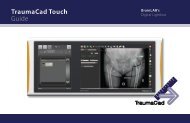
intra-<strong>operative</strong>ly by incorporating <strong>the</strong> Digital Lightbox © from BarinLAB.<br />
The picture can be edited and refined directly on a large touch-screen<br />
display. The TraumaCad template library contains more than 47,000<br />
templates, each with an anteroposterior (AP) and a lateral view, derived<br />
from more than 32 leading companies worldwide.<br />
<strong>Pre</strong>-<strong>operative</strong> <strong>Planning</strong> for Joint Replacement<br />
<strong>Pre</strong>-<strong>operative</strong> planning for joint replacement in <strong>the</strong> hip, knee, shoulder,<br />
elbow, and ankle has become an integral stage of surgical preparation.<br />
It is an effective tool for training <strong>the</strong> surgeon by pre-<strong>operative</strong>ly<br />
deciding <strong>the</strong> type and size of <strong>the</strong> implant, and probably reduces <strong>the</strong> intra<strong>operative</strong><br />
complication rate. In <strong>the</strong> past, pre-<strong>operative</strong> radiological<br />
planning was performed by applying <strong>the</strong> transparent template onto<br />
standard film. Progression to digitalized technology led to <strong>the</strong> need for<br />
software that can perform digitalized templating. One of <strong>the</strong> new<br />
programs is TraumaCad, a system that combines importing from and<br />
exporting properties to all kinds of digitalized imaging technologies.<br />
This allows <strong>the</strong> attainment of precise implant size and accurate<br />
© TOUCH BRIEFINGS 2010<br />
87
PACS and IT<br />
Figure 1: Anteroposterior Pelvic View of a Failed<br />
Total Hip Replacement<br />
<strong>Pre</strong>-<strong>operative</strong> planning and <strong>the</strong> post-<strong>operative</strong> result (from left to right).<br />
Figure 2: <strong>Pre</strong>-<strong>operative</strong> <strong>Planning</strong> of a Fracture of <strong>the</strong><br />
Distal Tibia<br />
The distal fragment was traced to <strong>the</strong> desired position and a nail with screws template was<br />
applied on <strong>the</strong> image.<br />
measurements, and <strong>the</strong> number of mismatches between <strong>the</strong> transparent<br />
template and <strong>the</strong> digital reprint is decreased. The TraumaCad system<br />
(see Figure 1) has <strong>the</strong> properties of digitalized radiography calibration<br />
and versatile templating software that can be adapted to accommodate<br />
various types and sizes of pros<strong>the</strong>ses.<br />
The earlier application of transparent templates on a standard<br />
non-digital film had a predicted accuracy of 62–99% of <strong>the</strong> acetabular<br />
cups and 78–99% within two sizes of <strong>the</strong> femoral stems. 3,6 Magnification<br />
differences were found to have affected <strong>the</strong> choice of <strong>the</strong> implant in<br />
17% of cases. 3 Similar or even better results—i.e. a predicted accuracy<br />
of 86–92% of <strong>the</strong> acetabular cups and 95–96% within two sizes of <strong>the</strong><br />
femoral stems—were achieved by shifting to <strong>the</strong> digitalized technology<br />
that uses digitalized radiographs and template software. 1,5,9,10<br />
A study conducted in our department using <strong>the</strong> TraumaCad software for<br />
total hip replacement found that <strong>the</strong> acetabular component, measured<br />
within ±1 size, was accurate in 65 patients (89%), and that <strong>the</strong> femoral<br />
stem design component was accurate in 70 patients (97%). 10 TraumaCad<br />
successfully predicted <strong>the</strong> sizes of femoral and acetabular components<br />
and was easily integrated with all PACS files. The same approach of<br />
pre-<strong>operative</strong> planning with TraumaCad can be applied for total knee<br />
replacement and o<strong>the</strong>r joints as well.<br />
<strong>Pre</strong>-<strong>operative</strong> <strong>Planning</strong> for Fracture Treatment<br />
Understanding <strong>the</strong> fracture pattern is a crucial step in <strong>the</strong> surgeon’s<br />
pre-<strong>operative</strong> planning of <strong>the</strong> proper approach and <strong>the</strong> type of<br />
hardware needed for fracture fixation. In addition to knowledge of <strong>the</strong><br />
mechanism of a given injury, appropriate imaging modalities will be<br />
needed in order to correctly assess <strong>the</strong> fracture type. A view of <strong>the</strong><br />
unaffected limb is recommended to serve as a reference for<br />
<strong>the</strong> surgeon during <strong>the</strong> pre-<strong>operative</strong> process as well as during <strong>the</strong><br />
operation. The image is processed by <strong>the</strong> TramaCad software to<br />
achieve a better fracture reposition and to determine <strong>the</strong> best<br />
placement of hardware type and size. The outlines of <strong>the</strong> various<br />
broken components are marked separately, after which each one is<br />
shifted to achieve a straight alignment of <strong>the</strong> bone. Estimating<br />
hardware size is critically important, especially in cases in which <strong>the</strong><br />
fracture is too close to <strong>the</strong> joint line, <strong>the</strong>reby limiting <strong>the</strong> amount of<br />
hardware to be inserted (see Figure 2).<br />
Limb deformities are mainly congenital or developmental and<br />
post-traumatic malunions. The first two etiologies will be discussed<br />
in <strong>the</strong> section on pediatric orthopaedics below. Accurate estimations in<br />
<strong>the</strong> pre-<strong>operative</strong> planning for <strong>the</strong> correction of a malunion deformity<br />
require AP and lateral views of <strong>the</strong> bone, CT scans, or MRI studies, and 3D<br />
reconstruction in order to differentiate between a simple and a complex<br />
deformity. Specifically, a simple deformity is visible in only one plane while<br />
a complex deformity occurs in at least two planes (e.g. a shortening,<br />
angular, and rotational deformity). Images of both <strong>the</strong> normal and <strong>the</strong><br />
deformed limb are needed to adjust <strong>the</strong> plan for performing <strong>the</strong><br />
osteotomy, repositioning as close as possible to that of <strong>the</strong> normal limb.<br />
TraumaCad integrates all of <strong>the</strong> recorded images and <strong>the</strong> pre-<strong>operative</strong><br />
planning is similar to that for fracture repair. The deformity outline is drawn<br />
and <strong>the</strong> bone is split into two fragments at <strong>the</strong> level of <strong>the</strong> desired<br />
osteotomy. The two fragments are <strong>the</strong>n lined up to obtain <strong>the</strong> required<br />
corrected position. The TraumaCad archive is used in <strong>the</strong> ensuing step for<br />
selecting <strong>the</strong> proper available fixation device (see Figure 3). When <strong>the</strong>se<br />
steps are completed to <strong>the</strong> surgeon’s satisfaction, <strong>the</strong> planned program is<br />
saved to be used at <strong>the</strong> time of <strong>the</strong> actual surgery.<br />
<strong>Pre</strong>-<strong>operative</strong> <strong>Planning</strong> for Lower Limb<br />
Deformities in <strong>the</strong> Pediatric Population<br />
Patient management in pediatric orthopaedic surgery relies on <strong>the</strong><br />
interpretation of radiographs and <strong>the</strong> measurement of skeletal<br />
anatomical sizes, angles, and indices as important supplements to <strong>the</strong><br />
clinical examination. Conditions that may appear at birth, such as bone<br />
dysplasia, developmental dysplasia of <strong>the</strong> hip (DDH), and scoliosis, or<br />
that develop later in life, such as Per<strong>the</strong>s’ disease, slipped capital<br />
femoral epiphysis, limb deformities due to trauma, infection, or<br />
metabolic condition, and o<strong>the</strong>rs are classified according to radiographic<br />
parameters. The severity of <strong>the</strong>se conditions, <strong>the</strong>ir natural history,<br />
indications for surgery, and <strong>the</strong> follow-up of surgical results are based<br />
on specific radiographic measurements between defined landmarks for<br />
assessing hip joint development, lower limb length differences and<br />
alignments, scoliosis curves, and more.<br />
The key point for performing all of <strong>the</strong>se measurements is defining <strong>the</strong><br />
anatomical landmarks necessary for producing <strong>the</strong> line drawings. 11<br />
Recent studies comparing inter- and intra-observer agreement for<br />
various measurements on conventional and digital radiographs showed<br />
digital measurements to be equal or more accurate. 12–14 The pediatric<br />
section of <strong>the</strong> TraumCad software (TraumCa version 2.2, OrthoCrat)<br />
was designed so that an illustration corresponding to various<br />
conventional measuring tools would appear at <strong>the</strong> bottom of <strong>the</strong> page<br />
88 US RADIOLOGY
<strong>Pre</strong>-<strong>operative</strong> <strong>Planning</strong> <strong>Using</strong> <strong>the</strong> <strong>Traumacad</strong> Software System<br />
Figure 3: <strong>Pre</strong>-<strong>operative</strong> <strong>Planning</strong> of a Malunion of a<br />
Distal Radius Fracture<br />
Figure 4: Standing Radiograph of <strong>the</strong> Lower Limbs in a<br />
Patient with Deformity of <strong>the</strong> Left Lower Limb<br />
A<br />
A simulated osteotomy of <strong>the</strong> distal tibia is shown in <strong>the</strong> image.<br />
Figure 5: Pelvic View of Developmental Dysplasia<br />
of <strong>the</strong> Right Hip and an Analysis and Measurement<br />
Table for <strong>the</strong> Patient<br />
B<br />
when <strong>the</strong> anatomy of <strong>the</strong> hip, long leg, spine, or foot and ankle was<br />
analyzed, <strong>the</strong>reby facilitating <strong>the</strong> location and positioning of markers at<br />
specific anatomical sites (see Figure 4). Some of <strong>the</strong> illustrations<br />
contain a short text for providing a more exact definition of <strong>the</strong><br />
anatomical landmarks.<br />
In addition, a dedicated wizard was developed to guide <strong>the</strong> marking of<br />
anatomical landmarks for carrying out <strong>the</strong> various measurements<br />
during acetabular, hip joint, lower limb length and angle, scoliosis, and<br />
foot analysis. This technique created a reproducible method of carrying<br />
out measurements on digital radiographs of various anatomical<br />
parameters (see Figure 5).<br />
<strong>Pre</strong>-<strong>operative</strong> planning (A) and intra-<strong>operative</strong> radiographs (B).<br />
Hip morphology analysis using TraumaCad can be performed for a<br />
non-, partially, or fully ossified femoral head. The acetabular index,<br />
central edge angle, Reimer subluxation index, and o<strong>the</strong>r more specific<br />
parameters can be measured for conditions such as DDH, cerebral<br />
palsy, Per<strong>the</strong>s’ disease, and o<strong>the</strong>r acquired hip pathologies. 15<br />
US RADIOLOGY<br />
89
PACS and IT<br />
The deformity wizard tool enables <strong>the</strong> surgeon to perform bone length<br />
and mechanical axis analysis, measure hip knee and ankle joint<br />
orientation in <strong>the</strong> frontal and sagittal planes, and trace <strong>the</strong> center<br />
of rotation angulation (CORA) for each bone segment. After<br />
<strong>the</strong> CORA has been defined, a simulated ‘osteotomy’ for deformity<br />
correction and leng<strong>the</strong>ning can be carried out on <strong>the</strong> radiographs<br />
(see Figure 4). 16<br />
Scoliosis analysis by <strong>the</strong> TraumaCad software is performed by placing<br />
<strong>the</strong> Cobb angle tool on <strong>the</strong> appropriate vertebrae, while o<strong>the</strong>r<br />
parameters such as frontal and sagittal spinal balance and pelvic<br />
inclination can be traced using special tools. All angle and length<br />
measurements as well as <strong>the</strong> final radiographs can be stored in <strong>the</strong><br />
patient’s PACS page or in any o<strong>the</strong>r file in <strong>the</strong> form of a report page for<br />
future reference. 15<br />
O<strong>the</strong>r Applications of <strong>the</strong> TraumaCad Software<br />
TraumaCad software can be used for various measurements of <strong>the</strong><br />
spine, such as <strong>the</strong> Cobb angle, pelvic radius angle, sacral obliquity,<br />
coronal balance spondylolis<strong>the</strong>sis, and kyphotic or lordotic angle. It<br />
provides <strong>the</strong> values required for foot and ankle surgery by using <strong>the</strong> foot<br />
osteotomy wizard for growth calculation, hallux valgus deformity<br />
correction, limb measurements, and talar tilt after ankle injury. New<br />
applications include <strong>the</strong> incorporation of an implant template into a 3D<br />
configuration using <strong>the</strong> TeraRecon’s Aquarius iNtuition program and<br />
intra-<strong>operative</strong> assistance by software system integration with BrainLAB<br />
navigation system.<br />
Conclusion<br />
The transition from hard-copy radiographic films to <strong>the</strong> digital technique<br />
has brought with it increasing numbers of software programs, some using<br />
applications similar to those of <strong>the</strong> TraumaCad. A considerable amount of<br />
time will be saved by using PACS-compatible software: <strong>the</strong> various built-in<br />
tools have been constructed according to common orthopaedic consensus<br />
and in collaboration with software developers, <strong>the</strong> PACS producers, and<br />
<strong>the</strong> orthopaedic community at large. The short learning curve, user-friendly<br />
features, accurate prediction of implant size, high versatility, and options in<br />
various fields of orthopaedic surgery toge<strong>the</strong>r with low-cost maintenance<br />
make TraumaCad software an especially attractive choice. n<br />
Ely L Steinberg, MD, is Director of <strong>the</strong> Orthopedic<br />
Trauma Service and Deputy Director of <strong>the</strong> Orthopedic<br />
Department at Sourasky Tel-Aviv Medical Center. He is<br />
also a Lecturer at <strong>the</strong> Sackler Faculty of Medicine at Tel<br />
Aviv University. Dr Steinberg is a member of <strong>the</strong><br />
Orthopedic Trauma Association and <strong>the</strong> American<br />
Academy of Orthopaedic Surgeons (AAOS) and is on <strong>the</strong><br />
Editorial Board of Injury.<br />
Eitan Segev, MD, FCS-ORTH, is Director of <strong>the</strong> Limb<br />
Leng<strong>the</strong>ning and Deformity Correction Service in <strong>the</strong><br />
Department of Pediatric Orthopedics at Dana Children's<br />
Hospital. He is <strong>Pre</strong>sident of <strong>the</strong> Israeli Pediatric<br />
Orthopaedics Society and a Senior Lecturer at <strong>the</strong> Sackler<br />
Faculty of Medicine at Tel Aviv University.<br />
1. Davila JA, Kransdorf MJ, Duffy GP, Surgical planning of total<br />
hip arthroplasty: accuracy of computer-assisted EndoMap<br />
software in predicting component size, Skeletal Radiol,<br />
2006;35:390–93.<br />
2. Eggli S, Pisan M, Müller ME, The value of pre-<strong>operative</strong><br />
planning for total hip arthroplasty, J Bone Joint Surg,<br />
1998;80:382–90.<br />
3. Knight JL, Atwater RD, <strong>Pre</strong>-<strong>operative</strong> planning for total hip<br />
arthroplasty. Quantitating its utility and precision,<br />
J Arthroplasty, 1992;(Suppl. 7):403–9.<br />
4. Krettek C, Blauth M, Miclau T, et al., Accuracy of<br />
intramedullary templates in femoral and tibial radiographs,<br />
J Bone Joint Surg, 1996;78:963–4.<br />
5. Wedemeyer C, Quitmann H, Xu J, et al., Digital templating in<br />
total hip arthroplasty with <strong>the</strong> Mayo stem, Arch Orthop Trauma<br />
Surg, 2008;128:1023–9.<br />
6. Della Valle AG, Slullitel G, Piccaluga F, Salvati EA, The<br />
precision and usefulness of pre-<strong>operative</strong> planning for<br />
cemented and hybrid primary total hip arthroplasty,<br />
J Arthroplasty, 2005;20:51–8.<br />
7. Noble PC, Sugano N, Johnston JD, et al., Computer<br />
simulation: how can it help <strong>the</strong> surgeon optimize implant<br />
position?, Clin Orthop Relat Res, 2003;417:242–52.<br />
8. Viceconti M, Lattanzi R, Antonietti B, et al., CT-based<br />
surgical planning software improves <strong>the</strong> accuracy of total<br />
hip replacement pre-<strong>operative</strong> planning, Med Eng Phys,<br />
2003;25:371–7.<br />
9. Kosashvili Y, Shasha N, Olschewski E, et al., Digital<br />
versus conventional templating techniques in pre<strong>operative</strong><br />
planning for total hip arthroplasty, Can J Surg,<br />
2009;52:6–11.<br />
10. Steinberg EL, Shasha N, Menahem A, <strong>Pre</strong>-<strong>operative</strong> planning<br />
of total hip replacement using <strong>the</strong> TraumaCadTM system,<br />
Arch Orthop Trauma Surg, 2010; Epub ahead of print.<br />
11. Nelitz M, Guen<strong>the</strong>r K, Gunkel S, Reliability of radiological<br />
measurements in <strong>the</strong> assessment of hip dysplasia in adults,<br />
Br J Radiol, 1999;72:331–4.<br />
12. Sailer J, Scharitzer M, Peloschek P, et al., Quantification of<br />
axial alignment of <strong>the</strong> lower extremity on conventional and<br />
digital total leg radiographs, Eur Radiol, 2005;15:170–73.<br />
13. Hankemeier S, Gosling T, Richter M, Computer-assisted<br />
analysis of lower limb geometry: higher intraobserver<br />
reliability compared to conventional method, Comput Aided<br />
Surg, 2006;11:81–6.<br />
14. Halanski MA, Noonan KJ, Hebert M, et al., Manual versus<br />
digital radiographic measurements in acetabular dysplasia,<br />
Orthopedics, 2006;29:724–6.<br />
15. Herring JH (ed.), Tachdjian's Pediatric Orthopaedics, 4th Edition,<br />
Philadelphia, Saunders Elsevier Inc., 2008:358–9.<br />
16. Paley D (ed.), Principles of Deformity Correction, 1st Edition, Berlin,<br />
Heidelberg, Springer-Verlag, 2002:8–9.<br />
90 US RADIOLOGY
European Medical Imaging Review<br />
Now available on subscription<br />
Published annually, European Medical Imaging Review<br />
endeavors to support radiologists and related healthcare<br />
professionals in continuously developing <strong>the</strong>ir knowledge,<br />
effectiveness, and productivity.<br />
Directed by an Editorial Board comprising internationally<br />
respected physicians, European Medical Imaging Review’s peerreviewed<br />
articles aim to assist time-pressured physicians to stay<br />
abreast of key advances and opinion within radiology.<br />
Ensure that researchers, students, and fellow physicians at<br />
your institution enjoy <strong>the</strong> educational benefits:<br />
European<br />
Medical Imaging<br />
Review<br />
V o l u m e 1<br />
BRIEFINGS<br />
• Neuroradiology<br />
• Cardiothoracic imaging<br />
• Breast imaging<br />
• Dentomaxillofacial radiology<br />
• Vascular imaging<br />
• Gastrointestinal imaging<br />
• Musculoskeletal imaging<br />
• Oncological imaging<br />
• Digital radiography<br />
• Concise review articles detail <strong>the</strong> most salient developments<br />
in technology and practice.<br />
• Latest opinion and practice guidelines.<br />
• Detailed bibliographies make it a valuable reference and<br />
research tool.<br />
• Breadth of coverage helps professionals to stay abreast<br />
of developments beyond <strong>the</strong>ir core specialties.<br />
Subscription Rates<br />
Online and print<br />
Online only<br />
Full Institutional (<strong>the</strong> Americas) $225 $210<br />
Full Institutional (Europe) €180 €170<br />
Full Personal (<strong>the</strong> Americas) $100 $85<br />
Full Personal (Europe) €80 €70<br />
www.touchbriefings.com<br />
Articles include:<br />
Evidence-based Medicine Meets Radiology<br />
Athanasios N Chalazonitis<br />
Imaging <strong>the</strong> Human Brain with Functional<br />
Computed Tomography<br />
Sotirios Bisdas, Zoran Rumboldt, Choon Hua Thng<br />
and Tong San Koh<br />
US Radiology<br />
Cardiac Dual-source Computed Tomography<br />
Volume 2 Evaluation in Heart Transplant Recipients<br />
• Issue 1<br />
Gorka Bastarrika, Florian Schwarz, Jesús C Pueyo,<br />
Balazs Ruzsics, Yeong Shyan Lee, Gregorio Rábago,<br />
Philip Costello and U Joseph Schoepf<br />
Novel Techniques in Breast Imaging?<br />
Erik Mittra and Andrei Iagaru<br />
Virtual Colonoscopy – 2008 Update<br />
Andrea Laghi and Cesare Hassan<br />
Acute Non-traumatic Knee Pain in Adults –<br />
The Role of Magnetic Resonance Imaging<br />
The Optimization of Dynamic<br />
Anastasia N Fotiadou and Apostolos H Karantanas<br />
and Blood-pool Magnetic<br />
Resonance Angiography<br />
Beyond <strong>the</strong> Eye – Medical Applications of<br />
Imaging with Gadofosveset in<br />
3D Rapid Prototyping Objects?<br />
Peripheral Vascular Disease<br />
Fabian Rengier, Hendrik von Tengg-Kobligk,<br />
Christian Zechmann, Hans-Ulrich Kauczor<br />
Winfried A Willinek, MD<br />
and Frederik L Giesel<br />
Looking for Fetal Heart<br />
Disease—Who Is Next?<br />
Fetal Echocardiogram and<br />
Screening Criteria<br />
Mert Ozan Bahtiyar, MD, et al.<br />
Ultrasound-guided Fine-needle<br />
Aspiration of Axillary Lymph<br />
Nodes in Patients with<br />
Breast Cancer—A Review<br />
Roshni Rao, MD, et al.<br />
Technical Advances in <strong>the</strong><br />
Fluoroscopic Imaging Chain—A<br />
Review of Technical Innovations<br />
and Radiation-saving Devices<br />
Pei-Jan Paul Lin, PhD and<br />
Meguru Watanabe, MD, PhD<br />
www.touchbriefings.com<br />
Print and online subscriptions cover one print edition per annum<br />
and full online access to <strong>the</strong> electronic version of <strong>the</strong> journal for<br />
a 12-month period.<br />
European radiologists and o<strong>the</strong>r professionals within this field<br />
qualify for free subscriptions.<br />
Order online or download a pdf subscription form at:<br />
www.touchbriefings.com/subscriptions


