Uterine Pathology Benign Disorders of the Uterus
Uterine Pathology Benign Disorders of the Uterus
Uterine Pathology Benign Disorders of the Uterus

Create successful ePaper yourself
Turn your PDF publications into a flip-book with our unique Google optimized e-Paper software.
ProSono Copyright 2006<br />
<strong>Uterine</strong> <strong>Pathology</strong><br />
<strong>Uterine</strong> Anomalies<br />
Congenital anomalies<br />
(See section on Mullerian duct anomalies)<br />
DES exposure:<br />
“T”- shaped uterus<br />
Constricting bands within <strong>the</strong> uterine cavity<br />
Hypoplastic uterus<br />
Intrauterine polypoid defects and synechiae. Synechiae are<br />
adhesions found within <strong>the</strong> uterine cavity.<br />
Acquired anomalies<br />
Asherman’s syndrome: obliteration <strong>of</strong> <strong>the</strong> endometrial cavity as a result<br />
<strong>of</strong> excessive or traumatic uterine instrumentation. Endometrial tissue is<br />
replaced with collagen resulting in development <strong>of</strong> uterine adhesions or<br />
synechiae.<br />
Clinical presentation: amenorrhea or hypomenorrhea; infertility;<br />
recurrent spontaneous abortions.<br />
<strong>Benign</strong> <strong>Disorders</strong> <strong>of</strong> <strong>the</strong> <strong>Uterus</strong><br />
Pyometra and hematometra: pus and/or blood filling <strong>the</strong> uterine cavity.<br />
Conditions are typically associated with outflow obstruction <strong>of</strong> <strong>the</strong> genital tract<br />
such as cervical or vaginal atresia, acquired cervical stenosis, imperforate<br />
hymen; and transverse vaginal septum.<br />
Clinical presentation: primary amenorrhea; dysmenorrhea;<br />
endometriosis, infertility.<br />
Endometritis: an inflammatory reaction <strong>of</strong> <strong>the</strong> endometrium. May be acute or<br />
chronic. Most common causes include: STD infections; instrumentation<br />
procedures that introduce organisms into <strong>the</strong> endometrial cavity.<br />
O<strong>the</strong>rs Include:<br />
Teratoma<br />
Lymphangioma<br />
Polypoid adenomas<br />
<strong>Uterine</strong> <strong>Pathology</strong> (1)
ProSono Copyright 2006<br />
Leiomyomas (fibroids, myomas)<br />
Incidence: Frequent and commonly appearing benign uterine tumor <strong>of</strong><br />
muscle cell origin. In one large study, 75% <strong>of</strong> women had some degree <strong>of</strong><br />
myomatous changes in <strong>the</strong> uterus; only 20-30% have clinically detectable<br />
myomas; most are asymptomatic. More common in:<br />
Black women: three times higher than in Caucasians<br />
Older women: incidence increases as a woman moves through<br />
reproductive years.<br />
Peri-menopausal status<br />
Obese women.<br />
<strong>Pathology</strong>: benign muscle tumor. Estrogens and progesterone stimulate<br />
myomatous tissue proliferation. Gross appearance is that <strong>of</strong> a white,<br />
swirling compact cellular tumor that creates a pseudocapsule by its<br />
expansion into <strong>the</strong> myometrium. Myomas may occupy any position within<br />
<strong>the</strong> uterus or Mullerian ductal system and <strong>the</strong>re development will distort <strong>the</strong><br />
uterine shape accordingly.<br />
Clinical signs and symptoms:<br />
Four major presenting symptoms:<br />
Abnormal uterine bleeding<br />
Heavy periods<br />
Alteration in normal menstrual flow<br />
Pelvic mass: palpable on pelvic exam<br />
Sensations <strong>of</strong> pressure in <strong>the</strong> pelvis<br />
Frequency <strong>of</strong> urination<br />
Dyspareunia (painful intercourse)<br />
Pain: may be caused by torsion or degeneration <strong>of</strong> a fibroid.<br />
Clinical complications may include:<br />
Torsion: <strong>the</strong> twisting <strong>of</strong> a pedunculated myoma on its pedicle.<br />
Results in interruption <strong>of</strong> blood flow to <strong>the</strong> mass and typically causes<br />
sudden onset, acute pelvic pain with tenderness localized to <strong>the</strong><br />
myoma. If <strong>the</strong> torsion is not reduced, ischemia <strong>of</strong> <strong>the</strong> myoma results.<br />
Prolapse: extrusion <strong>of</strong> a submucosal myoma through <strong>the</strong> vagina.<br />
Degeneration: inadequate perfusion to a fibroid can cause chronic<br />
ischemic changes that result in breakdown <strong>of</strong> <strong>the</strong> tissue (necrosis).<br />
This degeneration <strong>of</strong> <strong>the</strong> mass typically results in liquefaction <strong>of</strong> <strong>the</strong><br />
central portion <strong>of</strong> <strong>the</strong> fibroid. Calcific changes can also occur as<br />
calcium salts precipitate within <strong>the</strong> tumor. Ultrasound changes may<br />
reflect <strong>the</strong>se alterations in gross appearance <strong>of</strong> <strong>the</strong> fibroid.<br />
Pregnancy related complications: during pregnancy, myomas can<br />
change due to <strong>the</strong> altered hormonal status. About 20% increase in<br />
size; 20% decrease in size and 60% remain unchanged.<br />
<strong>Uterine</strong> <strong>Pathology</strong> (2)<br />
prgmea.com
ProSono Copyright 2006<br />
Dystocia: difficult vaginal delivery. Malpresentation <strong>of</strong> <strong>the</strong> fetus may<br />
occur if <strong>the</strong> myoma lies in <strong>the</strong> lower uterine segment. Lower uterine<br />
segment and/or cervical myomas may prevent normal cervical<br />
dilatation.<br />
Placental abruption: if a portion <strong>of</strong> <strong>the</strong> placenta is implanted over a<br />
submucous myoma, <strong>the</strong>re may be defective implantation resulting in<br />
an increased risk <strong>of</strong> premature separation <strong>of</strong> <strong>the</strong> placenta from <strong>the</strong><br />
uterine wall (placental abruption).<br />
Gross pathologic appearances: Myomas are usually multiple, discrete,<br />
and spherical, or irregularly lobulated. Although myomas have a false<br />
capsular covering, <strong>the</strong>y are clearly demarcated from <strong>the</strong> surrounding<br />
myometrium and can be easily and cleanly enucleated from <strong>the</strong> surrounding<br />
tissue. On gross examination in transverse section, <strong>the</strong>y are buff-colored,<br />
rounded, smooth, and usually firm. Generally <strong>the</strong>y are lighter in color than<br />
<strong>the</strong> myometrium. When a fresh specimen is sectioned, multiple, whorling<br />
tissue planes can be identified.<br />
Location <strong>of</strong> fibroids. Fibroids can occur anywhere in <strong>the</strong> uterus, cervix, or<br />
broad ligament but are most commonly<br />
found in <strong>the</strong> uterine corpus. Description<br />
<strong>of</strong> <strong>the</strong> location <strong>of</strong> a fibroid is based on its<br />
relationship to uterine layer and<br />
anatomical part.<br />
P–pedunculated: arising from a stalk<br />
I –intramural: interstitial location within <strong>the</strong><br />
myometrium<br />
M –submucosal: lying directly beneath <strong>the</strong><br />
endometrium and frequently projecting<br />
into <strong>the</strong> uterine cavity; most commonly<br />
produces symptoms, i.e., bleeding<br />
S –subserous: (subserosal) lying beneath<br />
<strong>the</strong> outer peritoneal surface <strong>of</strong> <strong>the</strong> uterus<br />
L–interligamentous: occurring within <strong>the</strong><br />
broad ligament<br />
C –cervical<br />
Exophytic:<br />
<strong>the</strong> uterus.<br />
growing out <strong>of</strong> and away from<br />
<strong>Uterine</strong> <strong>Pathology</strong> (3)<br />
prgmea.com
ProSono Copyright 2006<br />
Sonographic appearances: specific sonographic appearance depends on<br />
<strong>the</strong> size <strong>of</strong> <strong>the</strong> fibroid and type <strong>of</strong> degeneration present. Most common<br />
sonographic findings include:<br />
Well-circumscribed, hypoechoic masses<br />
Increased attenuation within <strong>the</strong> mass<br />
Calcification within or surrounding <strong>the</strong> mass<br />
Distortion <strong>of</strong> normal uterine contour<br />
Extrinsic compression <strong>of</strong> <strong>the</strong> posterior bladder wall.<br />
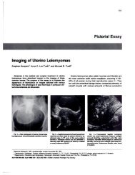
*<br />
Focal subserous fibroids () Hyperattenuation in a focal fibroid (*)<br />
Adenomyosis<br />
Incidence: With routine histological examination, adenomyosis is found in<br />
approximately 5-10% <strong>of</strong> postmenopausal women and 15% <strong>of</strong> women under<br />
<strong>the</strong> age <strong>of</strong> 40. It is associated with uterine fibroids 50% <strong>of</strong> <strong>the</strong> time and with<br />
endometriosis
ProSono Copyright 2006<br />
<strong>Uterine</strong> Neoplasia<br />
Endometrial Hyperplasia<br />
<strong>Pathology</strong>: Excessive proliferation <strong>of</strong> endometrial glandular tissue. There<br />
are two histologic types; each may produce symptoms clinically<br />
indistinguishable from endometrial carcinoma. Some types <strong>of</strong> endometrial<br />
hyperplasia are considered “pre-malignant”. Because <strong>of</strong> <strong>the</strong> similarityin<br />
clinical presentation and <strong>of</strong> <strong>the</strong> potential increased risk <strong>of</strong> endometrial<br />
carcinoma, careful management <strong>of</strong> <strong>the</strong>se patients is necessary. D&C with<br />
histologic evaluation <strong>of</strong> <strong>the</strong> tissue obtained is important.<br />
Etiology: Endometrial hyperplasia is associated with hyperestrogenic<br />
states: Common causes include:<br />
Unopposed estrogen administration (HRT)<br />
Estrogen producing tumors<br />
Persistent anovulatory cycles<br />
Polycystic ovarian disease (PCO)<br />
Clinical signs and symptoms:<br />
Vaginal bleeding: inter-menstrual, hypermenorrhea or<br />
postmenopausal<br />
Hyperstrogenism: (conditions with possible alterations in estrogen<br />
metabolism such as:<br />
• Ovarian granulosa cell tumor<br />
• Polycystic ovarian disease<br />
• Obesity<br />
• Late menopause<br />
• Hormone replacement <strong>the</strong>rapy (especially unopposed exogenous<br />
estrogen)<br />
• Tamoxifen use (used for treatment <strong>of</strong> breast cancer)<br />
Gross pathologic appearances: increased thickens <strong>of</strong> endometrial tissue,<br />
sometimes with cystic changes producing a classic “Swiss-cheese”<br />
appearance to <strong>the</strong> endometrium.<br />
Sonographic appearances:<br />
Increased thickness <strong>of</strong> <strong>the</strong> endometrial stripe<br />
Smooth, well defined borders<br />
Homogenous appearance <strong>of</strong> <strong>the</strong> endometrium; may be cystic<br />
changes present.<br />
Suspected in pre-menopausal women if EC >14mm<br />
<strong>Uterine</strong> <strong>Pathology</strong> (5)<br />
prgmea.com
ProSono Copyright 2006<br />
<strong>Uterine</strong> (endometrial) polyps<br />
Incidence: common in <strong>the</strong> endometrial cavity particularly at ages 29-59;<br />
greatest incidence occurs after age 50.<br />
<strong>Pathology</strong>: a mass <strong>of</strong> endometrial tissue that projects out or away from <strong>the</strong><br />
surface <strong>of</strong> <strong>the</strong> endometrium. They consist <strong>of</strong> an excessive localized growth<br />
<strong>of</strong> endometrial tissue with a stromal core and epi<strong>the</strong>lial and mucosal tissue<br />
surrounding it.<br />
May be single or multiple and range in size from small, 1mm<br />
excrescences to masses that fill or distend <strong>the</strong> uterine cavity.<br />
Most commonly arise in <strong>the</strong> fundal region<br />
May undergo malignant change<br />
Clinical signs and symptoms:<br />
Often asymptomatic<br />
Vaginal bleeding; ei<strong>the</strong>r inter-menstrual flow or heavy periods<br />
(menorrhagia).<br />
Infertility<br />
Occasionally cause postmenopausal bleeding<br />
Usually discovered incidentally during D&C<br />
Gross pathologic appearances: Smooth, red or<br />
brown ovoid body with a velvety texture.<br />
Sonographic appearances:<br />
Non-specific thickened endometrium, usually<br />
focal but occasionally diffuse<br />
Discrete mass in endometrium, possibly with a<br />
vascular stalk demonstrated with color Doppler<br />
May be indistinguishable form endometrial hyperplasia<br />
Hysterosonography is ideal for demonstrating polyp size and location<br />
Endometrial Carcinoma<br />
Incidence: Endometrial carcinoma is <strong>the</strong> most common type <strong>of</strong> gynecologic<br />
malignancy. It usually occurs in women 60 - 70 years <strong>of</strong> age.<br />
<strong>Pathology</strong>: There are three histologic types <strong>of</strong> uterine cancer:<br />
Adenocarcinoma MOST COMMON<br />
Adenoacanthoma<br />
Adenosquamous carcinoma<br />
<br />
Risk factors include:<br />
Obesity<br />
Hypertension<br />
Diabetes mellitus<br />
Strong familial history <strong>of</strong> uterine cancer<br />
<strong>Uterine</strong> <strong>Pathology</strong> (6)
ProSono Copyright 2006<br />
Natural history: Initially <strong>the</strong> tumor mass grows into <strong>the</strong> uterine cavity.<br />
Myometrial invasion is <strong>the</strong> first indication <strong>of</strong> continued spread <strong>of</strong> <strong>the</strong> disease.<br />
Without treatment, <strong>the</strong> malignancy may spread to <strong>the</strong> cervix, adnexa,<br />
fallopian tubes and ovaries. Distant metastases may occur if <strong>the</strong> pelvic<br />
lymphatic system is infiltrated.<br />
Clinical Signs:<br />
Vaginal bleeding; post-menopausal<br />
Hypermenorrhea, intermenstrual flow in patients still having periods<br />
Pain as <strong>the</strong> result <strong>of</strong> uterine distention<br />
Sonographic Findings:<br />
Alteration in size, shape and sonographic texture <strong>of</strong> <strong>the</strong> uterine<br />
parenchyma<br />
Increased uterine size<br />
Thickening <strong>of</strong> endometrial echoes ( >5 mm) especially in a postmenopausal<br />
woman (varies with patient's hormone status)<br />
Fluid in <strong>the</strong> endometrial cavity<br />
<strong>Uterine</strong> <strong>Pathology</strong> (7)
ProSono Copyright 2006<br />
References:<br />
Scott JR, et al. Danforth’s Obstetrics and Gynecology 8 th ed. Lippincott<br />
Williams and Wilkins, Philadelphia. 1999.<br />
DeCherny AH (ed). Current Obstetric and Gynecologic Diagnosis and<br />
Treatment<br />
8 th ed Lange Medical Books, 1994.<br />
Berman, M (ed). Diagnostic Medical Sonography: A Guide to Clinical Practice<br />
2 nd ed Lippincott, Philadelphia, 1997.<br />
<strong>Uterine</strong> <strong>Pathology</strong> (8)