Marginal placenta previa
Marginal placenta previa
Marginal placenta previa
- No tags were found...

Create successful ePaper yourself
Turn your PDF publications into a flip-book with our unique Google optimized e-Paper software.
Abnormal vaginal bleeding insecond half of pregnancyผศ.นพ.วัลลภ ปานพูนทรัพย์ภาควิชาสูติศาสตร์-นรีเวชวิทยาศูนย์การแพทย์สมเด็จพระเทพฯคณะแพทยศาสตร์ มศว.
Definition-Vaginal bleeding after 20 weeksgestational age (before delivery) maybe cause of maternal mortality andassociate with perinatal morbidity andmortalityIncidence5–10% of pregnancyprgmea.com
Obstetric cause-bloody show-<strong>placenta</strong> <strong>previa</strong>-<strong>placenta</strong>l abruption-uterine rupture-vasa <strong>previa</strong>prgmea.com
Gynecologic cause-cervicitis-cervical polyps-vaginal laceration, foreign bodies-varices-benign and malignant neoplasms of genitalorgansprgmea.com
Non obstetric-gynecologic causeHematologic diseases• Thrombocytopenia• Coagulopathy• Platelet dsyfunction• etc.prgmea.com
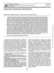
Placenta Previa
Definition and ClassificationThe <strong>placenta</strong> located over or very near theinternal os, classified as Total <strong>placenta</strong> <strong>previa</strong> Partial <strong>placenta</strong> <strong>previa</strong> <strong>Marginal</strong> <strong>placenta</strong> <strong>previa</strong> Low-lying <strong>placenta</strong>
Definition and Classification• Total <strong>placenta</strong> <strong>previa</strong>: the internal os iscovered completely by <strong>placenta</strong>• Partial <strong>placenta</strong> <strong>previa</strong>: the internal os ispartially covered by <strong>placenta</strong>• <strong>Marginal</strong> <strong>placenta</strong> <strong>previa</strong>: the edge of the<strong>placenta</strong> is at the margin of the internal os• Low lying <strong>placenta</strong>: the <strong>placenta</strong> isimplanted in the lower uterine segment,the<strong>placenta</strong> edge does not reach the internalos but is in close proximity to it
Etiology• defective vascularization of the decidua,inflammatory process or atrophic changes --multiparity- advancing age• previous cesarean delivery or uterine surgical scar• large <strong>placenta</strong>: multiple fetuses, erythroblastosis• others : smoking, cocaine use
Signs and symptoms-Painless hemorrhage usually nearthe end of the second trimester-Abnormal fetal presentationabout 1/3 of cases
Diagnosispainless bleedinguterus : soft, no tendernessfetus : may be in oblique or transverse liepresenting part not engagedUltrasonography - accuracy 95-98 %transvaginal ultrasound identifyinternal os better thantransabdominal ultrasound(double set up)
Fetal echoPlacentaBladderCervix
Management• Depend on severity of bleeding,gestational age, fetal maturity andfetal well-being• Principle of management– Hospitalization with hemodynamicstabilization– Obstetric evaluation– Do not PV or PR
Management Preterm fetus, good fetal well-being withno active bleeding• expectant, close observation• very sedentary lifestyle• avoidance of any intravaginal manipulation :vgdouch,PV,etc.• immediate availability of appropriate therapy
Management The fetus with poor fetal assessment ormore than 37 weeks gestation or severehemorrhage as to necessitate immediatedelivery of the fetus• Cesarean delivery: in most cases -transverse incisionor vertical uterine incision• Vaginal delivery: in cases of low-lying <strong>placenta</strong> ormarginal <strong>placenta</strong> <strong>previa</strong>
ComplicationMaternal- Postpartum hemorrhage- Infection- EmbolismFetus- Preterm (major cause ofperinatal death)- Intrauterine asphyxia- IUGR
Placental Abruption
Definition• The separation of the <strong>placenta</strong> from itssite of implantation in the uterus beforethe delivery of the fetus• abruption <strong>placenta</strong>e, ablatio <strong>placenta</strong>e,premature separation of the normallyimplanted <strong>placenta</strong>
Pathologyhemorrage from a decidual spiral artery into thedecidua basalis(the bleeding between the membranes anduterus,retro<strong>placenta</strong>l hematoma )
Classification-acute <strong>placenta</strong>l abruption and chronic<strong>placenta</strong>l abruptionor- external hemorrhage (revealedhemorrhage ~80-85%- no external hemorrage (concealedhemorrhage) ~15-20%- Mixed typeormarginal separation , partial separation,complete separation
Etiology• unknown
Risk factor• Prior abruption• hypertensive disorder in pregnancy• sudden decompression of the uterus: afteramniotomy, hydramnios• shortness of the umbilical cord: esp. inmultifetal pregnancy• uterine anomaly or tumor• pressure by the enlarged uterus on the inferiorvena cava• Increased age and parity• ethanol consumption, cigarette smoking, cocineuse, dietary deficiency• external trauma
PathologyLocal vascular injury in decidua basalisDecidual spiral artery rupture• (uterine venous pressure)Retro<strong>placenta</strong>l hematomaSeparation of <strong>placenta</strong>
Signs and symptoms* may be no clinical symptoms in early stage* profuse external bleeding per vagina* uterine tenderness, rapid uterine contractionor tetanic uterine contraction
Signs and symptoms* Hypotension, Shock out of proportion to theamount of hemorrhage* Fetal heart rate decelerations or no FHRbeen heard
Clinical findings• Classical : pain, uterine tenderness andrigidity, fetal distress or absent fetalheart sound (Decreased short-termvariability, increased baseline uterinetone, uterine hyperstimulation, andworsening variable decelerations)• Vaginal bleeding, shock• Shock not relate to vaginal bleeding• Idiopathic preterm labor
Clinical findings• Vaginal bleeding 78%• Uterine tenderness or back pain 66%• Fetal distress 60%• Preterm labor 22%• High frequency contraction, hypertonus 17%• Dead fetus 15%
Laboratory findings• Hct decrease• Thrombocytopenia• Hypofibrinogenemia (fibrinogen20 ug/ml• Abnormal coagulogram
Ultrasound findings• Retro<strong>placenta</strong>l hematoma• Exclude <strong>placenta</strong> <strong>previa</strong>Differential diagnosis• Placenta <strong>previa</strong>• Rupture of the uterus
Complication* - Comsumptive coagulopathy:hypofibrinogenemia(less than 150 mg/dlof plasma), elevated levels offibrinogen-fibrin degradation products,decrease of coagulation factors* - Renal failure may be from impaired renalperfusion* - Utero<strong>placenta</strong>l Apoplexy (CouvelaireUterus) : extravasations of blood intothe uterine musculature and beneath theuterine serosa* - feto-maternal hemorrhage* - Post-partum hemorrhage* - Death of the fetus
Utero<strong>placenta</strong>l Apoplexy(Couvelaire Uterus)
Managementbbb-evaluation for fetal well-being, maternalcirculation status-intense therapy with blood, coagulationfactor,plus electrolyte solution in case of seriousmaternal hemorrhage-prompt delivery (may be very close observationcoupled with facilities for immediate interventionin mild cases)
Route of delivery• Cesarean section- Fetal distress,unfavorable cervix- Uncontrol bleeding• Vaginal delivery with induction oflabor by oxytocin infusion andamniotomy- Dead fetus with control bleeding- Favorable cervix, rapid progress oflabor, stable maternalhemodynamic, no fetal distress
prognosis• Fetal mortality rate 60-80 %• Maternal mortality rate 1-5 %
Rupture of the uterusRupture of the uterus
Rupture of the uterus• Complete uterinerupture• Incomplete uterinerupture(uterine dehiscence)
Predisposing factors• Uterine injury or abnormality duringcurrent pregnancy– before deliverypersistent, intense, spontaneous contractions oruterine hyperstimulationexternal versionuterine overdistension: hydramnios– During deliveryobstructed laborinternal versiondifficult forceps deliverybreech extraction
Predisposing factors• Others:- <strong>placenta</strong> increta or percreta- previous surgery involving themyometrium: cesarean section- congenital anomaly- grand multiparity- coincidental uterine trauma
How to diagnosis-uterine scar-suprapubic pain and tenderness-vaginal bleeding-cessation of uterine contractions-pathological retraction ring (Bandl’ring)-disappearance of fetal heart sound-recession of the presenting part (lossof station)-easily palpated fetus-hypovolemic shock with hemoperitoneum
management-hemodynamic stabilization-emergency surgery:repair wound with or without internaliliac ligation or hysterectomy dependon maternal condition pathology ofthe uterus, need of child-bearing)
complication• amniotic fluid embolism• hypovolemic shock• DIC• ureteral injury• postoperative infection
prognosis• Perinatal mortality rate 40-70%• Maternal mortality rate 5-40%
Rupture of vasa <strong>previa</strong>Thank you for your attention
vasa <strong>previa</strong>- incidence 1:3000 – 1:5000 pregnancies- associated with- velamentous insertion (fetalvessels in the membranes cross theregion of the cervical os)- <strong>Marginal</strong> cord insertion- Bilobed or succenturiate-lobed<strong>placenta</strong>
vasa <strong>previa</strong>How to diagnosis- palpate or directly visualize fetalvessel in the membrane overlying thepresenting part of the fetus- ultrasonography : color doppler-Lab : fetal blood- bloody amniotic fluid duringamniotomy with FHR change
management-immedate vaginal delivery orcesarean sectionPrognosis-Fetal death 60-70%
Thank you for your attention