Beamline G3 at DESY: Materials X-ray Imaging
Beamline G3 at DESY: Materials X-ray Imaging
Beamline G3 at DESY: Materials X-ray Imaging

Create successful ePaper yourself
Turn your PDF publications into a flip-book with our unique Google optimized e-Paper software.
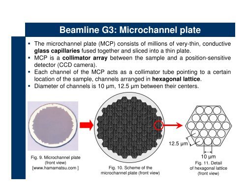
<strong>Beamline</strong> <strong>G3</strong>: Microchannel pl<strong>at</strong>e<br />
The microchannel pl<strong>at</strong>e (MCP) consists of millions of very-thin, conductive<br />
glass capillaries fused together and sliced into a thin pl<strong>at</strong>e.<br />
MCP is a collim<strong>at</strong>or ar<strong>ray</strong> between the sample and a position-sensitive<br />
detector (CCD camera).<br />
Each channel of the MCP acts as a collim<strong>at</strong>or tube pointing to a certain<br />
loc<strong>at</strong>ion of the sample, channels arranged in hexagonal l<strong>at</strong>tice.<br />
Diameter of channels is 10 µm, 12.5 µm between their centers.<br />
12.5 µm<br />
Fig. 9. Microchannel pl<strong>at</strong>e<br />
(front view)<br />
[www.hamam<strong>at</strong>su.com ]<br />
Fig. 10. Scheme of the<br />
microchannel pl<strong>at</strong>e (front view)<br />
10 µm<br />
Fig. 11. Detail<br />
of hexagonal l<strong>at</strong>tice<br />
(front view)