M E S '9 8 - University of Georgia College of Veterinary Medicine
M E S '9 8 - University of Georgia College of Veterinary Medicine
M E S '9 8 - University of Georgia College of Veterinary Medicine
- No tags were found...
You also want an ePaper? Increase the reach of your titles
YUMPU automatically turns print PDFs into web optimized ePapers that Google loves.
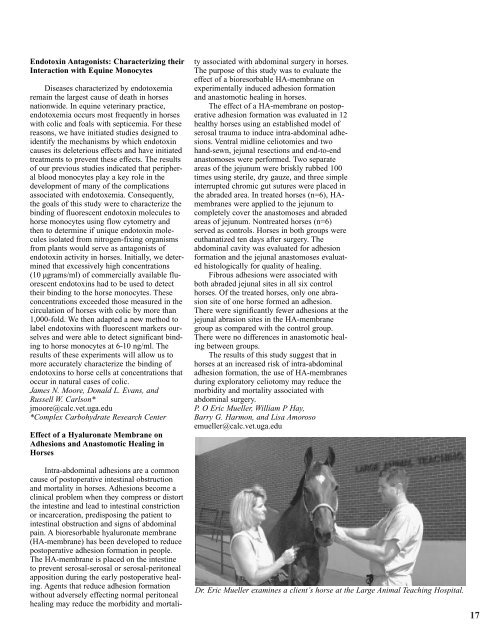
Endotoxin Antagonists: Characterizing theirInteraction with Equine MonocytesDiseases characterized by endotoxemiaremain the largest cause <strong>of</strong> death in horsesnationwide. In equine veterinary practice,endotoxemia occurs most frequently in horseswith colic and foals with septicemia. For thesereasons, we have initiated studies designed toidentify the mechanisms by which endotoxincauses its deleterious effects and have initiatedtreatments to prevent these effects. The results<strong>of</strong> our previous studies indicated that peripheralblood monocytes play a key role in thedevelopment <strong>of</strong> many <strong>of</strong> the complicationsassociated with endotoxemia. Consequently,the goals <strong>of</strong> this study were to characterize thebinding <strong>of</strong> fluorescent endotoxin molecules tohorse monocytes using flow cytometry andthen to determine if unique endotoxin moleculesisolated from nitrogen-fixing organismsfrom plants would serve as antagonists <strong>of</strong>endotoxin activity in horses. Initially, we determinedthat excessively high concentrations(10 µgrams/ml) <strong>of</strong> commercially available fluorescentendotoxins had to be used to detecttheir binding to the horse monocytes. Theseconcentrations exceeded those measured in thecirculation <strong>of</strong> horses with colic by more than1,000-fold. We then adapted a new method tolabel endotoxins with fluorescent markers ourselvesand were able to detect significant bindingto horse monocytes at 6-10 ng/ml. Theresults <strong>of</strong> these experiments will allow us tomore accurately characterize the binding <strong>of</strong>endotoxins to horse cells at concentrations thatoccur in natural cases <strong>of</strong> colic.James N. Moore, Donald L. Evans, andRussell W. Carlson*jmoore@calc.vet.uga.edu*Complex Carbohydrate Research CenterEffect <strong>of</strong> a Hyaluronate Membrane onAdhesions and Anastomotic Healing inHorsesIntra-abdominal adhesions are a commoncause <strong>of</strong> postoperative intestinal obstructionand mortality in horses. Adhesions become aclinical problem when they compress or distortthe intestine and lead to intestinal constrictionor incarceration, predisposing the patient tointestinal obstruction and signs <strong>of</strong> abdominalpain. A bioresorbable hyaluronate membrane(HA-membrane) has been developed to reducepostoperative adhesion formation in people.The HA-membrane is placed on the intestineto prevent serosal-serosal or serosal-peritonealapposition during the early postoperative healing.Agents that reduce adhesion formationwithout adversely effecting normal peritonealhealing may reduce the morbidity and mortalityassociated with abdominal surgery in horses.The purpose <strong>of</strong> this study was to evaluate theeffect <strong>of</strong> a bioresorbable HA-membrane onexperimentally induced adhesion formationand anastomotic healing in horses.The effect <strong>of</strong> a HA-membrane on postoperativeadhesion formation was evaluated in 12healthy horses using an established model <strong>of</strong>serosal trauma to induce intra-abdominal adhesions.Ventral midline celiotomies and twohand-sewn, jejunal resections and end-to-endanastomoses were performed. Two separateareas <strong>of</strong> the jejunum were briskly rubbed 100times using sterile, dry gauze, and three simpleinterrupted chromic gut sutures were placed inthe abraded area. In treated horses (n=6), HAmembraneswere applied to the jejunum tocompletely cover the anastomoses and abradedareas <strong>of</strong> jejunum. Nontreated horses (n=6)served as controls. Horses in both groups wereeuthanatized ten days after surgery. Theabdominal cavity was evaluated for adhesionformation and the jejunal anastomoses evaluatedhistologically for quality <strong>of</strong> healing.Fibrous adhesions were associated withboth abraded jejunal sites in all six controlhorses. Of the treated horses, only one abrasionsite <strong>of</strong> one horse formed an adhesion.There were significantly fewer adhesions at thejejunal abrasion sites in the HA-membranegroup as compared with the control group.There were no differences in anastomotic healingbetween groups.The results <strong>of</strong> this study suggest that inhorses at an increased risk <strong>of</strong> intra-abdominaladhesion formation, the use <strong>of</strong> HA-membranesduring exploratory celiotomy may reduce themorbidity and mortality associated withabdominal surgery.P. O Eric Mueller, William P Hay,Barry G. Harmon, and Lisa Amorosoemueller@calc.vet.uga.eduDr. Eric Mueller examines a client’s horse at the Large Animal Teaching Hospital.17


