

Studies on Earthworms. - Journal of Cell Science
Studies on Earthworms. - Journal of Cell Science
Studies on Earthworms. - Journal of Cell Science

You also want an ePaper? Increase the reach of your titles
YUMPU automatically turns print PDFs into web optimized ePapers that Google loves.
STUDIES ON EAETHWOEMS. 213<br />
<str<strong>on</strong>g>Studies</str<strong>on</strong>g> <strong>on</strong> <strong>Earthworms</strong>.<br />
By<br />
William Bla^land Benham, B>Sc,<br />
Dem<strong>on</strong>strator in the Zoological Laboratory <strong>of</strong> University College, L<strong>on</strong>d<strong>on</strong>.<br />
INTRODUCTION.<br />
IN this series <strong>of</strong> papers I intend to describe a number <strong>of</strong><br />
<strong>Earthworms</strong> from various parts <strong>of</strong> the world, which have been<br />
kindly put into my hands for the purpose by Pr<strong>of</strong>essor Eay<br />
Lankester. These include some new genera, with interesting<br />
variati<strong>on</strong>s from allied forms, and several new species <strong>of</strong> Perichasta.<br />
But before describing these new forms I shall give—•<br />
firstly, a c<strong>on</strong>densed historical review <strong>of</strong> the various works <strong>on</strong><br />
<strong>Earthworms</strong>, and a chr<strong>on</strong>ological record <strong>of</strong> the discovery <strong>of</strong> new<br />
facts about them.<br />
Sec<strong>on</strong>dly, I shall enumerate and briefly describe all known<br />
<strong>Earthworms</strong>.<br />
Thirdly, I shall take the various organs in order and point<br />
out their variati<strong>on</strong>s in different <strong>Earthworms</strong>, and the theories<br />
<strong>of</strong> various authors with regard to certain points.<br />
Having d<strong>on</strong>e this, I shall proceed to describe the new forms<br />
that I have studied myself.<br />
I wish to thank Pr<strong>of</strong>essor Lankester for his kind advice and<br />
assistance in this work, which was carried <strong>on</strong> in his Laboratory<br />
at University College, L<strong>on</strong>d<strong>on</strong>.<br />
I shall not give an exhaustive bibliography, since that will<br />
be found in Perrier's work <strong>on</strong> Urochssta (28), but I shall<br />
in all cases give references for any facts I menti<strong>on</strong> to a bibliography<br />
at the end <strong>of</strong> the third secti<strong>on</strong>, the figures in brackets<br />
referring to this bibliography.
214 WILLIAM BLAXLAND BBNHAM.<br />
I. HISTORICAL.<br />
Am<strong>on</strong>gst the earliest papers dealing with <strong>Earthworms</strong> anatomically<br />
are those <strong>of</strong> Savigny (1) in 1820, and <strong>of</strong> Duges (2) in<br />
] 828, who describe numerous species <strong>of</strong> Lumbricus, which will<br />
be menti<strong>on</strong>ed in Secti<strong>on</strong> II. Duges figures the prostomium <strong>of</strong><br />
some <strong>of</strong> these, and describes the genital organs; but his interpretati<strong>on</strong><br />
<strong>of</strong> the latter is wr<strong>on</strong>g, since he has, like so many<br />
<strong>of</strong> the earlier writers, c<strong>on</strong>fused the seminal reservoirs and<br />
the spermathecse, attributing each to the wr<strong>on</strong>g sex. Other<br />
authors followed him, who, whilst c<strong>on</strong>tradicting him, were no<br />
nearer the truth. V<strong>on</strong> Siebold (3), for instance, suggested<br />
that the ovary was invaginated into the seminal reservoirs.<br />
Even till quite recently the " seminal reservoirs " were spoken<br />
<strong>of</strong> as " testes." I may at <strong>on</strong>ce say that I shall use the former<br />
name for the three pairs <strong>of</strong> large white organs in Lumbricus<br />
which originate in somites x and xi and spread into the neighbouring<br />
somites, and for their homologues in other genera.<br />
The ovary was unknown till 1853, when d'Udekem (4)<br />
described it in Lumbricus agricolaj whilst in 1856 Hering<br />
(5) supplemented our knowledge <strong>of</strong> the genital organs by his<br />
figure <strong>of</strong> the ovary and his descripti<strong>on</strong> <strong>of</strong> its positi<strong>on</strong> <strong>on</strong> the<br />
posterior face <strong>of</strong> the septum between somites xn and XIII. He<br />
also showed that the oviduct was not in c<strong>on</strong>tinuity with the<br />
ovary, but that the ova fell into the body cavity, and were<br />
c<strong>on</strong>veyed thence to the exterior by the wide ciliated funnels <strong>of</strong><br />
the pair <strong>of</strong> short oviducts which pass through the posterior<br />
septum <strong>of</strong> somite XIII to the exterior in xrv. Hering described<br />
the process <strong>of</strong> copulati<strong>on</strong>, and thought that the spermatozoa<br />
passed from the sperm pore al<strong>on</strong>g a groove <strong>of</strong> the ventral surface<br />
to the spermathecse; but Dr. Fraisse, in 1882 (6), describes<br />
the spermatophores <strong>of</strong> various spcies <strong>of</strong> Lumbricus, and shows<br />
that the spermatozoa do not pass directly into the spermathecse,<br />
but are received in bodies secreted <strong>on</strong> somite xxvi. Previous<br />
authors had described as " testes" the large white sacs which<br />
are now known as " seminal reservoirs," but Hering, in this<br />
paper, describes and figures the true testes. Pr<strong>of</strong>essor A. G.
STUDIES ON EARTHWORMS. 215<br />
Bourne was the first to figure them in their true positi<strong>on</strong><br />
attached to the anterior septum <strong>of</strong> somites x and xi, as two<br />
pairs <strong>of</strong> small flat appendices. This figure and descripti<strong>on</strong> occur<br />
in a paper by J. E. Blomfield (7), who describes the development<br />
<strong>of</strong> the spermatozoa in the reservoirs.<br />
The sperm ducts were rightly described by Leo (8) in.<br />
1820, but Duges (2) wr<strong>on</strong>gly c<strong>on</strong>sidered them as oviducts.<br />
The nephridia or "segmental organs" also were err<strong>on</strong>eously<br />
interpreted by Dr. Williams in 1858 (40), being c<strong>on</strong>sidered<br />
as respiratory organs. Their true functi<strong>on</strong> was first<br />
suggested by d'Udekem (9) in his descripti<strong>on</strong> <strong>of</strong> Tubifex, whilst<br />
Gegenbaur (10) in 1853 published the well-known drawing <strong>of</strong><br />
this organ <strong>of</strong> Lumbricus agrieola, the histological structure<br />
<strong>of</strong> which was described by Claparede (11).<br />
In regard to the classificati<strong>on</strong> <strong>of</strong> <strong>Earthworms</strong>, that <strong>of</strong><br />
Claparede (12) in 1862 is usually followed. He divides the<br />
order Oligochseta into two families, Limicolse and Terricolffi,<br />
but the characters <strong>of</strong> the latter, as opposed to the<br />
former, were derived from the genus Lumbricus <strong>on</strong>ly, and<br />
now, since the investigati<strong>on</strong> <strong>of</strong> other genera, no l<strong>on</strong>ger hold<br />
true. These are his characters:<br />
a. The possessi<strong>on</strong> <strong>of</strong> two ventral blood-vessels.<br />
b. The presence <strong>of</strong> nephridia in the same somites with the<br />
sperm ducts and oviducts.<br />
c. The positi<strong>on</strong> <strong>of</strong> the clitellum far behind the male pores.<br />
d. The presence <strong>of</strong> a vascular network <strong>on</strong> the nephridia.<br />
Now, Perrier's genus P<strong>on</strong>todrilus (13) and Perichseta<br />
have no subneural blood-vessel [Microchseta 1 resembles<br />
these two genera in this respect], and very possibly others will<br />
also be found without this vessel.<br />
The positi<strong>on</strong> <strong>of</strong> the clitellum is now known to vary; sometimes<br />
it is in fr<strong>on</strong>t, sometimes around as well as behind the<br />
male pore.<br />
The truly distinctive characters <strong>of</strong> the Terricolas (or<br />
Lumbricinse, as Perrier calls them), as opposed to the<br />
Linricolss, are the following:<br />
1<br />
Names or sentences in square brackets refer to results <strong>of</strong> my own research.
216 WILLIAM BLAXLAND BENHAM.<br />
a. The presence <strong>of</strong> nephridia in the same somites with the<br />
genital ducts (except in some species <strong>of</strong> Perichseta and<br />
Pleurochseta (Megascolex), where nephridia are unknown<br />
in any somite, and in P<strong>on</strong>todrilus, in which the nephridia<br />
are said not to commence 1 till the hinder regi<strong>on</strong> <strong>of</strong> the sperm<br />
duct, so that there are n<strong>on</strong>e in the somite carrying the oviduct).<br />
b. The abundant vascular network <strong>on</strong> the nepridia and body<br />
wall.<br />
c. The almost universal presence <strong>of</strong> a gizzard (P<strong>on</strong>todrilus<br />
is again an excepti<strong>on</strong>).<br />
d. The much smaller size <strong>of</strong> the ova and the compactness <strong>of</strong><br />
the ovary.<br />
But even these charcters may have to be altered as new<br />
forms are studied.<br />
These Lumbricinas Perrier divides into four groups,<br />
taking as a basis the relati<strong>on</strong> <strong>of</strong> the clitellum to the male pore.<br />
1. The Anteclitelliani (Preclitelliani), in which the male<br />
pore is far in fr<strong>on</strong>t <strong>of</strong> the clitellum, include the genus Lumbricus,<br />
which Eisen (15) has lately subdivided into the genera<br />
Lumbricus, Allurus, Allolobophora, andDendroboena,<br />
as well as, doubtfully, Kinberg's (19) genera Alyattes and<br />
Eurydame, and Savigny's (1) Hypogse<strong>on</strong>. As these three<br />
latter genera are insufficiently described, it is doubtful whether<br />
the characteristics given by these authors justify the retenti<strong>on</strong><br />
<strong>of</strong> their names.<br />
Whilst this group c<strong>on</strong>tains <strong>on</strong>ly a few forms, the members<br />
<strong>of</strong> the other groups are numerous and mostly <strong>of</strong> extra-European<br />
origin.<br />
3. The Intraclitelliani, where the male pore is situated<br />
within the limits <strong>of</strong> the clitellum, include the genera Anteus,<br />
TJrochseta, Rhinodrilus, Microchseta, and perhaps<br />
Kinberg's Greogenia and Tritogenia.<br />
3. The Postclitelliani have the male pore behind the<br />
clitellum, and include Perichseta, Acanthodrilus, Eudri-<br />
1 It is not improbable that examinati<strong>on</strong> by means <strong>of</strong> microscopic secti<strong>on</strong>s<br />
would result in the discovery <strong>of</strong> nephridia in some cases where Perrier has<br />
failed to see them with the naked eye.
STUDIES ON EARTHWORMS. 217<br />
lus, Digaster, P<strong>on</strong>toscolex, P<strong>on</strong>todrilus, Plutellus,<br />
Peri<strong>on</strong>yx, Megascolex, and Pleurochasta.<br />
4. The group Aclitelliani is formed for the genus M<strong>on</strong>iligaster,<br />
which has no clitellum, although the <strong>on</strong>ly specimen<br />
studied had its genital organs fully mature, and, indeed, more<br />
complicated than any other form.<br />
The habitat <strong>of</strong> these forms is given later <strong>on</strong>, in Secti<strong>on</strong> II,<br />
where the names, &c, <strong>of</strong> all known <strong>Earthworms</strong> will be found.<br />
II. PREVIOUSLY DESCRIBED GENERA.<br />
In this secti<strong>on</strong> I shall menti<strong>on</strong>, in chr<strong>on</strong>ological order, and<br />
briefly notice, all <strong>Earthworms</strong> whose descripti<strong>on</strong> I have been<br />
able to find. In the case <strong>of</strong> the genus Lumbricus I have<br />
placed all the species together at the end <strong>of</strong> this secti<strong>on</strong>. The<br />
anatomy <strong>of</strong> the genus Lumbricus is sufficiently well known<br />
through the works <strong>of</strong> d'Udekem (16), Lankester (48), Claparede<br />
(11), and others, so that I will refer <strong>on</strong>ly to Eisen's work<br />
(15), where he subdivides the genus into three subgenera:<br />
1. Lumbricus, with the male pore in somite xv, and the<br />
prostomium embedded deeply in the first somite.<br />
2. Allolobophora, with the male pore in somite xv, and<br />
the prostomium embedded less deeply in the first somite; this<br />
includes Dendroboena, which Eisen at first separated, but<br />
now includes.<br />
3. Allurus, with the male pore in somite XHI.<br />
It seems to me that the character drawn from the prostomium<br />
is scarcely <strong>of</strong> generic importance, since forms otherwise similar<br />
have this difference (e.g. Lumbricus agricola and L. olidus),<br />
but the variati<strong>on</strong> in the positi<strong>on</strong>s <strong>of</strong> the male pore is a<br />
good sub-generic character.<br />
The earliest genus additi<strong>on</strong>al to Lumbricus was Rypogae<strong>on</strong>,<br />
formed by Savigny (1) in 1820, but, as in so many <strong>of</strong><br />
these earlier genera, a very poor descripti<strong>on</strong> is given, and <strong>on</strong>ly<br />
<strong>of</strong> external characters. Hypogse<strong>on</strong> has nine l<strong>on</strong>g sets in each<br />
somite, <strong>on</strong>e being in the dorsal mid-line; these setae do not<br />
alternate in c<strong>on</strong>secutive somites.
218 WILLIAM BLAXLAND BBNHAM.<br />
The clitellum occupies somites XXVII to xxxix, and the<br />
whole worm has 106 somites. This specimen came from Buenos<br />
Ayres and elsewhere, and the genus has since been studied by<br />
d'Udekem, Grube (18), and lastly by Kinberg (19), who, in his<br />
descripti<strong>on</strong> <strong>of</strong> two species, says nothing about the characteristic<br />
ninth seta, whilst no author has given a proper<br />
anatomical descripti<strong>on</strong> <strong>of</strong> any species.<br />
In 1844 Templet<strong>on</strong> (20) described a form resembling the<br />
wide-spread genus Perichseta, but differing from it in the presence<br />
<strong>of</strong> an interrupti<strong>on</strong> in the dorsal mid-line in the ring <strong>of</strong><br />
setse. He called the worm Megascolex ereruleusj he<br />
obtained it from Ceyl<strong>on</strong>; its length was from 20 to 40 inches<br />
by 1 to 11 inch broad; it c<strong>on</strong>tained 270 somites with a ring <strong>of</strong><br />
100 setse <strong>on</strong> each. This must I think be referred to the genus<br />
Perichseta.<br />
In 1845 H<strong>of</strong>fmeister (22) described and figured several<br />
species <strong>of</strong> Lumbricus (see below), as well as the following<br />
forms which are European.<br />
Phreoryctes, which is now placed am<strong>on</strong>gst theLimicolae.<br />
Criodrilus has four rows <strong>of</strong> paired setse, and is 8 to 12<br />
inches l<strong>on</strong>g, and c<strong>on</strong>sists <strong>of</strong> 300 somites. The male pore is <strong>on</strong><br />
somite xiv.<br />
Helodrilus is 2 to 5 inches l<strong>on</strong>g, c<strong>on</strong>tains 160 somites,<br />
with setae arranged as in the preceding. The male pore is <strong>on</strong><br />
somite xr.<br />
Neither <strong>of</strong> the latter has a clitellum.<br />
In 1848 Rapp (21) described a worm under the name<br />
Lumbricus microchetus, which is probably the same as<br />
Beddard's Microchaeta (33) from the Cape.<br />
In 1851 Grube (23) described a peculiar form which he<br />
called Lumbricus multispinus; its chief characteristic<br />
is the possessi<strong>on</strong> <strong>of</strong> four bundles <strong>of</strong> 5 setse in each somite;<br />
there was no trace <strong>of</strong> clitellum. The male pores are in<br />
somite XII, in a line with the most ventral group <strong>of</strong> setse,<br />
and each carries a papilla. Its habitat is not menti<strong>on</strong>ed,<br />
nor is the internal anatomy. Le<strong>on</strong> Vaillant (24) has<br />
founded a new genus for it, Echinodrilus. Judging from
STUDIES ON BASTHWOEMS. 219<br />
the forward positi<strong>on</strong> <strong>of</strong> the male pore it is an Anteclitellian<br />
form.<br />
Then followed Schmarda (25) in 1861, who described and<br />
formed the genera P<strong>on</strong>toscolex and Perichaeta. The<br />
former is from Jamaica and has seven setae <strong>on</strong>ly in each somite,<br />
which alternate with those <strong>of</strong> the next somites, giving fourteen<br />
rows <strong>of</strong> setss. In Perichasta the clitellum occupies somites<br />
xiv, xv, xvi; the female pore is single, median, and in xiv;<br />
the paired male pore is in xvm; the setss are numerous, and<br />
form a ring all round each somite. All the species described<br />
by Schmarda came from Ceyl<strong>on</strong>.<br />
P. brachycycla has no clitellum; is 88 mm. l<strong>on</strong>g, and<br />
3 mm. broad.<br />
P. leucocycla has a white ring round each somite (probably<br />
<strong>on</strong> this ring the setss were placed); it c<strong>on</strong>tains eightyeight<br />
somites. Length 300 mm., breadth 15 mm.<br />
P. viridis c<strong>on</strong>tains 209 somites, fifty setae to each. Length<br />
100 mm., breadth 4 mm.<br />
P. cingulata c<strong>on</strong>tains 100 somites, with forty setse to each.<br />
Length 130 mm., and breadth 6 mm.<br />
Besides these new genera, Schmarda describes two species <strong>of</strong><br />
Hypogse<strong>on</strong> (Sav.), but says nothing about the ninth seta.<br />
H. heterostich<strong>on</strong> came from Quito and Cuenca, with the<br />
setae diverging posteriorly. Its length is 220 mm., and<br />
breadth 11 mm. It has 263 somites.<br />
H. orthostich<strong>on</strong>, from New Zealand, has the setae parallel<br />
throughout the body, which c<strong>on</strong>sists <strong>of</strong> sixty somites. The clitellum<br />
is at somite xiv. The total length is 80 mm., and<br />
breadth 4 mm. The descripti<strong>on</strong> <strong>of</strong> these two is insufficient to<br />
give any c<strong>on</strong>fidence in their validity.<br />
Kinberg (19) added numerous new genera in 1866, most <strong>of</strong><br />
which are so insufficiently described that it is impossible to<br />
retain their names.<br />
Tritogenia is said to have no clitellum, and the male pores<br />
are between somites xvi and xvji. There are <strong>on</strong>ly six setae to<br />
each somite. (Habitat not given.)<br />
Mandane, from M<strong>on</strong>tevideo aud Patag<strong>on</strong>ia,has the clitellum
220 WILLIAM BLAXLAND BENHAM.<br />
<strong>on</strong> somites xn to xrv. There are four male pores situated in<br />
<strong>on</strong>e species <strong>on</strong> somites xvi, XVIII, and in the other <strong>on</strong> somites<br />
xsi and xxin.<br />
Geogenia, from Natal, has the clitellum <strong>on</strong> somites ix to<br />
XVIII ; the setae alternate anteriorly. There are two ventral<br />
pits, <strong>on</strong>e in xvi, the other in xvn (? male pores or copulatory<br />
pits), and the " lateral pores " (probably he refers to nephridiopores)<br />
are in a line with the dorsal setag.<br />
Alyattes, from Buenos Ayres, has the setae separated<br />
posteriorly.<br />
Eurydame,from St. Joseph, near Panama, has the anterior<br />
setae paired, but the posterior <strong>on</strong>es are further apart.<br />
Hegesipyle, from Natal, has all the setae wide apart, except<br />
the ventral <strong>on</strong>es anteriorly.<br />
Then follow five, which Perrier c<strong>on</strong>siders merely species <strong>of</strong><br />
Perichaeta, so that it will be best to use Kinberg's names<br />
specifically if they are to be retained.<br />
Amyntas, from Guam, with fifty or sixty setae per somite.<br />
Nitocris, from Rio Janeiro, with fifty-two setae per somite.<br />
Pheretima, from Tahiti and California, with fifty setae<br />
per somite.<br />
Rhodopis, from Java, has fifty to sixty setae per somite,<br />
has the clitellum <strong>on</strong> somites xn, xm, and the male pores between<br />
somites xiv andxv (so that this differs from Perichaetae where<br />
the clitellum and male pore is c<strong>on</strong>stant).<br />
Lorn pi to, from Mauritius, with forty-four setae per somite.<br />
P. catinus, from Oahu, with forty setae per somite.<br />
Kinberg also describes two worms which he puts into<br />
Savigny's genus Hypogae<strong>on</strong>, but denies the existence <strong>of</strong> the<br />
characteristic dorsal seta.<br />
H. havaicus, from Oahu, is 44 mm. l<strong>on</strong>g, c<strong>on</strong>tains 100<br />
somites, and has the clitellum <strong>on</strong> somites xxix and xxx.<br />
H. atys, from Buenos Ayres, is 30 mm. l<strong>on</strong>g, by 4 mm.<br />
broad, and c<strong>on</strong>tains 140 somites.<br />
Thus, in each <strong>of</strong> the species <strong>of</strong> " Hypogae<strong>on</strong>," in which a<br />
clitellum is menti<strong>on</strong>ed, it differs in positi<strong>on</strong>. In the absence<br />
<strong>of</strong> any record <strong>of</strong> anatomical detail, it is impossible to tell what
STUDIES ON BAETHWOEMS. 221<br />
genus Kinberg was dealing with, or indeed what significance is<br />
to be attributed to Savigny's Hypogse<strong>on</strong>.<br />
In 1869 Baird (26) described a Perichseta (though he<br />
called it at first Megascolex after Templet<strong>on</strong>) which he had<br />
obtained from South "Wales, whither it had apparently come<br />
with exotic plants.<br />
Perichseta diffringens is 4 to 5 inches l<strong>on</strong>g, c<strong>on</strong>tains<br />
104 somites, and has sixty setas to each <strong>of</strong> them.<br />
In 1869 Le<strong>on</strong> Vaillant (24) described two species <strong>of</strong> Perichaeta,<br />
where the " prostate " is described for the first time.<br />
One from Java, P. posthuma, is 18 cm. l<strong>on</strong>g, c<strong>on</strong>sisted <strong>of</strong><br />
100 somites, and has sixty-five to seventy-seven setee in each<br />
somite. The setss average about "25 mm. in length. It has a<br />
pair <strong>of</strong> " copulatory " papillae <strong>on</strong> the xvn, and a pair <strong>on</strong> the xix<br />
somite, in line with the male pores. The prostate occupies two<br />
somites. The spermathecse are four in number in the somites<br />
v, vi, vn, and vm, opening in the anterior regi<strong>on</strong>; each c<strong>on</strong>sists<br />
<strong>of</strong> a bilobed sac. Vaillant denies the existence <strong>of</strong> a gizzard and<br />
<strong>of</strong> intestinal caeca, but Perrier has c<strong>on</strong>tradicted him as to the<br />
first point, and regards this worm as the same as the P.<br />
affinis.<br />
The other species is P. cingulata, Sch., which came from<br />
Bourb<strong>on</strong>. Vaillant's descripti<strong>on</strong> does not corresp<strong>on</strong>d in some<br />
points with his figure, and Perrier reserves the name for the<br />
species figured, whilst he calls the <strong>on</strong>e described P. ro b u s t a. P.<br />
cingulata is 17'4 cm. l<strong>on</strong>g, c<strong>on</strong>sists <strong>of</strong> 114 somites, and has<br />
forty setae in each somite, the length <strong>of</strong> the setae being -36 mm.<br />
There are no " copulatory" papillae <strong>on</strong> or near the somite<br />
carrying the male pore. The prostate occupies <strong>on</strong>ly <strong>on</strong>e<br />
somite. The spermathecae have the same positi<strong>on</strong> as in P.<br />
posthuma.<br />
Apparently Vaillant included other species under the name<br />
P. cingulata; these Perrier has named and separated from<br />
this form.<br />
In his paper Vaillant numbers the somites differently from<br />
the way in which they are now reck<strong>on</strong>ed. He regards the first<br />
setigerous somite as the somite i (instead <strong>of</strong> n). He also regards<br />
VOL. XSVI.—NEW SEE. Q.
222 WILLIAM BLAXLAND BENHAM.<br />
the clitellum as occupying <strong>on</strong>ly <strong>on</strong>e somite, which he calls<br />
xin, instead <strong>of</strong> xiv, xv, xvi.<br />
We now come to the most important work <strong>of</strong> late years <strong>on</strong><br />
<strong>Earthworms</strong>, in which the first attempt is made to c<strong>on</strong>sider the<br />
relati<strong>on</strong>s <strong>of</strong> different forms from an anatomical standpoint.<br />
It is here that the <strong>on</strong>ly rati<strong>on</strong>al classificati<strong>on</strong> and generic<br />
grouping <strong>of</strong> <strong>Earthworms</strong> is first given. I refer to Edm<strong>on</strong>d<br />
Perrier's works.<br />
In 1872 he published his researches <strong>on</strong> various <strong>Earthworms</strong><br />
c<strong>on</strong>tained in the Paris museum (14). In this paper he describes<br />
nine new genera, two new species <strong>of</strong> Lumbricus, and<br />
several new species <strong>of</strong> Perichseta.<br />
The following are the new genera and their chief characters<br />
:<br />
Anteus (A. gigas, from Cayenne).—Its length is 1 met. 16<br />
cm., and breadth 3 cm. The clitellum occupies somites xv to<br />
xxix, and is not c<strong>on</strong>tinued across the ventral mid-line. The s et 33<br />
are iufour couples in each somite. The nephridiopores are<br />
in a line with the uppermost seta <strong>of</strong> the lateral couple (i.e. with<br />
the fourth seta from the ventral mid-line). No sperm ducts<br />
could be found, but the nephvidia in the somites xi to xix are<br />
short simple tubes, which Perrier c<strong>on</strong>siders as sperm ducts.<br />
No accessory copulatory organs nor ovaries are found. There<br />
is a single pair <strong>of</strong> spermathecse in somite vn. The anterior<br />
septa are very str<strong>on</strong>g, and cover the pharynx, gizzard, and<br />
seminal reservoirs.<br />
Titanus (T. brasiliensis, from Brazil).—Length 1 met. 26<br />
cm., and breadth 3 cm. The clitellum occupies somites xv to<br />
XXIII. The male pores are between somites XVIII and xix,<br />
and no nephridiopores exist in this somite. The setse are in<br />
four couples in each somite anteriorly, posteriorly become scattered,<br />
but do not alternate. The nephridiopores are in fr<strong>on</strong>t<br />
<strong>of</strong> the sec<strong>on</strong>d setae, reck<strong>on</strong>ing from the ventral mid-line, whether<br />
in couples or separate, that is to say, they are in line with the<br />
outer ventral setae. No nephridiopores exist anterior to somite<br />
XIII. The seminal reservoirs are very l<strong>on</strong>g and c<strong>on</strong>sist <strong>of</strong><br />
<strong>on</strong>ly <strong>on</strong>e pair, extending from somite xnto xxv. The sperm
STUDIES ON EAETHWOEMS. 223<br />
duct opens into an oval muscular pouch. No sperinatheeae<br />
nor ovaries were found.<br />
Rhinodrilus (R. paradoxus, from Venezuela).—Length<br />
15 cm., breadth 3 or 4 mm. Prostomium el<strong>on</strong>gated to form<br />
a proboscis 3 to 6 mm. l<strong>on</strong>g. The clitellum occupies somites<br />
xix, xx, xxi. The s e t as are in four couples, and are ornamented<br />
with two series <strong>of</strong> semicircular folds, with a c<strong>on</strong>cavity towards<br />
the free end. The nephridiopores are in a line with the<br />
lateral or outer couple <strong>of</strong> setas (the setae 3 and 4). The male<br />
pores are between somites xix and xx in a transverse groove<br />
No spermathecae were found.<br />
Eudrilus.—The clitellum occupies somites xin to xv, or<br />
XIII to xvm. The seta? are in four couples in each somite.<br />
The nephridiopores are in line with the lateral couple <strong>of</strong><br />
setae. The male pores in line with the ventral (1 and2) setse<br />
in xvn. There is a curved chitinous penis in a sac (modified<br />
penial seta) ; a prostate is present. The female genital organs<br />
are very peculiar : the ovary is fixed to the oviduct and to the<br />
spermatheca, according to Perrier's interpretati<strong>on</strong> <strong>of</strong> the parts.<br />
He describes three species, all about the size <strong>of</strong> the comm<strong>on</strong><br />
Earthworm:<br />
Eu. decipiens, from the West Indies.<br />
Eu. Lacazii, from Martinique.<br />
Eu. peregrinus, from Rio Janeiro.<br />
Acanthodrilus.—The clitellum occupies somites xiv to<br />
xvn, and completely surrounds the body. The setse are in<br />
four couples in each somite. The nephridiopores are in<br />
line with the lateral couple <strong>of</strong> setse (3, 4). The male pores<br />
are four in number in somites xvin and xx in line with setae 1<br />
and 2; at each pore is a penis formed <strong>of</strong> a bundle <strong>of</strong> setae in<br />
a sac. The genital organs are not altogether understood, and<br />
differ in each <strong>of</strong> the three species.<br />
Ac. obtusuSj from New Caled<strong>on</strong>ia.—Length 66 cm. Penial<br />
setae are blunt.<br />
Ac. ungulatus, from New Caled<strong>on</strong>ia.—Length 1 dcm.<br />
Penial setae are recurved.<br />
Ac. verticillatus, from Madagascar.—Length 350 mm..,
224 WILLIAM BLAXLAND BENHAM.<br />
breadth 8 mm. Penial setas are serrated. No clitellum was<br />
found.<br />
Disaster, from Australia.—Only <strong>on</strong>e species is described—<br />
D. lumbricoides. The clitellum occupies somites xiv, xv,<br />
xvi. The male pores are in somite xvm. The setse are in<br />
four couples. The nephridiopores are in line with the outer<br />
<strong>of</strong> the ventral couple <strong>of</strong> setse. There are two gizzards; <strong>on</strong>e in<br />
somite v, the sec<strong>on</strong>d in somite vn.<br />
Peri<strong>on</strong>yx.—The <strong>on</strong>ly species is P. excavatus, from<br />
Cochin China. The length is 120 mm., breadth 4 mm. The<br />
clitellum occupies somites XIII, xiv, xv, xvi, xvn. The<br />
setae are about 30 to each somite, and form a ring all round.<br />
The nepridiopores are not visible, though nephridia are<br />
present. The male pores are close together in a median<br />
ventral fossa in somite XVIII. The spermathecal pores are<br />
close to <strong>on</strong>e another <strong>on</strong> the ventral surface <strong>of</strong> the anterior<br />
edge <strong>of</strong> somites vm and ix. There are no intestinal caeca.<br />
The ovaries are not pedunculated.<br />
M<strong>on</strong>iligaster.—A single species, M. Deshayesii, from<br />
Ceyl<strong>on</strong> is described. Length 150 mm., breadth 6 mm. The<br />
clitellum is absent altogether. The setse are in four couples<br />
in each somite. The nephridiopores are in fr<strong>on</strong>t <strong>of</strong> the<br />
lateral couple. There are four male pores; two between<br />
somites vn and vm in line with setse 1 and 2, and two<br />
between somites X and xi dorsad <strong>of</strong> these setae. There is <strong>on</strong>e<br />
gizzard in somite vi, and a sec<strong>on</strong>d extends through somites<br />
XIII to xxii c<strong>on</strong>stricted into four nearly equal porti<strong>on</strong>s. The<br />
genital organs are very complicated. The anterior and posterior<br />
seminal reservoirs differ from <strong>on</strong>e another. The ovary is very<br />
excepti<strong>on</strong>al in that it is a l<strong>on</strong>g sac, lying above, and <strong>on</strong> each<br />
side <strong>of</strong> the alimentary tract, in somites xn, XIII, xiv, xv.<br />
Urochseia.—This is described in the same memoir as the<br />
preceding genera, and also in a separate memoir (28), where<br />
Perrier gives a very minute descripti<strong>on</strong> <strong>of</strong> it, as well as an<br />
exhaustive bibliography <strong>of</strong> the literature <strong>of</strong> the Lumbricinse at<br />
ths end <strong>of</strong> the paper. Only <strong>on</strong>e species is known—Urochasta<br />
hystrix, which has been found in Martinique, Gloria, Java,
STUDIES ON EARTHWORMS. 225<br />
and Brazil; showing thus a very wide distributi<strong>on</strong>. Length<br />
1 deeim., breadth 3 mm., it c<strong>on</strong>sists <strong>of</strong> 220 somites. The<br />
anterior extremity tapers gradually, but there is no prostomium.<br />
The clitellum occupies somites siv to xxm. The setge are<br />
in eight l<strong>on</strong>gitudinal rows anteriorly, all the setse being equidistant<br />
; but posteriorly they alternate giving sixteen rows.<br />
The setse themselves are notched at the extremity, which is<br />
excepti<strong>on</strong>al. The nephridiopores are in a line with the third<br />
seta counting from the ventral mid-line, but do not follow this<br />
seta in its displacement in the posterior somites. The male<br />
pores are in somite xs, and the sets <strong>of</strong> this somite are transversely<br />
ridged. The spermathecal pores are <strong>on</strong> the anterior<br />
edges <strong>of</strong> somites vii, VIII, ix, in a line with the nephridiopore.<br />
There is <strong>on</strong>ly a single pair <strong>of</strong> seminal reservoirs in somite<br />
XIII. In the posterior regi<strong>on</strong> <strong>of</strong> the body there is a pair <strong>of</strong><br />
peculiar " pyriform sacs," <strong>of</strong> unknown significance, in each<br />
somite, opening to the exterior between the nerve cord and<br />
seta 1; the nephridia are also present in these somites.<br />
The following are the new species <strong>of</strong> Perichaeta, described by<br />
Perrier. I shall divide these into two groups according to the<br />
presence or absence <strong>of</strong> papillae, <strong>on</strong> the somite XVIII, or <strong>on</strong> the<br />
neighbouring somites, or in relati<strong>on</strong> to the spermathecse. Each<br />
<strong>of</strong> these groups may then be subdivided according to the<br />
number <strong>of</strong> spermathecse; and probably some further subdivisi<strong>on</strong><br />
may be made in reference to the simple or complex structure<br />
<strong>of</strong> this organ. The arrangement <strong>of</strong> the species <strong>of</strong> this genus<br />
must be left till more have been studied. 1 have several species<br />
at present ready for descripti<strong>on</strong>, and I will give a more detailed<br />
synopsis <strong>of</strong> the species in a future paper.<br />
I. Perichsetse without papillae.<br />
a. With <strong>on</strong>e pair <strong>of</strong> spermathecse.<br />
h. With more than two pairs.<br />
IT. Perichsetss with papillae.<br />
a. With two pairs <strong>of</strong> spermathecse.<br />
b. With more than two pairs.
226 WILLIAM BLAXLAND B.ENHAM.<br />
I. Perichaetas without papillae.<br />
a. With <strong>on</strong>e pair <strong>of</strong> spermathecae.<br />
P. quadragenaria, from the East Indies.—Length 210<br />
mm., breadth 4 mm. The setse are about 40 to each somite.<br />
The spermathecae are <strong>on</strong>ly two in number, in the<br />
somite VIII ; their aperture is in the anterior part <strong>of</strong> this<br />
somite, and each c<strong>on</strong>sists <strong>of</strong> a globular sac in somite VIII, and<br />
narrow coiled appendage with enlarged extremity in the somite<br />
VII.<br />
P. el<strong>on</strong>gata, from Peru.—Length 355 mm., breadth 4 mm.<br />
The number <strong>of</strong> setse is not menti<strong>on</strong>ed. The spermathecEe<br />
are <strong>on</strong>ly two in number; their pores are between somites iv<br />
and v, but whether they lie in the somite iv or in v is not<br />
stated. They c<strong>on</strong>sist each <strong>of</strong> a single sac, and have no accessory<br />
parts.<br />
b. With more than two pairs <strong>of</strong> spermathecse.<br />
P! Houlleti, from Calcutta and Cochin China, is very fully<br />
described. Length 1 dcm.<br />
There are forty-five to fifty setse round each somite.<br />
There are three pairs <strong>of</strong> spermathecae, lying in somites vn,<br />
VIII, and is, and opening <strong>on</strong> the anterior edges <strong>of</strong> these<br />
somites. Each c<strong>on</strong>sists <strong>of</strong> three parts; a large ovoid sac, with<br />
a coiled tube opening into its duct, lying in the somites<br />
named; whilst a very much smaller sac, also opening at the<br />
same point, lies in the preceding somite in each case.<br />
[P. cingulata, Sch. will come in here.]<br />
II. Perichsetae with papillae.<br />
a. With two pairs <strong>of</strong> spermathecae.<br />
P. aspergillum (habitat unknown).—Length 370 mm.,<br />
breadth 10 mm. There are about eighty setse round each somite.<br />
The spermathecse are in two pairs, in somites VIII<br />
and ix, with their pores in the anterior regi<strong>on</strong>. Each is a<br />
simple, large, and somewhat globular sac; but there are numerous<br />
smaller sacs round it, each having a separate pore j<br />
some lying in the same somite as the spermathecse, others in<br />
the somite in fr<strong>on</strong>t. So that, both in fr<strong>on</strong>t and behind each
STUDIES ON EAETHWOEMS. 227<br />
spermathecal pore, is a line <strong>of</strong> four or five smaller pores.<br />
The male pores are in somite xvm, and each is situated<br />
<strong>on</strong> a papilla, which is studded by a row <strong>of</strong> smaller pores<br />
in fr<strong>on</strong>t and behind, each pore bel<strong>on</strong>ging to a small sac<br />
internally.<br />
P. robusta, from Bourb<strong>on</strong> and Manilla.—Its length is 150<br />
to 180 mm., breadth 6 mm. There are forty-five sete in each<br />
somite. There are two pairs <strong>of</strong> spermathecss in somites VIII,<br />
ix, opening anteriorly. Each c<strong>on</strong>sists <strong>of</strong> a large, ovoid sac,<br />
into the neck <strong>of</strong> which opens the duct <strong>of</strong> a narrower sac. Just<br />
behind these is a much smaller sac opening to the exterior <strong>on</strong><br />
a papilla just behind the pore <strong>of</strong> the spermatheca. Between<br />
the male pores are two papillae, each with a small pore. Nephridia<br />
are present as extremely delicate tubules attached to<br />
the septa.<br />
Perrier gives the name P. robusta to the worm described<br />
by Le<strong>on</strong> Vaillant (24), under the name P. cingulata, Sch.<br />
b. With more than two pairs <strong>of</strong> spermathecae.<br />
P. affinis from Cochin China.—Length 110 mm., breadth<br />
5 mm. The number <strong>of</strong> setae is not menti<strong>on</strong>ed. There are four<br />
pairs <strong>of</strong> spermathecae lying in somites vi, vn, VIII and is,<br />
opening anteriorly. These pores are quite lateral. Each c<strong>on</strong>sists<br />
<strong>of</strong> a large globular sac, with a smaller globular sac opening<br />
into its neck. There are no papillae near their pores.<br />
The male pores are <strong>on</strong> papillae, and there is in additi<strong>on</strong> a pair<br />
<strong>of</strong> papillae in somite xvir, and a pair in six.<br />
Perrier c<strong>on</strong>siders this worm to be the same as Vaillant's P.<br />
posthuma.<br />
(Horst's Perichaeta from Java bel<strong>on</strong>gs to this group (31).)<br />
In the same memoir Perrier describes two new species <strong>of</strong><br />
Lumbricus.<br />
L. am eric anus, from New York. Length 1 dcm. It<br />
seems to differ from L. agricola <strong>on</strong>ly in having the posterior<br />
pair <strong>of</strong> ciliated rosettes <strong>of</strong> the sperm duct rather larger<br />
than the anterior pair. Perrier menti<strong>on</strong>s that the ciliated<br />
rosettes are not enclosed in the seminal reservoirs, but that,<br />
I think, merely depends <strong>on</strong> the state <strong>of</strong> maturity <strong>of</strong> the worm.
228 WILLIAM BLAXLAND BENHAM.<br />
for in L. agrieola when about half ripe, and with large<br />
seminal reservoirs, the rosettes are free.<br />
The sec<strong>on</strong>d new species is named Lumbricus victoris;<br />
it was obtained from Damietta (West Africa); its size is nearly<br />
the same as the preceding. The clitellum commences at<br />
somite xxvn, and occupies eight somites. The ovaries are<br />
in somite xiv, and there are three pairs <strong>of</strong> spermathecse in<br />
somites ix, x, xi.<br />
The following are the species <strong>of</strong> Lumbricus described by<br />
H<strong>of</strong>fmeister (22) in 1842, with their various syn<strong>on</strong>yms,<br />
according to d'Udekem (16), Perrier (14), and Eosa (17).<br />
1. Lumbricus agrieola, H<strong>of</strong>fm.<br />
Syn. L. terrestris, Lin..<br />
L. herculeus, Dug.<br />
Two pairs <strong>of</strong> spermathecse, in somites ix and x; opening <strong>on</strong><br />
the posterior edge <strong>of</strong> somite.<br />
2. L. communis, H<strong>of</strong>fm.<br />
Syn. L. trapezoides, Dug.<br />
L. caliginosus, Sav.<br />
L. cyaneus, Sav.<br />
L. ictericus, Sav.<br />
Allolobophora turgida, Eisen (15), partim.<br />
„ „ mucosa, Eisen (15), partim.<br />
According to Rosa (17) the two species Allolobophora here<br />
named are partly syn<strong>on</strong>ymous.<br />
Two pairs <strong>of</strong> spermathecse, in somites x and xi; with apertures<br />
anteriorly.<br />
3. L. rubellus, H<strong>of</strong>fm., is adopted by the other writers. Two<br />
pairs <strong>of</strong> spermathecse, in somites ix and x; opening posteriorly.<br />
4. L. riparius, H<strong>of</strong>fm.<br />
Syn. L. chloroticus, Dug.<br />
Allolobophora riparia, Eisen (15).<br />
„ „ chlorotica, Rosa (17).<br />
Three pairs <strong>of</strong> spermathecse, in somites ix, x, xi; opening<br />
anteriorly.
STUDIES ON EARTHWOBMS. 229<br />
5. L. olid us, H<strong>of</strong>fm.<br />
Syn. L. fostidus, Dug.<br />
Enteri<strong>on</strong> rubidum, Sav.<br />
Allolobophora fostida, Eisen (15).<br />
Two pairs <strong>of</strong> spermathecse, in somites ix, x; opening posteriorly.<br />
6. Lumbricus stagnalis, H<strong>of</strong>fm.?—Seven pairs <strong>of</strong> spermathecae,<br />
in somites vi, vn, VIII, ix, x, si, xn; opening posteriorly.<br />
The setae <strong>of</strong> the four couples are wide apart.<br />
Syn. L. complanatus, Dug.<br />
Allolobophora eomplanata, Eisen.<br />
7. L. pictus ,H<strong>of</strong>fm.<br />
8. L. agilis, H<strong>of</strong>fm. Male pores in somite XIII.<br />
Syn. L. tetraedrus, Dug.<br />
L. amphisbaenaj Dug.<br />
Allurus.tetraedrus, Eisen 15.<br />
Other species menti<strong>on</strong>ed by Duges (2) that have not received<br />
syn<strong>on</strong>yms so far as I am aware, and have not been recognised<br />
or further characterised since his time, are the following :<br />
L. opimus, Sav.<br />
L. mollis, Dug.<br />
L. teres, Dug.<br />
L. Blainvilleus, Dug.<br />
L. tyrtoeus, Sav.<br />
L. festivus, Sav.<br />
L. roseus, Sav.<br />
L. dubius, Dug.<br />
L. mammalis, Sav.<br />
L. purus, Dug.<br />
L. pygmseus, Sav. "| Setae separated, male pore in soli,<br />
vetsedrus, Sav. J mite xv.<br />
L. phosphoreus, Dug. Setae separated, male pore in<br />
somite sin.<br />
As the external points <strong>on</strong>ly are menti<strong>on</strong>ed, it may probably<br />
turn out that some <strong>of</strong> these are known under other names.<br />
They seem all to be European, as Duges does not menti<strong>on</strong>, in<br />
most cases, where he obtained them.
230 WILLIAM BLAXLAND BBNHAM.<br />
The following are new forms <strong>of</strong> Lumbricus described by<br />
Eisen (15):<br />
Lumbricus puter.<br />
Syn. Allolobophora Boeckii, Rosa (17).<br />
Dendrobosna Bosckii, Eisen (15).<br />
L. purpureus.—Two pairs <strong>of</strong> spermathecae, in somites<br />
TX, x; opening posteriorly.<br />
Allolobophora subrubricunda.—One pair <strong>of</strong> spermathecae,<br />
in somite x; opening anteriorly.<br />
Rosa (17) has added the following European forms:<br />
Allolobophora c<strong>on</strong>stricta.<br />
All. minima.<br />
All. transpadana.—Five pairs <strong>of</strong> spermathecae, in somites<br />
vi, vn, VIII, ix, x; opening posteriorly.<br />
All. pr<strong>of</strong>uga.—Four pairs <strong>of</strong> spermathecse, in somites ix,<br />
x, xi, XII.<br />
All. alpina.—Two pairs <strong>of</strong> spermathecse, in somites ix, x.<br />
L. melibceus.—Two pairs <strong>of</strong> spermathecse, in somites ix, x;<br />
opening posteriorly.<br />
Kinberg (19) described several new species <strong>of</strong> Lumbricus,<br />
though probably some <strong>of</strong> them bel<strong>on</strong>g to other genera. They<br />
are extra-European:<br />
L. Josephinae, from St. Helena.<br />
L. infelix, from Port Natal.<br />
L. armatus, from Buenos Ayres.<br />
L. Novse-hollandise, from Sydney.<br />
L. Helense, from St. Helena.<br />
L. Hortensise, from St. Helena.<br />
L. Vineti, from Madeira.<br />
L. pampicola, from M<strong>on</strong>tevideo.<br />
L. tellus, from Buenos Ayres.<br />
L. Tahitana, from Tahiti.<br />
L. capensis, from Cape <strong>of</strong> Good Hope.<br />
L. Apii, from California.<br />
In 1851 Grube (23) described L. triannularis, and L.<br />
multispinus (Echinodrilus, Vaillant).
STUDIES ON EARTHWORMS. 231<br />
Hutt<strong>on</strong> (35) has described four Earth-worms which he refers<br />
to the genus Lumbricus, but they seem to bel<strong>on</strong>g to various<br />
genera. L. uliginosus has four male pores, two being in<br />
somite is and two in somite x. L. campestris has a pair<br />
<strong>of</strong> male pores in somite ix.<br />
In 1873 Perrier described a new genus (27), <strong>of</strong> which <strong>on</strong>ly<br />
<strong>on</strong>e species is known—Plutellus heteroporus, from Pennsylvania.<br />
Length 15 cm., breadth 3 mm. The clitellum<br />
occupies somites xrv, xv, rvi, xvn, and is complete. The<br />
setse are eight in each somite, at nearly equal distances apart,<br />
and do not alternate. The nephridiopores vary in positi<strong>on</strong>.<br />
The first four pores are in a line with the third seta from the<br />
ventral mid-line. The rest alternate in successive somites, <strong>on</strong>e<br />
series being in line with the sec<strong>on</strong>d seta, the other series being<br />
in line with the fourth seta. The spermathecse are five<br />
pairs in somites v, vi, vn, vm, ix, their pores being in the<br />
anterior regi<strong>on</strong> <strong>of</strong> these somites in line with the sec<strong>on</strong>d setae (i. e.<br />
with <strong>on</strong>e set <strong>of</strong> nephridiopores). Each spermatheca c<strong>on</strong>sists<br />
<strong>of</strong> a sac and a short, slightly swollen blind tube opening into<br />
its neck, resembling the arrangement in some Perichaetss. The<br />
male pores are, in somite xvni, dorsad <strong>of</strong> the first setas<br />
counting from the ventral mid-line. There are no papillae.<br />
The female pores are in somite x, in line with the first setse.<br />
The seminal reservoirs are a single pair <strong>of</strong> grape-like<br />
glands in somite xn. There is a prostate and a penis<br />
in somite xvm. The nephridia do not pass through the<br />
anterior septa, but lie wholly in <strong>on</strong>e somite.<br />
Perrier regards this genus as very likely the same as Kinberg's<br />
Hypogffi<strong>on</strong>, as opposed to the similarly named genus <strong>of</strong><br />
Savigny.<br />
In 1874 Perrier published an abstract in the 'Comptes<br />
Rendus' (13), and in 1881 a detailed account (29) <strong>of</strong> a worm<br />
which lives <strong>on</strong> the seashore at Marseilles. He named it<br />
P<strong>on</strong>todrilus Mari<strong>on</strong>is. Its length is 80 mm., and breadth<br />
4 mm. It c<strong>on</strong>sists <strong>of</strong> fifty somites. The clitellum occupies<br />
somites XIII to xvn. The setsa are rod-like. The ventral<br />
couple (1 and 2) are close together, but the other two (3 and
232 WILLIAM BLAXLAND BENHAM.<br />
4) are wide apart. The nephridiopores are in a line with<br />
seta 2, but <strong>on</strong>ly commence in somite xiv. The male pore<br />
is in somite XVIII, in line with seta 1. There is an ellipsoidal<br />
copulatory papilla in the ventral mid-line between somites xix<br />
and xx, and another between xx and xxi. The oviduct opens<br />
in somite xiv. There are two pairs <strong>of</strong> spermathecae in<br />
somites vm and ix, each having a small spherical appendage;<br />
their pores are <strong>on</strong> the anterior edge <strong>of</strong> the somite in line<br />
with seta 1. The ciliated rosettes <strong>of</strong> the sperm duct<br />
are in the somites preceding the seminal reservoirs, which lie<br />
in somites xi and xn. There is a large prostate in somite<br />
XVIII. There is no gizzard and no subneural blood-vessel.<br />
Grube (30) has described, under the name Lumbricus<br />
littor alis, a worm from Villafranca, which is probably another<br />
species <strong>of</strong> P<strong>on</strong>todrilus. This has three pairs <strong>of</strong> " copulatory<br />
papillae," in line with setae 2, in somites xix, xx, and xxi.<br />
In 1875 M. Perrier (32) described four species <strong>of</strong> Perichffita<br />
from the Philippines, and <strong>on</strong>e from Cochin China.<br />
P. bicincta has <strong>on</strong>ly two somites in the clitellum.<br />
P. biserialis has the usual three somites in the clitellum;<br />
there is a ventral median and a lateral break <strong>on</strong> each side in<br />
the ring <strong>of</strong> seta?, and the ventralmost seta <strong>on</strong> each side is<br />
larger than the other setae <strong>of</strong> the somite. There are several<br />
pairs <strong>of</strong> copulatory papillae behind the male pores.<br />
P. luz<strong>on</strong>ica has a similar arrangement <strong>of</strong> setae, but has<br />
the clitellum <strong>on</strong> four somites (xiv to xvn).<br />
P. coerulea also has the clitellum <strong>on</strong> three somites.<br />
P. Juliana (from Saig<strong>on</strong>) has a c<strong>on</strong>tinuous ring <strong>of</strong> setae<br />
<strong>on</strong> each somite. It has four pairs <strong>of</strong> spermathecse.<br />
Grube described (32A) an Earthworm from Rodriguez, and<br />
named it Perichaeta rodericensis.<br />
Lankester, in 1879, described an Acanthodrilus from Kerguelen<br />
(33), A. kerguelenensis, in which the setae are<br />
separated and form eight rows (but become paired in the genital<br />
somites); the male pores are in somites xvn and xix; the spermathecse<br />
are in somites vm and ix; the penial setae are notched.<br />
The nephridia <strong>of</strong> this genus are here menti<strong>on</strong>ed for the first time.
STUDIES ON EABTHWOEMS. 233<br />
Horst (31) has described a Perichssta from Java. Length<br />
120 mm., c<strong>on</strong>sisting <strong>of</strong> 100 somites. The spermathecss are<br />
four pairs in somites vi, vn, vm, and is, with their pores anteriorly.<br />
Each c<strong>on</strong>sists <strong>of</strong> a large ovoid sac with a neck, into<br />
which opens the duct <strong>of</strong> a much smaller, rather c<strong>on</strong>ical sac.<br />
The male pores are situated <strong>on</strong> papillse, but no other<br />
papillae are present. In other respects, it has the usual Perichsetal<br />
structure; but in somite x (in three individuals) there<br />
was, <strong>on</strong> the left side, an unpaired blood-vessel passing from<br />
the dorsal to the ventral trunk.<br />
In 1883 Horst described (34) nine <strong>Earthworms</strong> bel<strong>on</strong>ging to<br />
Schmarda's genus Perichssta, but he uses Templet<strong>on</strong>'s name<br />
<strong>of</strong> Megascolex as having a priority. As they all appear to<br />
have a c<strong>on</strong>tinuous ring <strong>of</strong> setae in each somite, and no<br />
deficiency in the dorsal mesial line, menti<strong>on</strong>ed by Templet<strong>on</strong><br />
for his worm (20), it seems copfusing to change the names:<br />
accordingly, in the list at the end <strong>of</strong> this secti<strong>on</strong> I have placed<br />
them in the genus Perichssta.<br />
M. indicus, from Sumatra, may, according to the author,<br />
be the same as Schmarda's Perichseta cingulata.<br />
M. sum at ranus has the male pore, as usual in this genus,<br />
<strong>on</strong> somite xvm, but here placed in a slight pit surrounded by<br />
a plicated wall.<br />
M. Hasseltii, from Sumatra, has the sets <strong>on</strong> each side <strong>of</strong><br />
the ventral mid-line placed closer together than elsewhere in<br />
the ring <strong>of</strong> setae.<br />
M. Sieboldii, from Japan, and M. musicus, from Java,<br />
have six pairs <strong>of</strong> intestinal caeca in somite xxvi. Horst further<br />
describes M. capensis, from the Cape <strong>of</strong> Good Hope, and<br />
M. annulatus, from the Malayan Archipelago.<br />
Hutt<strong>on</strong> has described (35) two species <strong>of</strong> Baird's Megascolex<br />
(that is, Schmarda's Perichseta) with a c<strong>on</strong>tinuous<br />
ring <strong>of</strong> setss in each somite, from New Zealand.<br />
M. sylvestris differs from all other species <strong>of</strong> the genus in<br />
having the male pores <strong>on</strong> somite SIXJ there are sixty setas<br />
round each somite, which are arranged in thirty couples.<br />
M. lineatus has a c<strong>on</strong>tinuous ring <strong>of</strong> setae in each
234 WILLIAM BLAXLAKD BENHAM.<br />
somite, and in other respects agrees with other species <strong>of</strong><br />
Perichseta.<br />
In 1883 Mr. P. E. Beddard (36) described a worm to<br />
which he gave the name Pleurochasta Moseleyi; but<br />
he has since formed the opini<strong>on</strong> that it bel<strong>on</strong>gs to Templet<strong>on</strong>'s<br />
genus Megascolex. It came from Kandy, in Ceyl<strong>on</strong>. Its<br />
length is 28 inches; there are 260 somites, and about 140 setse<br />
to each <strong>of</strong> them. The setse are not in a c<strong>on</strong>tinuous ring, but<br />
leave a space, ventrally and dorsally, al<strong>on</strong>g the median line]<br />
their length varies from '035 to •066 mm. The clitellum<br />
occupies somites XIII to xx. No nephridia were observed.<br />
The spermathecse are in somites vm and ix, with their<br />
pores in the anterior regi<strong>on</strong>. A pair <strong>of</strong> pores are present<br />
in each <strong>of</strong> the somites XIII, xvn, XVIII, and xix. Those <strong>of</strong><br />
somite XVIII bel<strong>on</strong>g to a pair <strong>of</strong> solid glands, which may perhaps<br />
be prostates, whilst the functi<strong>on</strong> <strong>of</strong> the other pores is quite<br />
unknown. The seminal reservoirs ("testes") are paired<br />
racemose glands in somite xn. Curious kidney-shaped glands<br />
open into the intestine in its anterior part.<br />
In 1883 Beddard described (37) some new <strong>Earthworms</strong> from<br />
India.<br />
Perichaeta armata (Megascolex, Templet<strong>on</strong>), from<br />
Calcutta, has the clitellum in somites xiv, xv, xvi, xvn.<br />
There is a narrow break in the ring <strong>of</strong> setse <strong>on</strong> the ventral<br />
surface <strong>of</strong> the somites. There are three pairs <strong>of</strong> spermathecse<br />
in somites vn, vni, ix. In somite XVIII, close to the male<br />
pore, <strong>on</strong> each side, is a sac c<strong>on</strong>taining a number <strong>of</strong> modified<br />
spiked setse (as in Acanthodrilus).<br />
Peri<strong>on</strong>yx M'Intoshii, from Burmah, is 15 inches l<strong>on</strong>g.<br />
The male pores are not quite so close together as in P.<br />
excavatuSj E. P.<br />
In this paper, as also in the same <strong>Journal</strong> for 1884, he<br />
discusses Horst's proposal as to the limitati<strong>on</strong> <strong>of</strong> the genera<br />
Perichffita and Megascolex; he also menti<strong>on</strong>s that his genus<br />
Pleurochseta is identical with Templet<strong>on</strong>'s Megascolex.<br />
A new genus is formed, Typhseus, for an intrachitellian<br />
worm from Calcutta.
STUDIES ON EAETHWOBMS. 235<br />
Ty. orientalis is ten inches l<strong>on</strong>g, and <strong>on</strong>e third <strong>of</strong> an inch<br />
broad, and is cylindrical throughout. The setae are in four<br />
couples, and are all <strong>on</strong> the ventral surface. The clitellum<br />
occupies somites xrv, xv, xvi, XVII. The male pores are in somite<br />
xvil, close together, <strong>on</strong> a flattened area. They are in a line<br />
with the ventral couples <strong>of</strong> setss. Copulatory papillee are<br />
present behind and in fr<strong>on</strong>t <strong>of</strong> the male pores, <strong>on</strong> the intersegmental<br />
grooves (as in P<strong>on</strong>todrilus).<br />
In somite xvm is a delicate sac c<strong>on</strong>taining modified penial<br />
setse.<br />
There is <strong>on</strong>ly <strong>on</strong>e pair <strong>of</strong> spermathecse, and they are in<br />
somite vin. There are five pairs <strong>of</strong> lobed glands, lying above<br />
the dorsal blood-vessel, about the middle <strong>of</strong> the intestinal<br />
regi<strong>on</strong>. Nephridia were observed <strong>on</strong>ly in the somites anterior<br />
to the clitellum.<br />
In 1884 Mr. Beddard menti<strong>on</strong>ed in 'Nature 3 (38), and subsequently<br />
at the Zoological Society (39), that he had received<br />
a gigantic Earthworm from the Cape <strong>of</strong> Good Hope, for which<br />
he proposed the name Microchseta. I shall describe below<br />
two specimens apparently bel<strong>on</strong>ging to the same species.<br />
In 1884 Horst described two species <strong>of</strong> Acanthodrilus<br />
from Liberia (43), A. Schlegelii, and A. Biittik<strong>of</strong>eri.<br />
In 1885 Beddard described some anatomical points observed<br />
in some species <strong>of</strong> Acanthodrilus from New Zealand (40),<br />
where the dorsal blood trunk is double, and where there are<br />
eight nephridia to each somite (41), <strong>on</strong>e corresp<strong>on</strong>ding to each<br />
seta; and another species where there are two alternating series<br />
<strong>of</strong> nephridia (42). Beddard also describes Ac. capensis<br />
(40) where the sets are in four couples anteriorly, but separated<br />
posteriorly; he here, for the first time in this genus, finds<br />
the ovary in somite xn, and the oviduct with its pore in somite<br />
XIII.
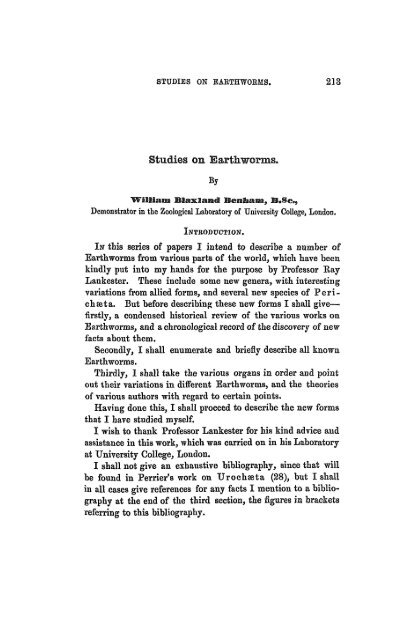
236 WILLIAM BLAXLAND BENHAM.<br />
A<br />
B<br />
C<br />
D EF<br />
G Perichffita, Sohm.<br />
H I<br />
K Geogenia, Kinb.<br />
I<br />
M<br />
N<br />
0<br />
GENUS.<br />
jumbricutf, Linn.<br />
lypogse<strong>on</strong>, Sav.<br />
tfegascolex, Temp.<br />
Driodrilus, H<strong>of</strong>fm.<br />
Helodrilus, H<strong>of</strong>fm.<br />
Echinodrilus, Vaill.<br />
P<strong>on</strong>toseolex, Schm.<br />
Tritogenia, Kinb.<br />
Mandane, Kinb.<br />
Alyattes, Kinb. .<br />
Eurydame, Kinb.<br />
Hegesipyle, Kinb.<br />
Bhodopis, Kinb.<br />
F Anteus, E. Perrier<br />
Q Titanus, E. Perrier<br />
Group.<br />
Ante.<br />
Ant.?<br />
Post.<br />
A.?<br />
A.?<br />
A.?<br />
Post.<br />
Post<br />
A.<br />
Post<br />
Intra<br />
p<br />
?<br />
?<br />
Post<br />
Intra<br />
Intra<br />
com-<br />
meuces at<br />
So-<br />
GliteUum<br />
mite.<br />
XXVII 6 to 10<br />
f<br />
• • •<br />
XIV<br />
XV<br />
*
STUDIES ON EAETHWOEMS.<br />
THE GENERA OJF EAKTHWOEMS.<br />
Ue marks<br />
<strong>on</strong><br />
Spermathecffl.<br />
From 2 to 10<br />
...<br />
... X<br />
• ••<br />
From 2 to 8,<br />
either simple<br />
or with appendages<br />
...<br />
...<br />
• ••<br />
er <strong>of</strong><br />
l<br />
Somite.<br />
\<br />
8<br />
9<br />
100<br />
8<br />
8<br />
20<br />
Very<br />
numerous<br />
7<br />
6<br />
8<br />
8<br />
8r<br />
8 L<br />
8<br />
50 to<br />
60<br />
8<br />
8<br />
Arrangement<br />
<strong>of</strong><br />
SeUe.<br />
4 couples<br />
Scattered<br />
In a ring,<br />
with dorsal<br />
break<br />
In 4 couples<br />
In 4 couples<br />
In 4 groups<br />
<strong>of</strong> 5<br />
Equidistant<br />
Alternating<br />
?<br />
Ventrally<br />
paired, dorsally<br />
scatterd<br />
In couples alternating<br />
anteriorly<br />
In couples,<br />
but scatter-<br />
ed posterior-<br />
J<br />
Scattered,<br />
except anteriorlyventrally<br />
Equidistant<br />
•4 couples<br />
4 couple3<br />
scattered<br />
posteriorly<br />
VOL, XXVI. —NEW SER.<br />
Positi<strong>on</strong><br />
<strong>of</strong><br />
Nephridiopore.<br />
In line with<br />
2nd setse<br />
?<br />
?<br />
?<br />
?<br />
?<br />
N<strong>on</strong>e<br />
f<br />
?<br />
?<br />
In line with<br />
3rd and 4th<br />
sete<br />
1 ?<br />
*<br />
In line with<br />
4th setae<br />
In line with<br />
2nd sete<br />
Length.<br />
8 to 12 inch.<br />
2 to 5 inch.<br />
80 to 370<br />
mm.<br />
70 mm.<br />
?<br />
62 to 80mm.<br />
85 mm.<br />
p<br />
58 mm.<br />
28 mm.<br />
75 mm.<br />
1 met.<br />
16 cm.<br />
1 met.<br />
26 cm.<br />
Habitat.<br />
4 to 6 inch. Europe and<br />
N. America<br />
30 to 40 mm. Buenos<br />
Ayres, and<br />
Sandwich<br />
Isles, &c.<br />
1820<br />
40 inches Ceyl<strong>on</strong> 1844<br />
Europe<br />
Europe<br />
...<br />
Various<br />
Jamaica<br />
?<br />
Patag<strong>on</strong>ia<br />
and M<strong>on</strong>tevideo<br />
Natal<br />
Buenos<br />
Ayres<br />
Panama<br />
Natal<br />
Java<br />
Cayenne<br />
Brazil<br />
1845<br />
1845<br />
1851<br />
1861<br />
1861<br />
1866<br />
1866<br />
1866<br />
1866<br />
1866<br />
1872<br />
1872<br />
ice to<br />
Refer<br />
aphy.<br />
Biblio<br />
1<br />
20<br />
22<br />
22<br />
23<br />
and<br />
24<br />
25<br />
25<br />
19<br />
19<br />
19<br />
19<br />
19<br />
14<br />
14<br />
A<br />
23?<br />
B<br />
C<br />
D<br />
E<br />
F<br />
6<br />
H<br />
I<br />
J<br />
K<br />
L<br />
1866 19 M<br />
1866 19 N<br />
0<br />
P<br />
Q
g<br />
1<br />
s 1 B<br />
£ I<br />
•-s CKJ<br />
f<br />
iB<br />
i tei<br />
tei a<br />
F<br />
tei<br />
I tei<br />
tetl<br />
1 3.<br />
Group.<br />
Clitellum commences<br />
at So-<br />
mite.<br />
3<br />
Number <strong>of</strong> Somites<br />
Clitellum<br />
extends througb<br />
03<br />
33<br />
S IN<br />
S co B s= P=;»<br />
pj «• ». a H — ^<br />
m.i<br />
O Bis-<br />
c 2 2<br />
C5 I<br />
(IF THE GENERA<br />
Remarks<br />
<strong>on</strong><br />
Spermathecae.<br />
t is united<br />
with ovary;<br />
opens in line<br />
with upper<br />
dorsal setse<br />
Withsmallappendage<br />
Simple<br />
?ores near median<br />
ventral<br />
line<br />
...<br />
With small<br />
spherical ap<br />
pendage<br />
Reniform.wit<br />
small lobed<br />
sac <strong>on</strong> eac<br />
side, openin<br />
into duct o<br />
spermathecsB<br />
Number <strong>of</strong><br />
Seiee per Somite.<br />
ornmened<br />
8<br />
8<br />
8<br />
30<br />
8<br />
8<br />
8<br />
8<br />
8<br />
8<br />
STUDIES ON EARTHWORMS. 239<br />
OF EARTHWORMS.—C<strong>on</strong>tinued.<br />
Arrangement<br />
<strong>of</strong> Setse.<br />
n 4 couples<br />
In 4 couples<br />
i couples, or<br />
8 separate<br />
4 couples<br />
Equidistant<br />
4 couples<br />
4 couples anteriorlyscattered<br />
and<br />
alternating<br />
posteriorly<br />
Separate<br />
1 and 2 are<br />
close toge<br />
ther, 3 anc<br />
4 separate<br />
4 couples<br />
4 couples<br />
Fo3iti<strong>on</strong><br />
<strong>of</strong><br />
ephridiopore<br />
n line with<br />
3rd and 4th<br />
setae<br />
n line with<br />
3rd & 4th<br />
setae<br />
n line with<br />
3rd & 4th<br />
setce<br />
n line with<br />
2nd setse<br />
n line with<br />
3rd and 4th<br />
seta;<br />
n line with<br />
3rd setae<br />
Alternate<br />
with 2nd<br />
and 4th<br />
setae<br />
In line wit]<br />
2nd setse<br />
In line wit<br />
2nd setse<br />
Length.<br />
15 cm.<br />
15 cm.<br />
0 to 35 cm.<br />
• >•<br />
120 mm.<br />
150 mm.<br />
1 dcm.<br />
15 cm.<br />
1 dcm.<br />
35to50mm<br />
10 inches<br />
Habitat.<br />
Venezuela<br />
ntilles,<br />
lartinique,<br />
Mo Janeiro<br />
ew Cale- 872<br />
d<strong>on</strong>ia and<br />
ladagascar<br />
Australia 872<br />
Cochin-<br />
China<br />
Ceyl<strong>on</strong><br />
Java, Brazil,<br />
&c.<br />
Pennsylvania<br />
France<br />
Europe<br />
Calcutta<br />
Date <strong>of</strong><br />
Descripti<strong>on</strong>.<br />
872<br />
872<br />
872<br />
872<br />
1872 14<br />
and and<br />
1874 28<br />
1873<br />
1874<br />
1874<br />
1883<br />
Reference to<br />
| Bibliography.<br />
14<br />
14<br />
14<br />
14<br />
14<br />
14<br />
27<br />
29<br />
15<br />
0<br />
r<br />
w<br />
Y<br />
Z<br />
AA<br />
37 BB
240 WILLIAM BLAXLAKD BENHAM.<br />
The following is a list <strong>of</strong> all <strong>Earthworms</strong> whose distributi<strong>on</strong><br />
is known, arranged according to Perrier's classificati<strong>on</strong>:<br />
I. Anteclitelliani.<br />
Lumbricus agricola, H<strong>of</strong>fm. . .<br />
„ trapezoides, Dug.<br />
„ rubellus, H<strong>of</strong>fm. .<br />
„ ehloroticus, Dug.<br />
„ olidus, H<strong>of</strong>fm.<br />
,, complanatus, Dug.<br />
„ tetraedrus, Dug.<br />
„ puter, Eis. .<br />
„ melibsBus, JRoaa .<br />
„ purpurens, Eis. ,<br />
„ Josephinse, Kin.<br />
„ Helens, .Kin.<br />
„ Hortensiee, Kin.<br />
„ infeliXj Kin.<br />
„ capensis, Kin.<br />
„ novas-hollandia?, Kin.<br />
j, Vineti, Kin.<br />
„ victoris, E. P.<br />
„ armatus, Kin.<br />
„ tellus, Kin. .<br />
„ pampicola, Kin. .<br />
„ Apii, Kin.<br />
„ tabitana, Kin.<br />
„ americanus, E. P.<br />
„ uliginosus, Hutt.<br />
„ campestris, Hutt.<br />
„ levis, Hutt.<br />
„ annulatus, Hutt.<br />
PAlyattes, Kinb.<br />
? Hypogse<strong>on</strong>, Sav.<br />
? „ Atys, Kin.<br />
? „ liavaious, Kin.<br />
? „ orthostioh<strong>on</strong>, Schtn.<br />
? „ lieterostich<strong>on</strong>, Schm.<br />
Habitat.<br />
Europe.<br />
Europe.<br />
Europe.<br />
Europe.<br />
Europe.<br />
Europe.<br />
Europe.<br />
Europe.<br />
Europe.<br />
Europe. •<br />
St. Helena.<br />
St. Helena.<br />
St. Helena.<br />
Port Natal.<br />
Cape <strong>of</strong> Good Hope.<br />
Sydney.<br />
Madeira.<br />
North Africa.<br />
Buenos Ayres.<br />
Buenos Ayres.<br />
M<strong>on</strong>tevideo.<br />
California.<br />
Tahiti.<br />
New York.<br />
New Zealand.<br />
New Zealand.<br />
New Zealand.<br />
New Zealand.<br />
Buenos Ayres.<br />
Philadelphia.<br />
Buenos Ayres.<br />
Oahu (Sandwich Isles).<br />
New Zealand.<br />
Quito and Cuenca.
STUDIES ON BAETHWOBMS. 241.<br />
II. Intraclitelliani.<br />
•<br />
Anteus gigas, E. P. .<br />
Tifcanus brasiliensis, E. P.<br />
Rhinodrilus paradoxus, E. P.<br />
Eudrilus decipiens, E.P.<br />
99 Lacazii, E. P. .<br />
1) peregriEus, E. P.<br />
Urochseta hystris, E. P. .<br />
Typhasus orientalis, Bedd.<br />
? Geogenia, Kin. . . • .<br />
III- Post elitelliani.<br />
PerichssUi<br />
leucocycla, Sch.<br />
» brachycycla, Sch. .<br />
9) viridis, Sch. .<br />
9) cingulata, Sch.<br />
• 99 posthuma, L. V.<br />
99 ?sp., Horst. . , .<br />
1) cingulata, Sph.<br />
99 robusta, E. P.<br />
99<br />
9><br />
39<br />
93<br />
95<br />
9*<br />
99<br />
99<br />
J)<br />
99<br />
9»<br />
99<br />
9)<br />
)t<br />
)9<br />
99<br />
99<br />
99<br />
)j<br />
99<br />
PNitocris,<br />
. Houlleti, E..P.<br />
affinis, E. P. .<br />
el<strong>on</strong>gata, E. P.<br />
quadragenaria, E.P.<br />
tahitensis, Gr.<br />
bicincta, E.P.<br />
biserialis, E. P. .<br />
luz<strong>on</strong>ica, E. P.<br />
crerulea, E. P.<br />
Juliana, E. P. .<br />
rodericensis, Gr. .<br />
sylvestris, Hutt.<br />
lineatus, Hutt.<br />
indicus, Horst.<br />
sumatranus, Horsl.<br />
Hasseltii, Horst. .<br />
Sieboldii, Horst.<br />
jap<strong>on</strong>icus, Horst. .<br />
musicus, Horst.<br />
capensisj Horst.<br />
annulatus, Horst. .<br />
Kin<br />
Habitat.<br />
Cayenne (South America).<br />
Brazil-.<br />
Venezuela.<br />
Antilles.<br />
Martinique.<br />
Rio Janeiro.<br />
Martinique, Gloria, Brazil, Java.<br />
Calcutta.<br />
Natal.<br />
Ceyl<strong>on</strong>.<br />
Ceyl<strong>on</strong>.<br />
Ceyl<strong>on</strong>.<br />
Ceyl<strong>on</strong>.<br />
Java.<br />
Java.<br />
Bourb<strong>on</strong>.<br />
Bourb<strong>on</strong> and Manilla<br />
(Philippines).<br />
Calcutta and Cochin China.<br />
Cochin China.<br />
Peru.<br />
East Indies.<br />
Tahiti.<br />
Philippines.<br />
Philippines.<br />
Philippines.<br />
Philippines.<br />
Cochin China.<br />
Rodsriquez.<br />
New Zealand.<br />
New Zealand.<br />
Sumatra.<br />
Sumatra.<br />
Sumatra.<br />
Japan.<br />
Japan.<br />
Java.<br />
Java.<br />
Malay.<br />
Rio Janeiro.
242 WILLIAM BLAXLANB BENHAM.<br />
PAmyntas, Kin<br />
PPheretima, Kin.<br />
Ffthodopis, Kin. . . . . .<br />
PLampito, Kin<br />
PMandane, Kin<br />
Megascolex eoeruleus, Temp. .<br />
„ arm at a, Bedd.<br />
Pleurochaeta Moseleyi, Bedd..<br />
Plutellus heteroporus, E. P. .<br />
P<strong>on</strong>todrilus Mari<strong>on</strong>is, E. P.. .<br />
Acanthodrilus obtusus, E. P.<br />
„ ungulatus, E.P.<br />
„ verticillatus,<br />
E. P..<br />
„ kerguelenensis,<br />
Lankester .<br />
„ capensis, Sedd.<br />
„ sp., Bedd. .<br />
„ sp., Horst. .<br />
„ sp., Horst.<br />
Digaster lumbricoides, E. P. .<br />
Peri<strong>on</strong>yx excavatus, E. P.<br />
„ M'Intoshii, Bedd.<br />
Habitat.<br />
Guam (East Indies.<br />
Tahiti and Ceyl<strong>on</strong>.<br />
Java.<br />
Mauritius.<br />
M<strong>on</strong>tevideo and Patag<strong>on</strong>ia.<br />
Ceyl<strong>on</strong>.<br />
Calcutta.<br />
Ceyl<strong>on</strong>.<br />
Pennsylvania.<br />
Europe.<br />
New Caled<strong>on</strong>ia.<br />
New Caled<strong>on</strong>ia.<br />
Madagascar.<br />
IV. Aclitelliani.<br />
M<strong>on</strong>iligaster Deshayesii, E. P. Ceyl<strong>on</strong>.<br />
1 Tritogenia, Kin.<br />
Criodrilus, H<strong>of</strong>fm.<br />
Europe.<br />
Helodrilus, H<strong>of</strong>fm.<br />
Europe.<br />
Kerguelen.<br />
Cape <strong>of</strong> Good Hope.<br />
New Zealand.<br />
West Africa.<br />
West Africa.<br />
New South Wales.<br />
Cochin China.<br />
Burmah.<br />
The genera marked ? are not distinctly enough characterised to be retained.<br />
I have not included in this tabular statement, and generally in this porti<strong>on</strong><br />
<strong>of</strong> the memoir, any details due to my own researches, which will be found in<br />
a subsequent secti<strong>on</strong>.
STUDIES ON EAETHWOHMS. 243-<br />
L THE VARIATIONS IN THE STRUCTURE OF "EARTHWORMS<br />
TREATED ACCORDING TO THE DIFFERENT SYSTEMS OF<br />
ORGANS.<br />
Tie Sets.—Glaparede (12) drew Ms characters for tlie<br />
Terricohe as opposed to the Limicolas, from Lumbricus, some<br />
<strong>of</strong> which characters are now known to hold <strong>on</strong>ly for that genus,<br />
others for <strong>on</strong>ly a few genera, and. am<strong>on</strong>gst them is the arrangement<br />
<strong>of</strong> the setae in four groups <strong>of</strong> two setes in each somite.<br />
This arrangement holds for many <strong>Earthworms</strong>, viz. Lumbri-<br />
CHSJ Anteus, Rhinodrilus, Eudrilus, Acanthodrilus,<br />
Digaster, and M<strong>on</strong>iligaster, including, therefore, forms<br />
from each <strong>of</strong> Perrier's groups (14).<br />
In Urocheeta, Titanus, and Acanthodrilus capensis,<br />
and in the doubtful Alyattes, and Eurydame <strong>of</strong> Kinberg,<br />
the same arrangement holds in the anterior part <strong>of</strong> the body,<br />
but varies posteriorly; the setas become scattered in Titanus,<br />
Ac. capensis, Alyattes, and Eurydame, but remain in line,<br />
whilst in U roc hast a they become scattered but alternate in<br />
c<strong>on</strong>secutive somites.<br />
In iEgesipyle, again <strong>on</strong>e <strong>of</strong> Kinberg's doubtful genera,<br />
they are scattered (that is, the two setae forming a couple<br />
become separated), except the anterior ventral couples.<br />
In Acanthodrilus kerguelenensis the setas are separate<br />
except in the genital somites.<br />
In P<strong>on</strong>todrilus throughout the body the ventral couple<br />
(setaa 1 and 2, counting from the mid ventral line) remain close<br />
together, but setse 3 and 4 are separated.<br />
Again, inPlutellus the eight setae are nearly equidistant,<br />
but do not alternate, whereas in P<strong>on</strong>toscolex there are <strong>on</strong>ly<br />
seven setee in each somite, which alternate in c<strong>on</strong>secutive<br />
somites throughout the body, and in Geogenia they alternate<br />
anteriorly <strong>on</strong>ly. In a form which I shall describe in a later<br />
paper, seven out <strong>of</strong> the eight alternate, whilst the ventralmost<br />
setas (No. 1) remain in line throughout the body.
2M WILLIAM BLAXLAND BENHAM.<br />
In Echinodrilus we have still four groups, but there are<br />
five setee to each group.<br />
In Hypogse<strong>on</strong>, Savigny described nine equidistant setae, <strong>of</strong><br />
which <strong>on</strong>e was said to be in the mid dorsal line.<br />
In Tritogenia, Kinberg, <strong>on</strong>ly six setse are present in each<br />
somite. The number is greatly increased in Perichseta,<br />
•where there may be as many as 100 to the somite, and in<br />
Peri<strong>on</strong>y x there are thirty. These in each case are equidistant<br />
and form a complete ring round each somite.<br />
In Megascolex, again, this arrangement is varied by an<br />
interrupti<strong>on</strong> in the ring in the mid dorsal line, whilst in Beddard's<br />
Pleurochseta, as well as this dorsal break, there is a<br />
similar break ventrally.<br />
The setse are not always simply pointed, as Clapar&de supposed,<br />
but in many cases are variously ornamented, as in Rhinodrilus,<br />
throughout the body; in the genital setse <strong>of</strong> Urochaata<br />
and <strong>of</strong> AJcanthodrilus; whilst in Urochseta all the<br />
setse are bifid at their free extremity. Moreover they are<br />
modified in certain parts <strong>of</strong> the body, as Hering (5) has shown,<br />
for copulati<strong>on</strong>: e. g. in Lumbricus, <strong>on</strong> clitellum, and <strong>on</strong> the<br />
somites xv and xxvi.<br />
Pores.—Perrier has pointed out a sort <strong>of</strong> relati<strong>on</strong>ship between<br />
the nephridiopores and the couples <strong>of</strong> setse; in some genera<br />
these pores are in fr<strong>on</strong>t <strong>of</strong> and slightly dorsad <strong>of</strong> the ventral<br />
couples (setse 1 and 2), as in Lumbricus, Titanus, P<strong>on</strong>todrilus,<br />
whilst in other genera the pores have a similar relati<strong>on</strong><br />
to the lateral couples (3 and 4), Ehinodrilus, Eudrilus,<br />
Acanthodrilus, AnteuSj M<strong>on</strong>iligaster [also in Microchseta].<br />
Plutellus is excepti<strong>on</strong>al at present in showing an alternati<strong>on</strong><br />
<strong>of</strong> the nephridiopores with the seta 2 and with seta 4 in<br />
c<strong>on</strong>secutive somites, whilst the first few pairs are in line with<br />
the third seta. Again, they are, in Urochseta, related to the<br />
third seta throughout the body. They remain in this line even<br />
when the setse alternate, though <strong>on</strong>e would expect, if there<br />
were any relati<strong>on</strong> between them and the setse, that the nephridiopores<br />
would also alternate.
STUDIES ON EARTHWORMS. 245<br />
This difference in the positi<strong>on</strong> <strong>of</strong> the nephridiopores helps to<br />
c<strong>on</strong>firm Pr<strong>of</strong>. Lankester's theory as to the original presence <strong>of</strong><br />
two pairs <strong>of</strong> nephridia in each somite, <strong>on</strong>e series <strong>of</strong> which has<br />
disappeared in <strong>on</strong>e set <strong>of</strong> worms, whilst the other series has<br />
g<strong>on</strong>e in the sec<strong>on</strong>d set, excepting in the genital somites, where<br />
they have been modified for the c<strong>on</strong>veyance <strong>of</strong> the genital products<br />
to the exterior. This is well seen in such forms as<br />
Budrilus, where the nephridiopore is in line with the lateral<br />
couple, and the male pore in line with ventral couple. In<br />
Anteus no sperm duct is known, but the nephridia are somewhat<br />
modified in the genital regi<strong>on</strong>, and may possibly serve as<br />
sperm ducts. It would be extremely important if it could be<br />
shown that such is really the case.<br />
The pores <strong>of</strong> the sperm athecse are always anteriorly placed •<br />
except in Eudrilus, where they are behind the seminal<br />
reservoirs [and in Microchseta, where they are still further<br />
back], and are rarely placed laterally as in Perichssta af finis,<br />
but more <strong>of</strong>ten in a line with the ventral setae, as in Plutellus,<br />
Digaster, P<strong>on</strong>todrilus, or as in Lumbricus, a<br />
little dorsad <strong>of</strong> these setae. In P<strong>on</strong>todrilus they are in a<br />
line with the first seta, in Plutellus they are in line with the<br />
sec<strong>on</strong>d seta; again, in Urochseta they are in line with the<br />
lateral third seta. InPeri<strong>on</strong>yx the pores are ventrad <strong>of</strong> the<br />
first seta.<br />
The pores are either <strong>on</strong> the anterior or <strong>on</strong> the posterior edge<br />
<strong>of</strong> the somites, so close to the edge that they appear to be <strong>on</strong><br />
the inter-segmental groove.<br />
The CliteUum.—The clitellum has been taken by Perrier<br />
as the basis <strong>of</strong> his classificati<strong>on</strong> <strong>of</strong> the Terricolous Oligochseta,<br />
and it is <strong>on</strong>ly in a very few cases that it is absent.<br />
M<strong>on</strong>iligaster has certainly no clitellum, although its<br />
genital organs are described as being fully developed.<br />
Helodrilus andCriodrilus were described byH<strong>of</strong>fmeister<br />
as having no clitellum, though sufficient details <strong>of</strong> the genital<br />
organs are not given by him, and the worms have not been<br />
studied recently.<br />
The various genera bel<strong>on</strong>ging to the four groups, Anteclitel-
246 WILLIAM BLAXLAND BENHASI.<br />
liani, Intraclitelliani, Postclitelliani, Aclitelliani, have already<br />
been given above.<br />
The extent <strong>of</strong> the clitellum varies c<strong>on</strong>siderably in the same<br />
genus, even in the different specimens <strong>of</strong> the same species<br />
when fully developed; for instance, in Lumbricus agricola<br />
it occupies six or seven somites, xxx to xxxvi; in L. rubellus,<br />
somites xxvi to XXXII (seven somites); in L. trapezoides,<br />
somites xxvn to xxxv (nine somites); whilst in L. (Allurus)<br />
tetraedrus it occupies <strong>on</strong>ly six somites, from xxn to XXVII.<br />
Am<strong>on</strong>gst the Intrachitellian forms the same variati<strong>on</strong> is to<br />
be noted : Anteus has as many as fifteen somites, xv to xxix,<br />
making up the clitellum; Titanus has nine somites, xv to<br />
xxmj Urochreta has ten somites, xiv to xxm; Geogenia<br />
has ten somites j whilst Rhinodrilus has <strong>on</strong>ly three somites<br />
iu this structure, xix, xx, xxi. It commences as far forwards<br />
as somite ix in Geogenia.<br />
In Eudrilus the three different species each have a different<br />
number <strong>of</strong> somites in the clitellum, E. decipiens having <strong>on</strong>ly<br />
three, XIII, xiv, xv; whilst E. Lacazii has six somites, XTII to<br />
XVIII, to form this organ. In Rhinodrilus the girdle is complete,<br />
whilst in the other forms it resembles Lumbricus in<br />
having a saddle shape.<br />
Am<strong>on</strong>gst the Postclitellian genera there are usually fewer<br />
somites occupied by the clitellum, which is a complete girdle,<br />
and nearly always commences with somite xiv (Perichseta,<br />
xiv, xv, xvi; Plutellus, xiv to XVII; Acanthodrilus, xiv<br />
to XVII, and others), or in XIII (Peri<strong>on</strong>yx, XIII to XVII, and<br />
P<strong>on</strong>todrilus, xni to XVII); in xn, in Kinberg's genera<br />
Rhodopis and Mandane, or in xv, in P<strong>on</strong>toscolex. (In<br />
Kinberg's genera, Nitocris, Amyntas, Lampito, Pheret<br />
i m a, the clitellum appears to commence at somite xiv, and<br />
this character with the numerous setse to each somite would<br />
justify us in c<strong>on</strong>sidering them as forms <strong>of</strong> Perichasta.)<br />
Rhodopis, if Kinberg's descripti<strong>on</strong> can be c<strong>on</strong>sidered <strong>of</strong><br />
any value, possesses the shortest clitellum, occupying <strong>on</strong>ly two<br />
somites, as is also the case in Perichseta bicincta.<br />
The positi<strong>on</strong> <strong>of</strong> the clitellum and the positi<strong>on</strong> <strong>of</strong> th'e gizzard
STUDIES ON EARTHWORMS, 247<br />
seem to have some sort <strong>of</strong> relati<strong>on</strong> to <strong>on</strong>e another, for in the<br />
Anteclitellian worms, where the clitellum commences somewhere<br />
about xxv or further back, the gizzard is behind the<br />
genital organs. But in the Postclitelliani and Intraclitelliani<br />
the gizzard passes forwards to somites vi or vu, and is usually<br />
in the same somites with or in fr<strong>on</strong>t <strong>of</strong> the seminal reservoirs.<br />
In the forms which have not a complete clitellum there is<br />
very frequently a glandular ridge <strong>on</strong> each side <strong>of</strong> the ventral<br />
surface, al<strong>on</strong>g or near the ventral edge <strong>of</strong> the clitellum. This<br />
is seen in Lumbricus, where Eisen (15) speaks <strong>of</strong> it as<br />
" tubercula pubertatis." Perrier figures the same sort <strong>of</strong> ridge<br />
in Rhinodrilus, but whether it occurs in other forms I do<br />
not know. It very probably does so as it would appear to have<br />
some functi<strong>on</strong> in copulati<strong>on</strong>.<br />
The true limits <strong>of</strong> the clitellum anteriorly and posteriorly<br />
are not always evident, as the thickening <strong>of</strong> the epidermis is<br />
gradual, and frequently is apparent <strong>on</strong> the dorsal surface before<br />
it is so ventrally.<br />
Dorsal Pore.—In many <strong>Earthworms</strong> the coelom is put into<br />
communicati<strong>on</strong> with the exterior by means <strong>of</strong> a series <strong>of</strong><br />
"dorsal pores" placed <strong>on</strong> the intersegmental grooves. In<br />
Lumbricus these pores occur in every somite after about<br />
somite vni. In Digaster and Peri<strong>on</strong>yx they commence<br />
just behind somite iv. In Plutellus behind somite vi. In<br />
Pleurochaeta andTyphoeus the pores are present <strong>on</strong>ly behind<br />
the clitellum. They are present in Acanthodrilus, and in<br />
many Perichsetse.<br />
The Alimentary Tract.—The main regi<strong>on</strong>s into which<br />
the digestive canal is divided are c<strong>on</strong>stant in all the <strong>Earthworms</strong>.<br />
There is a pharynx, oesophagus, gizzard (except in<br />
P<strong>on</strong>todrilus), and an intestine.<br />
The pharynx is a str<strong>on</strong>gly muscular organ and <strong>of</strong> glandular<br />
appearance, though in Lumbricus and others I can find no<br />
glands in the wall. Perrier has found glands in the pharynx <strong>of</strong><br />
P<strong>on</strong>todrilus. The anterior part <strong>of</strong> this organ has quite<br />
thin walls, and is in some at least capable <strong>of</strong> slight protrusi<strong>on</strong>.<br />
This thin-walled regi<strong>on</strong> is the " buccal " regi<strong>on</strong>,
248 WILLIAM BLAXLAND BENHAM.<br />
and extends back as far as the circumpharyngeal nerve commissure.<br />
In Perichseta the pharynx is provided with three pairs <strong>of</strong><br />
glands, which open into it, in M<strong>on</strong>iligaster also there are<br />
small glands in this regi<strong>on</strong>. In other cases, e.g. Urochaata,<br />
the first pair <strong>of</strong> nephridia are much modified, their tube enlarged<br />
and glandular, and the coils are flattened against the<br />
walls <strong>of</strong> the pharynx so as to become more or less buried in its<br />
muscles, but they have no opening into the pharynx.<br />
The pharynx usually occupies four or five somites, though,<br />
owing to the somewhat infundibulate septa, it appears to<br />
occupy more somites. Lumbricus is <strong>of</strong>ten described as<br />
having the pharynx extending through seven somites, whereas<br />
really it occupies <strong>on</strong>ly five. InP<strong>on</strong>todrilusit occupies <strong>on</strong>ly<br />
three somites.<br />
The (Esophagus varies much in length, occupying twelve<br />
somites in Lumbricus (including the excepti<strong>on</strong>al " proventriculus"<br />
<strong>of</strong> this form) whilst in Eudrilus it <strong>on</strong>ly extends<br />
through two somites. In Digaster, owing to the presence<br />
<strong>of</strong> a sec<strong>on</strong>d gizzard, there is a sec<strong>on</strong>d oesophagus between<br />
them.<br />
In the greater number <strong>of</strong> genera there are no glands in this<br />
porti<strong>on</strong> <strong>of</strong> the alimentary tract; e.g. in Eudrilus, Rhinodrilus,<br />
Acanthrodilus, Digaster, Peri<strong>on</strong>yx, and others.<br />
But in Lumbricus there are three pairs <strong>of</strong> enlargements<br />
known as " cesophageal" or calciferous glands, two being in<br />
somite xn, the third in somite XIII. The two anterior c<strong>on</strong>tain<br />
a milky liquid; the last c<strong>on</strong>tains solid carb<strong>on</strong>ate <strong>of</strong> lime.<br />
Anteus also possesses "ossophageal glands." But it is<br />
in Perichseta that the glandular appendices <strong>of</strong> the alimentary<br />
tract are most fully developed. Besides the three pairs <strong>of</strong><br />
pharyngeal glands there are three different sorts <strong>of</strong> glands to<br />
the ossophagus. In each <strong>of</strong> the somites, vi, vn, vm, is a pair<br />
<strong>of</strong> tufts <strong>of</strong> glandular tubules. In somite vi there is a pair <strong>of</strong><br />
large ovoid, solid-looking glands, each <strong>of</strong> which opens by a<br />
distinct pore into the (Esophagus, whilst in somite vn there is<br />
a pair <strong>of</strong> grape-like glands.
- STUDIES ON EABTHWOEMS. 249<br />
The great development <strong>of</strong> these glands in Perichseta seems<br />
related to the extremely small size (? absence in some) <strong>of</strong> the<br />
nephridia. Perrier has suggested that they act in some way<br />
as excretory glands, the excreti<strong>on</strong>, however, being used as a<br />
digestive fluid, instead <strong>of</strong> being passed directly to the exterior,<br />
just as the liver <strong>of</strong> Vertebrates is in a way an excretory gland.<br />
The gizzard is present in all but P<strong>on</strong>todrilus. The walls<br />
are very muscular, and the lining epithelium secretes a chitinous<br />
lining, which forms the crushing apparatus.<br />
The gizzard is situated very far back in Lumbricus, where<br />
it occupies somites XVII and SVIII. Here it is quite behind all<br />
the genital organs even behind the male pore. InPeri<strong>on</strong>yx<br />
the gizzard is in somite XII, in the same somite as the posterior<br />
seminal reservoir.<br />
In all the other forms it is in fr<strong>on</strong>t <strong>of</strong> the seminal reservoirs,<br />
being usually in somite vi (Anteus, Titanus, and Plutellus),<br />
or in somite vn (Urochaeta, Eudrilus, and<br />
Rhinodrilus). In some cases it occupies <strong>on</strong>ly <strong>on</strong>e somite,<br />
in others it occupies more than <strong>on</strong>e. In Anteus the anterior<br />
septa are very thick and infundibuliform, covering the gizzard;<br />
but frequently the septum immediately in fr<strong>on</strong>t <strong>of</strong> this organ<br />
is thinner than the neighbouring <strong>on</strong>es. In Perichseta the<br />
somite is c<strong>on</strong>tains the gizzard. Digaster is so named from<br />
its possessing two gizzards, the anterior being in somite v, the<br />
posterior in vn, the porti<strong>on</strong> <strong>of</strong> the alimentary tract between<br />
them being the sec<strong>on</strong>d oesophagus. A further complicati<strong>on</strong> I<br />
have found in a worm from St. Thomas's, where there are<br />
three separate gizzards. M<strong>on</strong>iligaster again has a gizzard<br />
in somite vi <strong>of</strong> the usual form, whilst an el<strong>on</strong>gated gizzard<br />
occupies somites XIII to xxn inclusive, being c<strong>on</strong>stricted into<br />
four porti<strong>on</strong>s. Here, therefore, the first gizzard is in fr<strong>on</strong>t <strong>of</strong><br />
the true genital organs, the sec<strong>on</strong>d being behind them.<br />
The Intestine following the gizzard is frequently separated<br />
into two regi<strong>on</strong>s, the porti<strong>on</strong> directly behind the gizzard being<br />
the "tubular," and following this a more or less "sacculated"<br />
regi<strong>on</strong> where the typhlosole is developed.<br />
In Lumbricus this tubular porti<strong>on</strong> occupies <strong>on</strong>ly two
250 WILLIAM BLAXLAM) BENHAM.<br />
somites, xix and xs; whilst behind this comes the sacculated,<br />
typhlosolar regi<strong>on</strong>, with thin walls covered externally by the<br />
large, yellow coslomie epithelium; in this form the typhlosole<br />
is well developed, and, as usual, presents a blood-vessel in<br />
frequent c<strong>on</strong>necti<strong>on</strong> with the dorsal trunk.<br />
In other forms the tubular porti<strong>on</strong> has a greater extent; for<br />
instance, in Anteus it extends from about somite vm to xvin,<br />
in Titanus from somite vi to xv. In Acanthodrilus,<br />
judging by Perrier's figures, <strong>on</strong>e species has n<strong>on</strong>e, in another<br />
this regi<strong>on</strong> extends to somite xx, and in Urochseta as far as<br />
somite xv. In all cases where a structure is said to extend to<br />
a certain somite, it is understood to exist in that somite as well<br />
as in the preceding somites.<br />
In these forms ossophageal glands are absent, but their analogue<br />
seems to be frequently present in the form <strong>of</strong> <strong>on</strong>e or more<br />
pairs <strong>of</strong> glands, which c<strong>on</strong>tain carb<strong>on</strong>ate <strong>of</strong> lime, situated <strong>on</strong><br />
the tubular regi<strong>on</strong> <strong>of</strong> the intestine.<br />
In Urochseta there are three pairs <strong>of</strong> such glands, el<strong>on</strong>gated<br />
and ovoid, in somites vm, ix, x; in Plutellus three<br />
pairs <strong>of</strong> glands <strong>of</strong> the same nature occur in somites x, xi, xu,<br />
but are reniform.<br />
In Titanus is a single pair <strong>of</strong> white, nearly spherical glands,<br />
in somite xnr, which Perrier mistook for a part <strong>of</strong> the vascular<br />
system. He describes it as a " ventricle." I have had the<br />
good fortune <strong>of</strong> dissecting a Titanus, and have seen distinctly<br />
the large openings <strong>of</strong> these glands into the intestine.<br />
One <strong>of</strong> the characteristics <strong>of</strong> the genus Perichseta appears<br />
to be the presence <strong>of</strong> a pair <strong>of</strong> el<strong>on</strong>gated caeca, springing<br />
from the ventral surface <strong>of</strong> the sacculated intestine in somite<br />
xxvi or xxvn. In P. Sieboldii, Horst, there are six pairs <strong>of</strong><br />
coeca in somite xxvi. Although Peri<strong>on</strong>yx resembles Perich<br />
set a in many points these caeca are absent in it.<br />
The typhlosole is usually a sub-cylindrical l<strong>on</strong>gitudinal<br />
valve produced by an involuti<strong>on</strong> al<strong>on</strong>g the dorsal wall <strong>of</strong> the<br />
sacculated intestine; but in Titanus this organ is flattened<br />
from side to side, whilst in P<strong>on</strong>todrilus the vessel al<strong>on</strong>e<br />
exists. Pie urochseta has no typhlosole and possesses a
STUDIES ON EARTHWORMS. 251<br />
series <strong>of</strong> reniform glands in the posterior part <strong>of</strong> the tract.<br />
Typhaeus (37) possesses lobed glands <strong>on</strong> the middle regi<strong>on</strong> <strong>of</strong><br />
<strong>of</strong> the intestine.<br />
The saccnlated porti<strong>on</strong> <strong>of</strong> the intestine appears to be similar<br />
in all these worms, and is c<strong>on</strong>tinued to near the end <strong>of</strong> the body<br />
when the typhlosole disappears, and the regi<strong>on</strong> is called rectum.<br />
The structure <strong>of</strong> the glands, whether they occur <strong>on</strong> the<br />
cesophagus or <strong>on</strong> the tubular intestine, is very similar in<br />
some <strong>of</strong> those that have been investigated.<br />
Claparede (11) has described and figured a secti<strong>on</strong> <strong>of</strong> the<br />
oesophageal gland <strong>of</strong> Lumbricus, where it c<strong>on</strong>sists <strong>of</strong> numerous<br />
alternate blood-vessels and glandular tubules placed<br />
radially.<br />
Perrier describes the "glandes de Morren" <strong>of</strong> Urochseta<br />
as having the same structure, but his figure does not quite agree<br />
•with the text, as he figures no blood-vessels. InTitanus the<br />
gland has the same structure [and I shall show that in a<br />
•worm (Microchsata), to be described further <strong>on</strong>, the structure<br />
<strong>of</strong> the intestinal gland resembles that <strong>of</strong> the oesophageal<br />
gland <strong>of</strong> Lumbricus' very closely]. But the various ossophageal<br />
glands in Perichseta each have a distinct structure,<br />
and n<strong>on</strong>e seem, from Perrier's figures, to resemble the abovementi<strong>on</strong>ed<br />
glands.<br />
In all the above glands (except Perichaeta) carb<strong>on</strong>ate <strong>of</strong><br />
lime has been found; usually solid, but sometimes in the form<br />
<strong>of</strong> a milky fluid. Hence we have analogous glands in various<br />
regi<strong>on</strong>s <strong>of</strong> the alimentary tract, and in different somites. So<br />
also the gizzard occurs in different somites. Perrier has<br />
suggested that these may not <strong>on</strong>ly be analogous but even<br />
homologous organs: the worm being made up <strong>of</strong> somites,<br />
each <strong>of</strong> which somites was originally exactly alike, in <strong>on</strong>e somite<br />
<strong>of</strong> <strong>on</strong>e worm a part <strong>of</strong> the alimentary tract becomes a gizzard,<br />
whilst in a sec<strong>on</strong>d worm the modificati<strong>on</strong> occurs in a different<br />
somite. But each <strong>of</strong> these gizzards is a modificati<strong>on</strong> <strong>of</strong> an.<br />
originally homologous organ, therefore the gizzards are homologous.<br />
In the same way the glands are modificati<strong>on</strong>s and<br />
swellings at different parts <strong>of</strong> the alimentary tracts, which were
2 52 WILLIAM BLAXLAND BENHAM.<br />
originally homologous; therefore these glands, whether ossophageal<br />
or intestinal, are homologous. This view cannot, however,<br />
be held if we apply the <strong>on</strong>ly true test <strong>of</strong> homology, that<br />
<strong>of</strong> comm<strong>on</strong> origin from a comm<strong>on</strong> ancestor. It is quite clear<br />
that a gland which is in somite xxiv cannot be the same thing<br />
as a gland which existed in somite XIII <strong>of</strong> an ancestor, or vice<br />
versa. If we are to suppose that similar parts have been<br />
similarly modified for similar wants in different somites, <strong>of</strong><br />
two genera <strong>of</strong> <strong>Earthworms</strong> compared, then the case is <strong>on</strong>e, not<br />
<strong>of</strong> homology, but <strong>of</strong> " homoplasy." (See Lankester, 44.)<br />
The Hervous System.—This seems very similar in all the<br />
worms studied, c<strong>on</strong>sisting <strong>of</strong> a pair <strong>of</strong> supra-pharyngeal<br />
ganglia, and a series <strong>of</strong> ventral ganglia, united by cords; besides<br />
these, in at least some forms (Urochseta, Perichseta, Lumbricus,<br />
and others), there is a visceral system <strong>of</strong> cords and<br />
ganglia <strong>on</strong> the pharynx, and probably c<strong>on</strong>tinued farther backwards:<br />
these originate partly from the supra-pharyngeal ganglia<br />
and partly from the circum-pharyngeal commissures.<br />
The presence <strong>of</strong> the " three great fibres " in the ventral cord<br />
appears pretty c<strong>on</strong>stant; but the sub-neural vessel is not<br />
so universal as Claparede supposed; for in P<strong>on</strong>todrilus and<br />
Perichseta Houlleti, Perrier has shown that this vessel is<br />
absent [as I shall show later <strong>on</strong> to be the case in at least <strong>on</strong>e<br />
other worm Microchasta], and as Beddard has shown to be<br />
the case in Pleurochseta. Perrier c<strong>on</strong>siders that the supra<br />
pharyngeal ganglia are always in somite m, but he is wr<strong>on</strong>gj<br />
for in Titanus these ganglia lie in somite n, although<br />
dragged back by the muscles <strong>of</strong> the pharynx; the first ventral<br />
gangli<strong>on</strong> lies in somite HI, and it is usual to find that gangli<strong>on</strong><br />
in the somite following that in which the supra-pharyngeal<br />
ganglia lie.<br />
[In Microchseta and other <strong>Earthworms</strong> which I shall<br />
describe below, the supra-pharyngeal ganglia lie in somite i<br />
distinctly.] Undoubtedly, most frequently it is the somite in<br />
that in the adult is occupied by the supra-pharyngeal ganglia,<br />
although their seat in embryological origin is the prostomium.<br />
The Vascular System.—In its simplest form, as in Peri-
STUDIES ON EAETHWOEMS 253<br />
chseta, the closed vascular system c<strong>on</strong>sists <strong>of</strong> a dorsal and<br />
a ventral l<strong>on</strong>gitudinal trunk, together with a typhlosolar<br />
trunk in the intestinal regi<strong>on</strong>. In the anterior regi<strong>on</strong> <strong>of</strong> the<br />
body, paired commissural vessels or " lateral hearts " c<strong>on</strong>nect<br />
the dorsal with the ventral trunk. In P<strong>on</strong>todrilus, a pair<br />
<strong>of</strong> l<strong>on</strong>gitudinal lateral trunks (" intestino-tegumentary " vessels<br />
<strong>of</strong> Perrier) are added to these three : these lateral trunks rise<br />
from a capillary network <strong>on</strong> the alimentary tract and pass<br />
forward to a similar network <strong>on</strong> the wall <strong>of</strong> the pharynx:;<br />
these lateral trunks are nowhere in direct communicati<strong>on</strong> with<br />
either the dorsal or the ventral trunk. The lateral l<strong>on</strong>gitudinal<br />
trunks occur in Lumbricus, where they rise as a pair <strong>of</strong><br />
branches from the dorsal trunk in somite x. In Urochaeta<br />
[in Microchsata], and possibly in Pleurochssta, they have<br />
the same arrangement as in P<strong>on</strong>todrilus. These trunks<br />
have not been described in other genera. There is a subneural<br />
trunk in all forms, except in Perichaeta, Pleurochaeta,<br />
P<strong>on</strong>todrilus [and Microchasta].<br />
From these l<strong>on</strong>gitudinal trunks, <strong>of</strong> which the dorsal and<br />
ventral are chiefly c<strong>on</strong>tractile, paired vessels pass to the septa,<br />
nephridia, body wall, and alimentary tract; in these organs<br />
they break up into networks <strong>of</strong> capillaries, whence the blood<br />
is again collected by vessels which returns it to the main<br />
trunks. In Perichseta and Peri<strong>on</strong>yx Beddard has shown<br />
that there are capillaries in the epidermis itself similar to<br />
those known in the leech. Howes has figured in the 'Biological<br />
Atlas' a small "infra-intestinal" vessel, closely<br />
attached to the ventral wall <strong>of</strong> the intestine in Lumbricus;<br />
this I have not myself seen, nor is it menti<strong>on</strong>ed by M. Jaquet<br />
(49) in his descripti<strong>on</strong> and figures <strong>of</strong> injected specimens <strong>of</strong> this<br />
geuus. But Mr. Beddard has informed me that he has seen it<br />
in Acanthodrilus. Besides the "lateral hearts" in the<br />
anterior somites, there may be also commissural vessels in the<br />
intestinal regi<strong>on</strong>. These in Lumbricus pass from the dorsal<br />
to the subneural trunk. I have found that the organ in<br />
Titanus, which Perrier regarded as part <strong>of</strong> a lateral heart<br />
and called " ventricle/' is really an intestinal gland.<br />
VOL. XXVI. NEW SEK. S
254 WILLIAM BLAXLAND BENHAM.<br />
The dorsal trunk is always muscular. It may be simply<br />
tubular in Lumbricus, M<strong>on</strong>iligaster, Perichasta, Titanus,<br />
and others, or it may be ampullate and valvular, as<br />
in Urochseta throughout the body; or tubular posteriorly<br />
and ampullate anteriorly as in. Anteus; in both these last<br />
forms the dorsal trunk is bent to <strong>on</strong>e side in a loop, just before<br />
the regi<strong>on</strong> where it gives <strong>of</strong>f the lateral hearts.<br />
In Pleurochseta the dorsal trunk divides in some <strong>of</strong> the<br />
anterior somites to form a double vessel, the halves <strong>of</strong> which<br />
unite again; there appears also to be a sec<strong>on</strong>d dorsal vessel below<br />
the large c<strong>on</strong>tractile trunk. Beddard (40) has described a<br />
somewhat similar arrangement in a species <strong>of</strong> Acanthodrilus<br />
and in MicrochEeta.<br />
L<strong>on</strong>gitudinal lateral trunks appear at each side <strong>of</strong> the<br />
alimentary tract anteriorly in some worms. In Lumbricus<br />
these are formed by a branch from the dorsal trunk <strong>on</strong> each<br />
side in somite x. In Urochseta and P<strong>on</strong>todrilus [and in<br />
Microchseta] (and doubtfully in Pleurochseta) they spring<br />
from a network in a part <strong>of</strong> the alimentary tract and run<br />
through eight or nine somites to the pharynx.<br />
The lateral hearts usually occupy the somites in which<br />
the seminal reservoirs are, and those just anterior to them.<br />
In Urochseta and Anteus there are three pairs, in somites<br />
vm, ix, x; in Plutellus, in somites x, xi, xu; in Rhinodrilus,<br />
in somites xvn, xvin, xix; whilst in Lumbricus,<br />
Digaster, and Titanus, they exist in somites VIII to xu; in<br />
T. forguesii, in somites xi, xu, the hearts communicate with<br />
dorsal and typhlosolar trunks. In Pleurochseta and Perichseta<br />
the lateral hearts are in somites x to xni, and in<br />
P<strong>on</strong>todrilus they are in somites v to xi. These lateral<br />
hearts pass from the dorsal to the ventral trunk, but other<br />
vessels exist in some forms, passing from the typhlosolar to the<br />
ventral trunk. These "intestinal hearts " are present in<br />
somites xx, xxi, xxii in Rhinodrilus just behind the lateral<br />
hearts, and in Urochseta in somites xni and XIVJ in this<br />
latter worm they are very much larger than the lateral<br />
hearts, whilst in Poutodrilus the two intestinal hearts
STUDIES ON EABTHWOBMS. 255<br />
in somites xn and XIII communicate both with the dorsal and<br />
with the typhlosolar trunks <strong>on</strong> the <strong>on</strong>e hand and with the<br />
ventral trunk <strong>on</strong> the other. Thus in the Anteclitelliani the<br />
hearts are anterior to, whilstfin the other groups they are either<br />
posterior to the gizzard or in its neighbourhood.<br />
The subneural trunk is absent in Pleurochagta, Perichseta,<br />
and P<strong>on</strong>todrilus [and in Microchaeta].<br />
The Course <strong>of</strong> the Blood.—The blood passes forwards<br />
al<strong>on</strong>g the dorsal (and typhlosolar) trunks and backwards<br />
al<strong>on</strong>g the ventral (and neural) trunks. The intestinal vessels<br />
rising from the dorsal trunk carry blood to the wall <strong>of</strong> the<br />
intestine, where there is a very close network <strong>of</strong> vessels,<br />
usually made up <strong>of</strong> l<strong>on</strong>gitudinal and circular branches; from<br />
this capillary network the blood is carried into the ventral<br />
trunk. Prom the ventral trunk a pair <strong>of</strong> vessels in each<br />
somite carries blood to the septa, nephridia, and body wall,<br />
where it is distributed in delicate loops and collected again by<br />
vessels which enter the dorsal trunk. This seems to be the<br />
course <strong>of</strong> the blood, as determined in Microchseta, where<br />
valves are placed in the dorsal trunk, at the exits and entrances<br />
<strong>of</strong> the vessels.<br />
Anteriorly the blood is distributed over the wall <strong>of</strong> the<br />
pharynx and collected from the network into the ventral<br />
trunk. In the lateral hearts the blood passes downwards from<br />
the dorsal to the ventral trunk.<br />
In the l<strong>on</strong>gitudinal lateral trunks the course <strong>of</strong> the blood<br />
seems to vary. In Lumbricus, it is evident that it passes<br />
forwards to be distributed over the pharynx, whence it is collected<br />
by the branches going to the ventral trunk. In Urochseta,<br />
Perrier c<strong>on</strong>sidered this forward directi<strong>on</strong> preferable;<br />
while in P<strong>on</strong>todrilus he thinks it probable that the blood<br />
has not the same directi<strong>on</strong> as in the dorsal trunk.<br />
The Blood, by which I refer to the red liquid in the closed<br />
system <strong>of</strong> vessels, is a liquid, coloured red by haemoglobin, in<br />
which float obl<strong>on</strong>g colourless corpuscles, as has been shown by<br />
Lankester in 1878 to be the case in Lumbricus (45), and<br />
by Bourne and Blomfield in 1881 for the Polychseta (46).
256 WILLIAM BLAXLAND BENHAM.<br />
These corpuscles can readily be seen by killing a piece <strong>of</strong><br />
tissue, such as a septum or a nephridium, with -j^- per cent,<br />
osmic, and then staining in picrocarmine.<br />
The Eephridia.—These have been figured by Gegenbauer<br />
(10) and described histologically by Claparede (11) for Lnmbricus.<br />
Each nephridium or "segmental organ" <strong>of</strong> Dr.<br />
Williams (47) is a more or less coiled tubule with an internal<br />
funnel-shaped opening at <strong>on</strong>e end, and an external pore at<br />
the other. The tube itself is divisible into three regi<strong>on</strong>s,<br />
the innermost leading from the funnel is cilated internallythis<br />
leads to a glandular regi<strong>on</strong>, and this to a short, muscular,<br />
slightly enlarged " vesicular " regi<strong>on</strong>. The lumen <strong>of</strong> the first<br />
two regi<strong>on</strong>s is intracellular, whilst that <strong>of</strong> the vesicle is intercellular<br />
and surrounded by muscle-fibres. The histology <strong>of</strong><br />
the nephridia has not been minutely studied in any form,<br />
except in Lumbricus.<br />
Nephridia are at present known in nearly all the forms<br />
whose internal anatomy has been described.<br />
In Digaster Perrier appears not to have detected the organ<br />
or its pore. Beddard did not find them in Pleurochseta.<br />
In most <strong>of</strong> the Perichsetas they are so small as to have<br />
led to the impressi<strong>on</strong> that they are absent, but in P. robusta<br />
and P. affinis delicate tubules are attached to the septa, but<br />
Perrier gives no details. [In a Perichseta from the Philippines<br />
I have found numerous small nephridia in each<br />
somite by means <strong>of</strong> secti<strong>on</strong>s. Beddard informs me that he<br />
has made a similar observati<strong>on</strong>.]<br />
In the worms which possess nephridia the internal funnel<br />
is usually situated in the somite anterior to that in which the<br />
tubule lies, but in Plutellus the whole organ lies in <strong>on</strong>e<br />
somite, and it has a large vesicular porti<strong>on</strong>.<br />
In Typhaeus the nephridia have <strong>on</strong>ly been observed in the<br />
anterior somites.<br />
In Titanus the nephridia do not commence till somite xiv.<br />
In Anteus those <strong>of</strong> the clitellar regi<strong>on</strong> are shorter, wider,<br />
and less coiled than the others, and are supposed by Perrier<br />
to functi<strong>on</strong> as sperm ducts.
STUDIES ON EARTHWORMS. 257<br />
In Urochseta tlie first pair <strong>of</strong> nephridia are much modified<br />
and c<strong>on</strong>sist <strong>of</strong> a rosette <strong>of</strong> tubules opening into a large vesicle,<br />
and closely applied to the pharynx.<br />
[The greatest development seems to take place in Mierochasta,<br />
where all the nephridia have the structure <strong>of</strong> the first<br />
nephridium <strong>of</strong> Urochseta, but have an immense vesicular<br />
regi<strong>on</strong>. Other <strong>Earthworms</strong>, that I shall describe later, also<br />
have large el<strong>on</strong>gated vesicles, and comparatively short glandular<br />
regi<strong>on</strong>.]<br />
In P<strong>on</strong>todrilus the anterior nephridia seem to be simple<br />
tubes, but in those posterior to the genital organs an immense<br />
compact glandular regi<strong>on</strong> is added, whilst the free porti<strong>on</strong> <strong>of</strong><br />
tubule is short.<br />
In M<strong>on</strong>iligaster the nephridia are rather smaller in the<br />
genital somites than, elsewhere.<br />
As to the pores <strong>of</strong> the nephridia, the positi<strong>on</strong> <strong>of</strong> which<br />
relative to the sets is an important generic character, we<br />
have more informati<strong>on</strong>.<br />
Kinberg, as a rule, makes no menti<strong>on</strong> <strong>of</strong> them, but in<br />
describing Geogenia he says the " lateral pores " are in a line<br />
with the dorsal setas.<br />
I have already menti<strong>on</strong>ed the positi<strong>on</strong> <strong>of</strong> these pores in the<br />
various genera in describing the external features. As a rule,<br />
they are placed in the anterior regi<strong>on</strong> <strong>of</strong> the somites, just in<br />
fr<strong>on</strong>t and slightly dorsad <strong>of</strong> either seta 4 or seta 2 (the upper<br />
seta <strong>of</strong> the lateral or <strong>of</strong> the ventral couple). But in Urochasta<br />
they are in this relati<strong>on</strong> to seta 3 (the lower seta <strong>of</strong> the lateral<br />
couple), whilst in Plutellus they alternate between setss 4<br />
and 2, and the first five are in line with seta 3.<br />
In a species <strong>of</strong> Acanthodrilus from New Zealand, Beddard<br />
has described (42) the positi<strong>on</strong> <strong>of</strong> the nephridia as alternating<br />
from somite to somite with the two couples <strong>of</strong> setaa; coinciding<br />
in <strong>on</strong>e somite with the lateral, in the nest with the ventral, and<br />
again, in the succeeding somite with the lateral couple <strong>of</strong> setae.<br />
Moreover, these two sets <strong>of</strong> nephridia are different from <strong>on</strong>e<br />
another; the ventral nephridium has a large diverticulum,<br />
whilst the dorsal <strong>on</strong>e has a very small diverticulum. This fact,
258 WILLIAM BLAXLAND BEUHAM.<br />
as he points out, is additi<strong>on</strong>al evidence in favour <strong>of</strong> Lankester's<br />
theory <strong>of</strong> there having been originally two pairs <strong>of</strong> nephridia to<br />
each somite.<br />
The nephridia in Lumbricus and others that have been studied<br />
are very well supplied with blood-vessels, which from time to •,<br />
time have dilatati<strong>on</strong>s <strong>on</strong> them, in which are frequently numerous<br />
blood-corpuscles; these have been figured by Lankester (48)<br />
and Claparede (11). These dilatati<strong>on</strong>s, however, are not c<strong>on</strong>fined<br />
to the nephridia, they occur <strong>on</strong> the vessels <strong>of</strong> the septa<br />
[and are particularly abundant <strong>on</strong> the grape-like pharyngeal<br />
glands <strong>of</strong> a new genus Trigaster].<br />
Genital System.—Throughout this descripti<strong>on</strong>, as also in<br />
future papers, I use the word " seminal reservoir" for the<br />
organs that Perrier and others call " testes " or " testicules."<br />
The male generative organs c<strong>on</strong>sist <strong>of</strong> <strong>on</strong>e or more pairs <strong>of</strong><br />
seminal reservoirs, the sperm ducts and their "ciliated rosettes"<br />
(or funnels). The true testes are probably always hidden in a<br />
mature worm within the substance <strong>of</strong> the seminal reservoirs,<br />
though this is <strong>on</strong>ly known to be the case in Lumbricus [an<br />
Microchffita]. Besides these more important organs various<br />
accessory copulatory appendages are known.<br />
The Testes.—In Lumbricus the testes are four small<br />
plate-like masses <strong>of</strong> large cells attached to the anterior septa<br />
<strong>of</strong> somites x and xi, close to the nerve cord in a similar positi<strong>on</strong><br />
to that <strong>of</strong> the ovary in somite xm. They can <strong>on</strong>ly be<br />
observed in a worm in which the clitellum is undeveloped.<br />
[In Microchseta the two pairs <strong>of</strong> testes exist in two<br />
pairs <strong>of</strong> horn-like, hollow prol<strong>on</strong>gati<strong>on</strong>s <strong>of</strong> the anterior<br />
median part <strong>of</strong> the seminal reservoirs; these prol<strong>on</strong>gati<strong>on</strong>s<br />
have thick walls, and I think they are very probably<br />
permanent.]<br />
The Seminal Reservoirs.—As a rule these are sac-like,<br />
more or less compact, whitish bodies.<br />
InTitanus there is <strong>on</strong>ly a single pair <strong>of</strong> seminal reservoirs<br />
<strong>of</strong> the same general appearance as the two pairs in Lumbricus,<br />
but they are prol<strong>on</strong>ged through twelve or fifteen somites<br />
<strong>on</strong> each side <strong>of</strong> the alimentary tract. [I shall describe another
STUDIES ON BAETHWOBMS. 259<br />
worm, Urobenus, n. g.} in. which the single pair <strong>of</strong> seminal<br />
reservoirs extends through thirty or more somites.]<br />
In TJrochseta, too, there is but a single el<strong>on</strong>gated pair,<br />
occupying somites XIII,XIV, xv, as is also the casein Typhosus.<br />
In Rhinodrilus there is but <strong>on</strong>e pair, spherical in shape.<br />
But the most usual number seems to be two pairs, and though<br />
Perrier describes three pairs in Eudrilus decipiens (for instance),<br />
I fancy that the third pair are really <strong>on</strong>ly prol<strong>on</strong>gati<strong>on</strong>s<br />
<strong>of</strong> the other reservoirs such as we have in Lumbricus.<br />
Two pairs occur in somites ix and x in Anteus and<br />
Eudrilusj in somites x, xi in Lumbricus; 1 in somites<br />
xi, XII in P<strong>on</strong>todrilus, Peri<strong>on</strong>yx, in Periehseta and<br />
Acanthodrilus ungulatus. In all these forms the reservoirs<br />
are very like those <strong>of</strong> Lumbricus, but in M<strong>on</strong>iligaster<br />
there are two pairs <strong>of</strong> seminal reservoirs, <strong>of</strong> which those in<br />
somite vm are very small and globular, whilst those in somite<br />
x are much larger; the whole generative system in this worm<br />
is very complicated and curious. In Digaster are two pairs<br />
<strong>of</strong> grape-like organs in somites x, xi, which Perrier c<strong>on</strong>siders<br />
as " testicules," but whether they are testes or seminal reservoirs<br />
is not apparent.<br />
In Plutellus in somite xn, and in Acanthodrilus<br />
obtusus in somite XIII, there is a pair <strong>of</strong> grape-like organs,<br />
which, c<strong>on</strong>tain bundles <strong>of</strong> developing spermatozoa. In Pleurochseta<br />
there are several enigmatical structures in the genital<br />
regi<strong>on</strong>, but whether the grape-like organs in somite xn are<br />
the same thing as the smooth-looking seminal reservoirs <strong>of</strong><br />
better-known worms seems to me uncertain, as Beddard says<br />
nothing <strong>of</strong> their structure.<br />
1 In Lumbricus the rudiments <strong>of</strong> the seminal reservoirs appear <strong>on</strong> each<br />
side as an anterior growth <strong>of</strong> the septum between the somites rx and x, as<br />
a similar pair <strong>of</strong> anterior outgrowths <strong>on</strong> the septum between x and xi, and a<br />
further similar pair <strong>of</strong> posterior outgrowths <strong>on</strong> the septum between xi and<br />
XII. There are thus three pairs <strong>of</strong> saccular rudiments, <strong>on</strong> each side = <strong>of</strong> which<br />
the two anterior pairs unite to form a single organ in somite x, whilst<br />
a similar uni<strong>on</strong> takes place between those <strong>of</strong> the right and left sides in<br />
somite xi.
260 WILMAM BLAXLAND BBNHAM.<br />
The sperm duct in <strong>Earthworms</strong> opens internally by a funnel,<br />
or " ciliated rosette." Usually the sperm duct, which is single<br />
at the external pore, becomes double anteriorly, and ends in a<br />
ciliated rosette beneath each seminal reservoir situated in the<br />
same somite; this ciliated rosette is usually enclosed in the<br />
reservoir, and though Perrier describes it as sometimes free,<br />
e.g. in Perichseta Houlleti, yet it is probably so <strong>on</strong>ly in<br />
the immature worm.<br />
Only in <strong>on</strong>e case, P<strong>on</strong>todriltis Mari<strong>on</strong>is, are the ciliated<br />
rosettes in fr<strong>on</strong>t <strong>of</strong> the seminal reservoirs, being here in somites<br />
x, xi, whilst the reservoirs are in somites xi, xn. Another<br />
variati<strong>on</strong> comes about by the presence <strong>of</strong> four separate sperm<br />
ducts, each with its external pore, as in Acanthodrilus and<br />
M<strong>on</strong>iligaster. In Anteus no sperm ducts are known, and<br />
the nephridia <strong>of</strong> the clitellar regi<strong>on</strong> are supposed by Perrier<br />
to functi<strong>on</strong> as sperm ducts.<br />
Accessory Organs.—It is <strong>on</strong>ly in a few genera that the<br />
sperm duct is without a gland or enlarged porti<strong>on</strong> near the<br />
pore, e. g. Lumbricus, am<strong>on</strong>gst the Anteclitelliani, Urochseta<br />
[as well asMicrochseta, and other forms that I shall<br />
describe later <strong>on</strong>], am<strong>on</strong>gst the Intraclitelliani.<br />
In Titanus the sperm duct opens into an enlarged, flat,<br />
muscular sac, which does not seem capable <strong>of</strong> protrusi<strong>on</strong>.<br />
There is no gland, or " prostate " as it is usually called.<br />
In most forms, whose setas have been examined, it is found<br />
that those <strong>on</strong> the somite at which the male pore opens are more<br />
or less modified. Thus, in Lumbricus they are slightly<br />
modified; in Rhinodrilus they are el<strong>on</strong>gated and ornamented,<br />
as they are also in Urochseta.<br />
In Acanthodrilus this modificati<strong>on</strong> is carried further. At<br />
each <strong>of</strong> the four male pores is a bundle <strong>of</strong> setse, usually<br />
recurved, enclosed in a sac opening close to the male pore, and<br />
the distal porti<strong>on</strong> <strong>of</strong> the doubtful prostate is muscular, and probably<br />
protrusible. The most complete " penis" is found in<br />
Eudrilus, where a str<strong>on</strong>g, recurved chitinous hook is enclosed<br />
in a spherical sac <strong>on</strong> each side <strong>of</strong> somite xvn, where the male<br />
pore opens. Opening into this sac, besides the sperm duct is an
STUDIES ON BABTHWOEMS. 261<br />
el<strong>on</strong>gated "prostate" (which Perrier calls "seminalvesicle").<br />
Beddardhas shown similar sacs <strong>of</strong> modified setss in Typhaeus<br />
and Perichseta (Megascolex) aimata (37).<br />
In Periehseta and Peri<strong>on</strong>yx a prostate is present in the<br />
form <strong>of</strong> a more or less lobed or incised gland, opening by a duct<br />
into the sperm duct, whilst M<strong>on</strong>iligaster, P<strong>on</strong>todrilus,<br />
and Plutellus have a t<strong>on</strong>gue-shaped prostate similarly situated,<br />
and the comm<strong>on</strong> duct is muscular, curved, and probably protrusible.<br />
In Pleurochaeta each <strong>of</strong> two pores in somite xvin leads<br />
into a solid white gland, which from its positi<strong>on</strong> appears to be<br />
a prostate.<br />
The male pore is situated, usually, about the somite xvn or<br />
xvin, but in Lumbricus it is in somite xv. As has been<br />
menti<strong>on</strong>ed above, the relati<strong>on</strong> <strong>of</strong> this pore to the clitellum is<br />
used to group the <strong>Earthworms</strong>, and the absence or presence <strong>of</strong><br />
papilla at or near these pores may be used to subdivide some<br />
<strong>of</strong> the genera. Lumbricus has its pores <strong>on</strong> wide but not<br />
prominent papillae.<br />
In most Perichaetae there are similar copulatory papillae.<br />
Thus, in P. robusta a pair <strong>of</strong> papillae are placed <strong>on</strong> somite<br />
xvni between the male pores. P. affinis has the male pores<br />
<strong>on</strong> papillae, as well as a pair <strong>of</strong> papillae in somite xvn and a<br />
pair in somite xix. In P. aspergilium the papillae carry,<br />
besides the male pores, numerous smaller pores corresp<strong>on</strong>ding<br />
to small, internal, globular sacs. Similarly, round the pores <strong>of</strong><br />
the spermathecae (in this species) are other pores opening into<br />
sacs internally.<br />
In P. Houlleti, quadragenaria, and el<strong>on</strong>gata no copulatory<br />
papillae are described.<br />
In Megascolex sylvestris (Hutt<strong>on</strong>) the male pore is in<br />
somite xix.<br />
In Peri<strong>on</strong>yx, P<strong>on</strong>todrilus, and Pleurochaeta the pores<br />
are situated in pits in somite xvin, which in the case <strong>of</strong><br />
Peri<strong>on</strong>yx are close together near the middle line, and not, as<br />
usual, latero-ventrally. In Typhaeus the male pore is in<br />
somite xvn, and copulatory papillae are present. In Rhino-
262 WILLIAM BLAXLAND BBNHAM.<br />
drilus the pores <strong>of</strong> the sperm ducts are in pits, the edges <strong>of</strong><br />
which are produced backwards as a ridge, <strong>on</strong> each side <strong>of</strong><br />
somite xx.<br />
There are four male pores in M<strong>on</strong>iligaster, <strong>on</strong>e pair between<br />
somites vn and VIII, and <strong>on</strong>e pair between somites x and<br />
xi; similarly inAcanthodrilus <strong>on</strong>e pair is <strong>on</strong> somite SVIII,<br />
and <strong>on</strong>e pair <strong>on</strong> somite xx. From the sides <strong>of</strong> each <strong>of</strong> the latter<br />
the bunches <strong>of</strong> penial setae project.<br />
The Female Organs.—These c<strong>on</strong>sist <strong>of</strong> the ovaries,<br />
oviducts, and spermathecse.<br />
The ovaries are usually <strong>of</strong> small size and are frequently<br />
overlooked; for instance, in Titanus brasiliensis Perrier<br />
could find n<strong>on</strong>e, but he menti<strong>on</strong>s a pair in somite XVIII in T.<br />
forguesii (29, p. 235).<br />
The ovaries are a single pair; they are always placed behind<br />
the seminal reservoirs, but in Plutellus a pair <strong>of</strong><br />
grape-like organs, supposed to be the ovaries, are situated in<br />
somite x, in fr<strong>on</strong>t <strong>of</strong> the male organs.<br />
In shape they are pyriform in Lumbricus (xin), grape-like<br />
in Plutellus (x), P<strong>on</strong>todrilus (xin), Peri<strong>on</strong>yx (xin),<br />
[Microchseta (XIII)], t<strong>on</strong>gue-shaped in M<strong>on</strong>iligaster (xn<br />
to xv), though it is doubtful whether these excepti<strong>on</strong>ally large<br />
organs in the last genus are ovaries.<br />
In Perichseta they are flat, pedunculated structures situated<br />
in somite xin.<br />
In Acanthodrilus it is doubtful whether the lobed organ<br />
described by Perrier in somite ix is an ovary or not; but<br />
Beddard has described (40) the ovary <strong>of</strong> Ac. cap en sis in<br />
somite xnt.<br />
In Eudrilus the ovary, according to Perrier, is very excepti<strong>on</strong>al<br />
; it is a globular sac, which is sessile <strong>on</strong> a supposed<br />
" spermatheca " in somite xn or xiv. The ovary has not been<br />
found in Anteus, Rhinodrilus, Digaster, nor in Urochseta,<br />
and is very doubtful in Pleurochseta and some <strong>of</strong><br />
the above genera.<br />
The oviduct is known in still fewer forms, though its pore<br />
is sometimes noticeable. In Lumbricus it is a short, rather
STUDIES ON EAETHWOEMS. 263<br />
wide duct, opening interiorly into somite XIII and exteriorly in<br />
somite xrv; it is similar and similarly placed in P<strong>on</strong>todrilus<br />
and Perichssta, and Acanthodrilns capensis; in Plutellus<br />
it is in somite x. In Eudrilus the neck <strong>of</strong> the socalled<br />
" spermatheca " acts as oviduct, according to Perrier.<br />
In M<strong>on</strong>iligaster the oviduct is excepti<strong>on</strong>al in being in c<strong>on</strong>tinuity<br />
with the ovary and opening in somite XII. In other<br />
forms it is unknown, though frequently, as in Pleurochssta<br />
[and in Microchseta], funnels are known which may have<br />
this functi<strong>on</strong>. The pores <strong>of</strong> the oviducts are in line with the<br />
ventral sets, except in Perichseta and Peri<strong>on</strong>yx, where<br />
they are median.<br />
It is interesting to note that the oviduct in Plutellus opens<br />
interiorly and exteriorly in the same somite, as the nephridia<br />
<strong>of</strong> the same animal do, whilst in other forms both organs pass<br />
through a septum.<br />
The Spermathecse.—These organs, being large and frequently<br />
complicated, are well known in nearly all the genera,<br />
and they may be used to subdivide the genera. They are in<br />
all cases but Eudrilus [and Microchasta, where they are<br />
twenty or more horse-shoe shaped organs, in 4 pairs <strong>of</strong> groups<br />
opening in a row] in fr<strong>on</strong>t <strong>of</strong> the seminal reservoirs. In the<br />
former they c<strong>on</strong>sist <strong>of</strong> a pair <strong>of</strong> pyriform sacs seated <strong>on</strong> a l<strong>on</strong>g<br />
coiled duct, in somites XII or xiv. The spermathecse are<br />
usually globular or pyriform, and open laterally, dorsad <strong>of</strong> the<br />
ventral setse, though they sometimes, as in P<strong>on</strong>todrilus, are<br />
in a line with the ventral (1 and 2) setag, and sometimes quite<br />
lateral as in Perichseta affinis. Even in the same genus<br />
they vary a great deal in number and in shape; Lumbricus<br />
subrubicunda has <strong>on</strong>ly a single pair in somite x, opening at the<br />
anterior edge <strong>of</strong> the somite, whilst L. complanatus has seven<br />
pairs, in somites vi to xn, opening at the posterior edge; L. agricola<br />
has two pairs in somites ix, x, opening posteriorly, and L.<br />
chlorotica three pairs, in somites ix, x, xi, opening anteriorly;<br />
there are other variati<strong>on</strong>s in this genus, but the spermatheese<br />
themselves are all simple, globular, sessile sacs. InTyphssus<br />
there is <strong>on</strong>ly a single pair <strong>of</strong> spermathecss in somite VIII.
264 WILLIAM BLAXLAND BENHAM.<br />
In Anteus, Titanus, Rhinodrilus, no spermathecse are<br />
known, nor are they certainly known in M<strong>on</strong>iligaster, but<br />
the genital system is so complicated and peculiar in this form<br />
that it demands further study.<br />
In Acanthodrilus, P<strong>on</strong>todrilus, Digaster, Pleurochseta,<br />
there are two pairs <strong>of</strong> spermathecse in somites vin and<br />
ix, opening anteriorly; in the last form are they bilobed,<br />
each having a smaller sac-like protrusi<strong>on</strong> from its side as in<br />
Perichseta. In Peri<strong>on</strong>yx, the spermathecas are two pairs<br />
<strong>of</strong> simple sacs, in somites vn and vin, open to the exterior close<br />
to the middle line.<br />
In Urochasta the spermathecse are also simple pyriform<br />
sacs, six in number, lying in somites VIII, ix, x, and opening<br />
in a line with the third setse.<br />
In Plutellus the spermathecal pores are in a line with the<br />
third setae ; the spermathecse are in somites v, vi, vn, VIII, and<br />
ix, open anteriorly, and each c<strong>on</strong>sists <strong>of</strong> a sac and a coiled<br />
accessory porti<strong>on</strong>.<br />
It is in the genus Perichseta that these organs become most<br />
complicated. In P. el<strong>on</strong>gata there is <strong>on</strong>ly a single pair, opening<br />
between somites iv and v; each c<strong>on</strong>sists <strong>of</strong> a simple sac. In<br />
P. affinis, P. cingulata, and P. posthuma, each spermatheca<br />
has a small sec<strong>on</strong>dary globular sac at its side, and there<br />
are four pairs placed in somites vi to ix. In P. quadragenaria<br />
and in Horst's species from Java, there is a narrow coiled<br />
tube opening into the neck <strong>of</strong> the spermathecal sac; in the<br />
former there is <strong>on</strong>ly <strong>on</strong>e pair <strong>of</strong> spermathecse, and they are<br />
placed in somite vm.<br />
In P. Houlleti the spermatheca c<strong>on</strong>sists <strong>of</strong> three parts, a<br />
large sac, a coiled tube, and a smaller sac.<br />
In all these cases these parts have a comm<strong>on</strong> external pore,<br />
but in P. robusta there are these three parts, <strong>of</strong> which the<br />
small sac-like porti<strong>on</strong> opens separately by means <strong>of</strong> a pore, just<br />
behind the true spermathecal opening. Again in P. aspergillum<br />
the spermatheca has numerous very small sacs around<br />
it, each having a separate pore to the exterior.<br />
It seems to me that the numerous species <strong>of</strong> Perichseta may
STUDIES ON EAETHWOEMS. 265<br />
be c<strong>on</strong>veniently grouped, firstly by the presence or absence<br />
<strong>of</strong> "copulatory papillae" at or near the genital pore, and<br />
then again grouped according to the number <strong>of</strong> their spermathecas.<br />
The number <strong>of</strong> setae per somite is perhaps rather<br />
variable in individuals <strong>of</strong> the same species—at any rate, they<br />
differ in the anterior and posterior somites.<br />
Evidence is c<strong>on</strong>tinually accumulating for Lankester's theory<br />
<strong>of</strong> the presence originally <strong>of</strong> two pairs <strong>of</strong> nephridia in each<br />
somite, and the modificati<strong>on</strong> <strong>of</strong> those <strong>of</strong> <strong>on</strong>e series, in the genital<br />
somites, to serve as genital ducts.<br />
In Lumbricus, Titanus, P<strong>on</strong>todrilus, the ventral series<br />
<strong>of</strong> nephridia persist, their apertures being in relati<strong>on</strong>- to the<br />
ventral setae. In these forms, then, the dorsal series <strong>of</strong> nephridia<br />
has disappeared, except in the genital regi<strong>on</strong>; that is, the<br />
oviducts are modified dorsal nephridia, the sperm ducts, and<br />
perhaps also the spermathecas, are also dorsal nephridia which<br />
have shifted their positi<strong>on</strong>.<br />
In Khinodrilus, Eudrilus, Anteus, Urochaeta, M<strong>on</strong>iligaster<br />
[and Microchaeta], the ventral series, except in the<br />
genital somites, have disappeared, the dorsal series remaining as<br />
nephridia. In Anteus it seems probable that a very slight<br />
modificati<strong>on</strong> <strong>of</strong> some <strong>of</strong> the dorsal nephridia in the clitellar<br />
regi<strong>on</strong>s enables them to perform the functi<strong>on</strong> <strong>of</strong> a sperm duct,<br />
as no distinct sperm duct is known.<br />
In <strong>on</strong>e species <strong>of</strong> Acanthodrilus the dorsal and ventral<br />
series <strong>of</strong> nephridia are alternately suppressed (42), as Beddard<br />
has shown, whilst the same arrangement has been shown by<br />
Perrier to be present in Plutellus.<br />
So far as the oviduct is c<strong>on</strong>cerned, the homology is fairly<br />
obvious, since it opens internally in <strong>on</strong>e somite, passes through<br />
the posterior septum and opens externally in the next, just as<br />
an ordinary nephridium does; and in Plutellus, where the<br />
nephridium does not pass through a septum, but lies wholly in<br />
<strong>on</strong>e somite, so does the oviduct.<br />
The modificati<strong>on</strong> which the nephridium undergoes to form<br />
a genital duct c<strong>on</strong>sists either in— •<br />
(e) A fusi<strong>on</strong> <strong>of</strong> a series <strong>of</strong> nephridia; or
266 WILLIAM BLAXLAND BENHAM.<br />
(b) A disappearance <strong>of</strong> a part <strong>of</strong> the nephridium ; or<br />
(c) A shifting <strong>of</strong> the positi<strong>on</strong> <strong>of</strong> the pore.<br />
In the case <strong>of</strong> the male duct each <strong>of</strong> these modificati<strong>on</strong>s is<br />
exhibited. In the somites, in which the ciliated rosettes are,<br />
the external extremity <strong>of</strong> the nephridium has disappeared; in<br />
the somites carrying the male pore, the funnel regi<strong>on</strong> <strong>of</strong> the<br />
nephridium is absent, whilst in the intervening somites both<br />
these regi<strong>on</strong>s have aborted, and a fusi<strong>on</strong> <strong>of</strong> these various parts<br />
has taken place to form the more or less el<strong>on</strong>gated duct.<br />
We are not warranted in supposing that these changes have<br />
actually occurred in the development <strong>of</strong> these forms; it is<br />
possible that the appearance exhibited is the retenti<strong>on</strong> <strong>of</strong> a<br />
c<strong>on</strong>diti<strong>on</strong> such as is seen in Hatschek's Polygordius larva.<br />
In the oviduct the intermediate porti<strong>on</strong> between the funnel<br />
and the pore has become very much shortened and widened.<br />
Again, in the case <strong>of</strong> the spermatheca, the greater part <strong>of</strong><br />
the organ has aborted, whilst the remnant has swollen to form<br />
a sac in most cases, though in those Perichsetse, where the<br />
coiled appendix is present, this may perhaps represent the<br />
tubular, whilst the sac represents the vesicular, porti<strong>on</strong> <strong>of</strong> the<br />
original nephridium. But it seems to me that there is much<br />
greater doubt and difficulty in the case <strong>of</strong> the spermatheca.<br />
[For in Microchaeta, as will be seen, there are six or eight<br />
spermathecse to a segment, each being separate and opening in<br />
a transverse line, more like the small sacs surrounding the<br />
spermatheca in Perichseta aspergillum.]<br />
It is possible that there is a great distincti<strong>on</strong> between the<br />
vesicular porti<strong>on</strong> and the rest <strong>of</strong> the structure; the former<br />
may be merely the invaginati<strong>on</strong> <strong>of</strong> the integument to meet the<br />
glandular secretory tube which itself has had a very different<br />
origin, as suggested by Lang in his researches <strong>on</strong> Planarians.<br />
The spermatheca would corresp<strong>on</strong>d then with this n<strong>on</strong>-essential<br />
vesicular porti<strong>on</strong> <strong>of</strong> the normal nephridium.
STUDIES ON EAETHWOEMS. 267<br />
PART II.<br />
DESCRIPTIONS OF NEW OK LITTLE-KNOWN EARTHWORMS.<br />
I. Microchseta Rappi, Beddard (Lumbricus microchetus,<br />
Rapp).<br />
INTRODUCTION.—Last year Pr<strong>of</strong>. Ray Lankester received<br />
from Dr. George Romanes, F.R.S., two very large <strong>Earthworms</strong><br />
which had been sent to him from South Africa, by Dr. J. W.<br />
Stroud, <strong>of</strong> Port Elizabeth, Algoa Bay. He kindly placed them<br />
in my hands for dissecti<strong>on</strong>. They were living when they arrived,<br />
so that I was able to make some observati<strong>on</strong>s in the fresh state.<br />
They were then placed in chromic acid, and afterwards in 30 per<br />
cent, spirit, being ultimately preserved for secti<strong>on</strong>s in str<strong>on</strong>g<br />
spirit. The examinati<strong>on</strong> <strong>of</strong> their external characters proved<br />
them to bel<strong>on</strong>g to a species which had been described by Mr.<br />
Beddard in the autumn, in a paper read before the Zoological<br />
Society.<br />
A. EXTERNAL ANATOMY.—The worms measured about three<br />
feet six inches in length, and averaged about three quarters <strong>of</strong><br />
an inch in width, though wider anteriorly. A life-sized, and<br />
naturally-coloured drawing was made by Miss St<strong>on</strong>e. The<br />
surface <strong>of</strong> the body is in colour, a beautiful iridescent,<br />
greenish brown dorsally and laterally, whilst ventrally it is<br />
<strong>of</strong> a pink tint. The clitellum is deep green, with a bright<br />
pink orange under-surface. The anterior and posterior extremities<br />
are very obtuse, whilst the body is nearly cylindrical,<br />
not being much flattened.<br />
The prostomium is a very small lobe and not embedded in<br />
the first or buccal somite.<br />
The somites themselves are not by any means as distinct<br />
as in Lumbricus, but each c<strong>on</strong>sists <strong>of</strong> a number <strong>of</strong> annuli, so that,<br />
from the exterior it is difficult to limit the anterior somites.<br />
The number <strong>of</strong> annuli to a somite in this regi<strong>on</strong> varies; but in<br />
the clitellum there are three annuli to a somite, and posteriorly
268 WILLIAM BLAXLAND BENHAM.<br />
three or four, <strong>on</strong>e <strong>of</strong> the annuli being sometimes subdivided.<br />
It was by tracing the nephridiopores that it was possible to ascertain<br />
the limits <strong>of</strong> the somites. The setae are much too minute<br />
to be <strong>of</strong> any assistance, as they are extremely difficult to see,<br />
whilst the nephridiopores are very evident. By this means,<br />
and subsequent dissecti<strong>on</strong>, I have drawn up the following table.<br />
I count the first nephridiopore as being in the first annulus <strong>of</strong><br />
somite n.<br />
Somite I c<strong>on</strong>sists <strong>of</strong> 3 annuli (exclusive <strong>of</strong> the prostomium).<br />
II „ 7 „<br />
HI „ 6 „<br />
IV „ 7 .,<br />
V „ 6<br />
VI „ 6<br />
VII „ 6<br />
„ VIII „ 4<br />
„ IX „ 3<br />
)> X „ 3<br />
and in all subsequent somites 3<br />
"Whether these numbers are c<strong>on</strong>stant for every individual I<br />
cannot say.<br />
There is a great thickening <strong>of</strong> the body in somites iv, v, vi,<br />
vn, due to the thickness <strong>of</strong> the muscular layers <strong>of</strong> the body wall,<br />
more especially to that <strong>of</strong> the l<strong>on</strong>gitudinal muscles.<br />
The clitellum is very noticeable <strong>on</strong> account <strong>of</strong> its green<br />
colour; it is further forwards than in Lumbricus. It extends<br />
over the somites sin to xxv inclusive. (PL XV, fig. 1). Its<br />
boundaries, however, are not very distinct, since both anteriorly<br />
and posteriorly it merges into the neighbouring somites; it<br />
does not extend completely round the body, as is the case in<br />
Perichaeta, but resembles that <strong>of</strong> Lumbricus. In histological<br />
structure it differs somewhat from the latter Earthworm, as<br />
will be seen below.<br />
Setae.—The setae are arranged, as in Lumbricus, in four<br />
couples in each somite, the two setae forming a couple being<br />
very close together. One pair <strong>of</strong> couples is quite lateral (s. I.)<br />
the other pair latero-ventral (s. v.). (Plate XV, fig. 1 and 16).<br />
The setae are very minute, whence, the name <strong>of</strong> the genus
STUDIES ON EAETHWOEMS. 269<br />
given by Mr. Beddard. The ventral setas in the posterior<br />
regi<strong>on</strong> <strong>of</strong> the body are '52 mm. l<strong>on</strong>g, and have a str<strong>on</strong>gly curved<br />
embedded porti<strong>on</strong> whilst the free end is <strong>on</strong>ly slightly curved;<br />
there is the usual thickening in the middle regi<strong>on</strong> (PI. XVI,<br />
fig. 29). The lateral setss are three quarters this size, *4 mm.<br />
l<strong>on</strong>g, and I found it impossible to extract them : it is <strong>on</strong>ly by<br />
means <strong>of</strong> secti<strong>on</strong>s or by teasing up a piece <strong>of</strong> body wall that<br />
<strong>on</strong>e can see them, although by means <strong>of</strong> a lens their pits can be<br />
seen superficially.<br />
In the anterior regi<strong>on</strong> the ventral setss are rather l<strong>on</strong>ger, and<br />
differ in shape : the thickened regi<strong>on</strong>, usually about the middle<br />
in the ordinary setas, is here just below the free end, giving<br />
the appearance <strong>of</strong> a spear-head to the seta. (PL XVI, fig. 28).<br />
The free ends in many cases were much worn.<br />
External Apertures.—The mouth is nearly terminal,<br />
being overlapped by the small prostomium; it is large and<br />
circular, and the surrounding "buccal" somite seems to<br />
be able to be used as a sucker, from what I saw <strong>of</strong> it when<br />
the animal was alive.<br />
The anus is a sub-terminal, horiz<strong>on</strong>tal slit.<br />
The dorsal pores are absent; I could see n<strong>on</strong>e either by<br />
means <strong>of</strong> a lens, or in a series <strong>of</strong> transverse secti<strong>on</strong>s through<br />
the posterior regi<strong>on</strong> <strong>of</strong> the body.<br />
The nephridiopores are very evident (PL XV, fig. 1).<br />
Each appears as a l<strong>on</strong>gitudinal depressi<strong>on</strong> in a line with the<br />
lateral setas. Since the width <strong>of</strong> a pore is as great as the<br />
space between the two setss forming the lateral couple, it is<br />
difficult to say whether they are in a line with the upper or<br />
lower seta <strong>of</strong> a couple. Thus they differ in positi<strong>on</strong> from those<br />
in Lumbricus, where they are in a line with the ventral setae.<br />
Each nephridiopore (ne. o.) occupies the anterior edge <strong>of</strong> the<br />
first annulus <strong>of</strong> a somite, and it was by counting the number<br />
<strong>of</strong> annuli between the pores that I was able to ascertain the<br />
number <strong>of</strong> annuli that go to make up a somite.<br />
The first nephridiopore is in the fourth annulus, and as there<br />
is never a nephridium in the first somite I regard this annulus<br />
as the commencement <strong>of</strong> the sec<strong>on</strong>d somite. Thus somite<br />
"VOL. XXVI. NEW SEE. T
270 WILLIAM BLAXLAND BENHAM.<br />
i c<strong>on</strong>sists <strong>of</strong> three annuli. The sec<strong>on</strong>d pore is placed in the<br />
thirteenth annulus, thus giving seven annuli to somite n, and<br />
so <strong>on</strong>.<br />
The oviducalpore was not to be found, nor could I find an<br />
oviduct internally; but since the ovary is in somite xin, as<br />
in Lumbricus, the oviduct is presumed to be in that neighbourhood.<br />
The spermathecal pores are very numerous and minute;<br />
it was <strong>on</strong>ly after dissecti<strong>on</strong> that I found where they are situated,<br />
since the spermathecae have the unusual positi<strong>on</strong> <strong>of</strong> being<br />
behind the ovary; they open <strong>on</strong> the anterior edge <strong>of</strong> somites<br />
xn, xm, xiv, xv, outside, i. e. dorsad, <strong>of</strong> the lateral setae, and<br />
vary from <strong>on</strong>e to four in each case. (PI. XV, fig. 1, spth. p.)<br />
The sperm-pores are not evident superficially, as there<br />
are no papillje or other marks in their neighbourhood ; but by<br />
tracing down the sperm duct I found it to end in somite xix,<br />
(fig. 1, nip.*). Thus the worm resembles the large forms from<br />
America, Titanus and Anteus, in being Intraclitellian.<br />
Although the worm was mature I could find no capsulogenous<br />
glands. I hope to return to the subject <strong>of</strong> capsulogenous<br />
glands in a subsequent paper.<br />
The Body Wall.—The general structure and arrangement <strong>of</strong><br />
the various layers is the same as in Lumbricus, but there are<br />
various small details in which a secti<strong>on</strong> through the body wall<br />
<strong>of</strong> the <strong>on</strong>e differs from that through the other, both in the<br />
clitellar regi<strong>on</strong> and in other parts <strong>of</strong> the body. (Plate XVI<br />
bis, figs. 39 and 40.)<br />
The ordinary epidermis c<strong>on</strong>sists <strong>of</strong> the usual elements, viz.<br />
columnar cells (col.) and goblet cells (gb.)<br />
The columnar cells appear to be more squeezed together,<br />
if possible, towards their inner ends, than is the case in Lumbricus<br />
; the nuclei <strong>of</strong> these cells are rather nearer the surface<br />
than in that form.<br />
The cuticle (cu.) is traversed by strise in two directi<strong>on</strong>s, and<br />
shows the numerous pores from the goblet cells, each at the<br />
juncti<strong>on</strong> <strong>of</strong> two strise. (PI. XV, fig. 1 a.)<br />
The goblet cells are rather more numerous than in
STUDIES ON EAETflWOEMS.<br />
Immbricus; they are filled with the same large globular<br />
granules, but the nucleus, instead <strong>of</strong> being near the base <strong>of</strong> the<br />
cell, is sometimes in the centre <strong>of</strong> the cell, rarely so close to<br />
the base as in Lumbricus. Some <strong>of</strong> these cells seem to be<br />
emptied <strong>of</strong> their granules, leaving a network <strong>of</strong> protoplasm<br />
(fig. 40, b). Frequently a goblet cell appears to have more than<br />
<strong>on</strong>e nucleus, but a faint line can generally be seen (a), and I<br />
take it that the goblet cells being very closely packed, a part <strong>of</strong><br />
a neighbouring cell and nucleus is included in the secti<strong>on</strong>.<br />
The basement membrane is fairly thick (bm.).<br />
The circular muscular layer (muse, tire.) is very much<br />
thicker compared to the epidermis than in Lumbricus, nor are<br />
the strands <strong>of</strong> muscle so closely packed nor so regularly circular<br />
as in that worm. In Microchseta some <strong>of</strong> this layer appears to<br />
be rather oblique, and the muscle-fibres to be grouped in strands,<br />
and separated more frequently by c<strong>on</strong>nective tissue than is the<br />
case with Lumbricus. Between the circular and the l<strong>on</strong>gitudinal<br />
layers <strong>of</strong> muscle is a fairly thick layer <strong>of</strong> c<strong>on</strong>nective<br />
tissue (ct.)<br />
The l<strong>on</strong>gitudinal muscles (mus.lff.) present the chief<br />
point <strong>of</strong> difference between the structure <strong>of</strong> the body wall <strong>of</strong><br />
Microchseta and <strong>of</strong> Lumbricus. In the latter form there are<br />
groups <strong>of</strong> muscles, where the strands are arranged <strong>on</strong> each side<br />
<strong>of</strong> a radial piece <strong>of</strong> c<strong>on</strong>nective tissue, each group appearing more<br />
or less separate from the neighbouring group and thus having<br />
a bipinnate arrangement. But in Microchsta we have no such<br />
grouping. The muscle-strands are all packed together in<br />
c<strong>on</strong>nective tissue. The secti<strong>on</strong> <strong>of</strong> the strands is circular, or<br />
elliptical—not linear as in Lumbricus—and we have two or<br />
three strands grouped together and surrounded by c<strong>on</strong>nective<br />
tissue; so that this layer, when the secti<strong>on</strong> is stained ia<br />
Borax carmine, appears as a number <strong>of</strong> more deeply stained,<br />
more or less circular masses, each in a setting <strong>of</strong> c<strong>on</strong>nective<br />
tissue. A similar difference in the breaking up <strong>of</strong> the l<strong>on</strong>gitudinal<br />
muscular layer is found to obtain am<strong>on</strong>gst various<br />
genera and siibgenera <strong>of</strong> Sipunculid Gephyreans.<br />
The whole depth <strong>of</strong> this layer is very great, being five or six
272 WILLIAM BLAXLAND BBNHAM.<br />
times that <strong>of</strong> the epidermis; both muscular layers are well<br />
supplied with blood-vessels, which, however, do not penetrate<br />
the epidermis.<br />
Embedded in this layer are, here and there, more deeplystained<br />
masses <strong>of</strong> cells, with well-marked nucleus in each<br />
(mlr. gl.); from these masses a duct may be traced towards the<br />
surface, but wfien it reaches the epidermis this duct has become<br />
very fine, so that, though doubtless it passes between the cells, it<br />
is very difficult to follow. I regard these as multicellular glands,<br />
which open to the surface.<br />
In the regi<strong>on</strong> <strong>of</strong> the body between somites vi and ix, the<br />
l<strong>on</strong>gitudinal muscular layer is enormously thick—that is, in the<br />
regi<strong>on</strong> <strong>of</strong> the str<strong>on</strong>g septa—giving a very much greater rigidity<br />
and strength to the animal here than elsewhere.<br />
Beneath the l<strong>on</strong>gitudinal layer is another layer <strong>of</strong> fibrous<br />
c<strong>on</strong>nective tissue, thicker than the layer outside the l<strong>on</strong>gitudinal<br />
muscles, but similar to it, and c<strong>on</strong>tinuous with it by the intermediati<strong>on</strong><br />
<strong>of</strong> the tissue around the muscle-strands.<br />
The parietal porti<strong>on</strong> <strong>of</strong> the c 03 lomic epithelium {cce.ep.pa.)<br />
c<strong>on</strong>sists, as in Lumbricus, <strong>of</strong> flat cells whose nuclei are evident<br />
in secti<strong>on</strong>s.<br />
The Clitellum.—In the clitellum <strong>of</strong> Lumbricus we have<br />
the three following chief c<strong>on</strong>stituents Claparede (11), and<br />
Mojsisovics (50):—<br />
1. Ordinary columnar cells, but usually shorter than in<br />
the other regi<strong>on</strong>s <strong>of</strong> the body.<br />
2. Narrow, el<strong>on</strong>gated cells, with globular granules similar<br />
to those found in the goblet cells <strong>of</strong> other parts <strong>of</strong> the<br />
epidermis; but these cells are much l<strong>on</strong>ger than the<br />
columnar cells, being nearly three times their length.<br />
3. Very l<strong>on</strong>g cells, swollen at their inner end, and c<strong>on</strong>taining<br />
densely packed minute granules; these are<br />
the club-shaped cells. They are five or six times<br />
the length <strong>of</strong> the columnar cells.<br />
These all have branched bases, and in the two last varieties<br />
the nucleus is found near the base <strong>of</strong> the cell.<br />
In Microchseta (PI. XVI bis, fig. 41) we have a fourth
STUDIES OTS EABTHWOBMS. 273<br />
element in the presence <strong>of</strong> goblet cells similar to those found<br />
in the other regi<strong>on</strong>s <strong>of</strong> the epidermis. Another difference<br />
presents itself in that the cells No. 2 are not so l<strong>on</strong>g as, but<br />
much narrower than, those in Lumbrieus.<br />
Another point <strong>of</strong> interest is a network <strong>of</strong> c<strong>on</strong>nective tissue<br />
at the base <strong>of</strong> the epidermic cells. This loose network (nt.)<br />
is c<strong>on</strong>tinuous with radial strands which separate*the groups <strong>of</strong><br />
club-shaped cells, and are themselves c<strong>on</strong>tinuous with the<br />
c<strong>on</strong>nective tissue between, the muscle strands. Accompanying<br />
the c<strong>on</strong>nective tissue and ramifying between the groups <strong>of</strong><br />
cells are numerous capillary blood-vessels.<br />
c. INTERNAL ANATOMY.—The points which strike <strong>on</strong>e most<br />
<strong>on</strong> opening the worm are, the immense thickness <strong>of</strong> the anterior<br />
septa, even str<strong>on</strong>ger than those figured by Perrier for<br />
Titanus and other large worms ; the distance <strong>of</strong> these septa<br />
from <strong>on</strong>e another; their funnel-like shape and freedom from<br />
any overlapping; the large size and curious shape <strong>of</strong> the<br />
nephridia, with their large vesicle and pinkish rosette <strong>of</strong> tubules<br />
(these are shown in PL XV, fig. 2); and, lastly, the rich cherryred<br />
colour <strong>of</strong> the eoelomic epithelium which clothes the intestine<br />
(instead <strong>of</strong> the yellow colour <strong>of</strong> this tissue in Lumbricus).<br />
The Septa.—The first septum is between the somites that I<br />
c<strong>on</strong>sider as m and rv, since there are two pairs <strong>of</strong> nephridia<br />
in fr<strong>on</strong>t <strong>of</strong> it, the first pair bel<strong>on</strong>ging to the sec<strong>on</strong>d somite. It<br />
is very thick, and its central porti<strong>on</strong> is carried some distance<br />
hack, though not so far as the next septum, closely adherent to<br />
the (esophagus, whilst peripherally it spreads out, and is fixed<br />
to the body wall by means <strong>of</strong> its own substance and also by<br />
means <strong>of</strong> muscle-strands (mb) which pass from its posterior side<br />
outwards and backwards to the body wall. The sec<strong>on</strong>d, fourth,<br />
and fifth septa are similar to this. Perrier has suggested that<br />
the use <strong>of</strong> these str<strong>on</strong>g septa is to give firmness as well as<br />
strength to this regi<strong>on</strong> <strong>of</strong> the body for the purpose <strong>of</strong> burrowing.<br />
The third septum, that immediately in fr<strong>on</strong>t <strong>of</strong> the gizzard, is<br />
very much thinner, in fact not much str<strong>on</strong>ger than the posterior<br />
septa; this probably allows the gizzard some freedom <strong>of</strong> movement<br />
forwards and backwards. The sixth, seventh, and eighth
274 WILLIAM BLAXLAND BENHAM.<br />
septa are slightly thinner than this third <strong>on</strong>e, whilst the septa<br />
subsequent to these are much weaker.<br />
The Alimentary Tract.—The digestive tube c<strong>on</strong>sists <strong>of</strong><br />
(a) Buccal regi<strong>on</strong>, (b) Pharynx, (c) (Esophagus, (d)<br />
Gizzard, (e) tubular intestine with gland, (/) sacculated<br />
intestine, and (g) Rectum (PI. XV, figs. 2<br />
and 3).<br />
(a) The Buccal regi<strong>on</strong> (B in the figure) immediately<br />
follows the mouth; it is a short, thin-walled regi<strong>on</strong>, and seems<br />
to be slightly protrusible, thus bringing the muscular pharynx<br />
into direct c<strong>on</strong>tact with the food, which can then be clasped by<br />
this organ. This regi<strong>on</strong> seems frequently to be omitted in<br />
descripti<strong>on</strong>s <strong>of</strong> <strong>Earthworms</strong>; it is certainly very limited in<br />
extent, but seems, according to Perrier (14), to be pretty c<strong>on</strong>stant,<br />
and. its posterior limit is marked by the circumpharyngeal<br />
nerve commissure. In Microchseta it <strong>on</strong>ly extends to the<br />
third annulus, i. e, through <strong>on</strong>e somite.<br />
(#) The Pharynx (PA.) then follows, and, as in other<br />
worms, is a very muscular organ, and besides its intrinsic<br />
muscles is held to the body wall and first septum by numerous<br />
radiating muscles (r.m.), some <strong>of</strong> which are very thick. The<br />
pharynx does not quite reach the first septum, and thus <strong>on</strong>ly<br />
occupies somite n and part <strong>of</strong> somite m. As a rule the pharynx<br />
extends through four or five somites, but here the somites,<br />
although <strong>on</strong>ly two in number, are very l<strong>on</strong>g, and the pharynx<br />
has an extent <strong>of</strong> nearly <strong>on</strong>e inch. The limit <strong>of</strong> the pharynx is<br />
<strong>of</strong>ten incorrectly stated, e. g. that <strong>of</strong> Lumbricus is put down as<br />
reaching to somite vn whilst really it extends <strong>on</strong>ly to the end<br />
<strong>of</strong> somite v; this error is due to the infundibulate shape <strong>of</strong> the<br />
septa. As for the vexed questi<strong>on</strong> <strong>of</strong> glands in the pharynx,<br />
which Perrier seems inclined to c<strong>on</strong>sider present, 1 could find<br />
n<strong>on</strong>e, either by dissecti<strong>on</strong> or by means <strong>of</strong> transverse secti<strong>on</strong>s;<br />
Claparede figures n<strong>on</strong>e in his paper <strong>on</strong> the histology <strong>of</strong> Lumbricus<br />
(11).<br />
(c) The (Esophagus (Oe.) commences just in fr<strong>on</strong>t <strong>of</strong> the<br />
first septum, and passes through the somites iv and v. The<br />
wall is thin, and close in fr<strong>on</strong>t <strong>of</strong> the septa through which it
STUDIES ON EABTHWOBMS. 275<br />
passes there is a mass <strong>of</strong> brown pigment. I expected to find<br />
glands <strong>of</strong> some sort here, but secti<strong>on</strong>s showed that the pigment<br />
was in the c<strong>on</strong>nective tissue round the (Esophagus; the wall<br />
internally is raised into numerous circular and l<strong>on</strong>gitudinal<br />
villi (PI. XVI, fig. 27).<br />
(d) The Gizzard (G.) then follows in somite vi. It thus<br />
lies very much further forward than in Lumbricus, though in<br />
other forms it has nearly as forward a positi<strong>on</strong> as in Microchssta.<br />
Perrier has noticed that this forward positi<strong>on</strong> <strong>of</strong> the gizzard<br />
accompanies the forward positi<strong>on</strong> <strong>of</strong> the clitellum in Postclitellian<br />
and Intraclitellian worms.<br />
The gizzard has the same appearance as in Lumbricus, being<br />
rather shiny, vascular, and hard; but in this case there is a<br />
c<strong>on</strong>stricti<strong>on</strong> near its hinder end, so that we have a large anterior<br />
porti<strong>on</strong>, which al<strong>on</strong>e has the characteristic chitinous lining<br />
secreted by the epithelium, and a smaller posterior porti<strong>on</strong> which<br />
is simply muscular; thus the anterior divisi<strong>on</strong> is the functi<strong>on</strong>al<br />
crushing organ.<br />
(e) The tubular intestine (int. t.) leads from the gizzard<br />
to the sacculated intestine in somite XIII. It is cylindrical, has<br />
a diameter about two thirds that <strong>of</strong> the oesophagus, has a fairly<br />
thick muscular wall, and its internal epithelium is raised into<br />
l<strong>on</strong>gitudinal ridges, as is the case in that <strong>of</strong> the oesophagus. In<br />
somite ix there is a hemispherical swelling (gl. int.) <strong>on</strong> each<br />
side, the intestinal gland, which in the fresh state is red<br />
in colour owing to the highly vascular character <strong>of</strong> the wall.<br />
These glands are merely saccular enlargements <strong>of</strong> the lumen <strong>of</strong><br />
the intestine.<br />
In secti<strong>on</strong> they have somewhat the appearance <strong>of</strong> Claparede's<br />
figure <strong>of</strong> the cesophageal gland <strong>of</strong> Lumbricus. The wall c<strong>on</strong>sists<br />
<strong>of</strong> a number <strong>of</strong> tubular glands radiating away from the intestine;<br />
each gland c<strong>on</strong>sists <strong>of</strong> a lumen surrounded by squarish, granular<br />
cells, and between each <strong>of</strong> these closely-packed tubes is a broad<br />
space, c<strong>on</strong>tinuous with the vascular network <strong>on</strong> the surface <strong>of</strong> the<br />
whole gland, and enlarging as it nears the lumen <strong>of</strong> the intestine.<br />
I tested for carb<strong>on</strong>ate <strong>of</strong> lime in a porti<strong>on</strong> <strong>of</strong> the gland and<br />
was successful in obtaining an effervescence proving the exist-
276 WILLIAM BLAXLAND BENHAM.<br />
ence <strong>of</strong> this substance, crystals <strong>of</strong> which could be seen in the<br />
secti<strong>on</strong>; so that in structure and c<strong>on</strong>tents it agrees with the<br />
ossophageal gland <strong>of</strong> Lumbricus.<br />
Such " intestinal glands " have been described as occurring in<br />
Urochseta (28), where Perrier calls them " glandes de Morren,"<br />
and c<strong>on</strong>siders them as having the same functi<strong>on</strong> as the calciferous<br />
or ossophageal glands <strong>of</strong> Lumbricus. But while in this latter<br />
form they are in fr<strong>on</strong>t <strong>of</strong> the gizzard, in Urochseta and in<br />
Microchseta they are posterior to it.<br />
(/) The Sacculated Intestine (int.s.).—The tubular intestine<br />
gradually widens in somite xn, till in the next somite<br />
it has increased to about three times its previous diameter, and<br />
in the succeeding somites retains this diameter, being, however,<br />
c<strong>on</strong>stricted as it passes through the septa; this gives this regi<strong>on</strong><br />
a sacculated appearance. It has very thin walls, which, in the<br />
fresh state, have a cherry-red colour, due partly to the vascular<br />
network <strong>on</strong> its wall, and partly to the brown red granules in<br />
the large cells <strong>of</strong> coalomic epithelium covering the wall—similar<br />
cells to those <strong>on</strong> the intestine <strong>of</strong> Lumbricus.<br />
This regi<strong>on</strong> differs from the tubular intestine in the possessi<strong>on</strong><br />
<strong>of</strong> a typhlosole (ty.), which commences in somite<br />
XIII, and is c<strong>on</strong>tinued nearly to the posterior third <strong>of</strong> the<br />
body. The typhlosole is cylindrical, and very large, nearly<br />
filling the lumen <strong>of</strong> the intestine; the brown-red vesicular<br />
ccelomic cells are c<strong>on</strong>tinued into it, and fill it, being traversed<br />
by the irregular typhlosolar trunk.<br />
A secti<strong>on</strong> across the wall <strong>of</strong> the intestine (PI. XVI bis, fig. 42)<br />
shows a c<strong>on</strong>diti<strong>on</strong> very similar to that in Lumbricus. The intestinal<br />
epithelium {int. ep.) is thrown into ridges, and the cells<br />
are l<strong>on</strong>ger <strong>on</strong> the typhlosole than <strong>on</strong> the outer wall. The majority<br />
<strong>of</strong> the cells are ciliated, and between them are some cells<br />
with granular c<strong>on</strong>tents which are stained by borax carmine;<br />
these are probably glandular {gl. c.) digestive cells.<br />
The muscular layers are more largely represented than in<br />
Lumbricus; and between the circular muscles and the epithelium<br />
are the great blood sinuses, which form a close network<br />
<strong>on</strong> the wall. The large coelomic epithelium cells have the same
STUDIES ON EABTHWOEMS. 277<br />
club shape with coloured granular c<strong>on</strong>tents as In Lumbricus<br />
(c. e. vis.)<br />
(g) The Rectum differs from the sacculated intestine chiefly<br />
in the absence <strong>of</strong> the typhlosole, and the absence <strong>of</strong> the coloured<br />
granules in coslomic epithelium.<br />
The main points <strong>of</strong> difference then between the alimentary<br />
tract in Microchseta and in Lumbricus, is in the absence in the<br />
former <strong>of</strong> glands <strong>on</strong> the (Esophagus (a very usual difference);<br />
the absence <strong>of</strong> a proventriculus; the positi<strong>on</strong> <strong>of</strong> the gizzard in<br />
somite vi, instead <strong>of</strong> in somites XVII, XVIII, and in tie presence<br />
<strong>of</strong> an intestinal gland.<br />
It is noteworthy that the septum immediately in fr<strong>on</strong>t <strong>of</strong><br />
the gizzard is very thin; similarly in Lumbricus, the septum<br />
between somites xvn and XYIII, occupied by the gizzard, is<br />
nearly deficient; evidently this is related to the necessity or<br />
habit <strong>of</strong> moving the gizzard during feeding.<br />
The Genital Organs.—The genital organs were all well<br />
developed in the two worms opened, and are formed in the same<br />
type as in Lumbricus; and, as in that form, there is no penis,<br />
or prostate. The most interesting organs are the spermathecas,<br />
both in their positi<strong>on</strong>, their number, and their small size. (PL<br />
XV, fig. 4.)<br />
The genital organs c<strong>on</strong>sist <strong>of</strong> the following:<br />
A. Male: (a) Seminal reservoirs; (b) testis; (c) sperm<br />
ducts and ciliated rosettes.<br />
B. Female: (a) Ovaries; (b) spermathecss.<br />
c. Certain structures <strong>of</strong> unknown functi<strong>on</strong>s in somite XII,<br />
which, being in the genital regi<strong>on</strong>, I shall describe<br />
here, though whether they have or have not any<br />
relati<strong>on</strong> to the generative organs, I do not know.<br />
A. Male Organs.—a. The seminal reservoirs, or "testicular<br />
sacs " {sem. res.), are four in number; a pair <strong>of</strong> nearly<br />
spherical, light brownish sacs in somite x, and a pair in somite<br />
xi. They are placed, as usual, close to the intestine, which they<br />
partly overlap j but they are not so irregular as in Lumbricus,<br />
and are much firmer. In secti<strong>on</strong> (PL XVI, fig. II) they are seen<br />
to be made up <strong>of</strong> trabecula <strong>of</strong> c<strong>on</strong>nective tissue, traversed by
278 WILLIAM BLAXLAND BENHAM.<br />
blood-vessels; and in the spaces between the trabeculse are the<br />
developing spermatozoa. This is the same structure as in<br />
Lumbricus, but much more compact, and the spermatospheres<br />
are fewer in number.<br />
Each sac is united to its fellow <strong>of</strong> the opposite side, but the<br />
anterior pair remain quite distinct from the posterior pair.<br />
We may take the sac in somite x for further examinati<strong>on</strong>.<br />
Each seminal reservoir is c<strong>on</strong>stricted as it passes forward<br />
through the septum into somite ix into a very narrow neck ;<br />
in this somite it swells out again into a globular sac, which<br />
lies in the posterior half <strong>of</strong> the somite; this c<strong>on</strong>tains the ciliated<br />
rosette <strong>of</strong> the sperm duct, and so it may be called the " rosettesac<br />
" (r. si). This latter sac unites across the middle line with<br />
its fellow <strong>of</strong> the opposite side, so that the cavity <strong>of</strong> the two<br />
seminal reservoirs <strong>of</strong> somite x are c<strong>on</strong>tinuous in somite ix.<br />
The sacs in somite xi have the same arrangement.<br />
b. Testis.—From the rosette-sac,.close to the middle line,<br />
there springs a cylindrical horn-like process (t.pr.), which runs<br />
outwards and forwards, and. abuts by an obtuse, rounded end<br />
against the anterior septum <strong>of</strong> somite ix; there are two <strong>of</strong><br />
these in somite ix, <strong>on</strong>e from each <strong>of</strong> the rosette-sacs. This prol<strong>on</strong>gati<strong>on</strong><br />
is hollow, and its wall c<strong>on</strong>sists <strong>of</strong> a thick layer <strong>of</strong><br />
c<strong>on</strong>nective tissue; there springs from a line al<strong>on</strong>g its outer<br />
side a mass <strong>of</strong> cells with large nuclei, am<strong>on</strong>gst which bloodvessels<br />
run; this mass more or less fills the lumen, and is a testis<br />
(PI. XVI, figs. 12, 13). The cells are like the sperm mothercells<br />
<strong>of</strong> testis <strong>of</strong> Lumbricus. This arrangement is repeated for<br />
the pair <strong>of</strong> rosette sacs in somite x. Thus there are two<br />
pairs <strong>of</strong> seminal reservoirs in somites x, xi; two pairs <strong>of</strong> rosettesacs<br />
in somites ix, x, and two pairs <strong>of</strong> the above prol<strong>on</strong>gati<strong>on</strong>s<br />
with enclosed testes in somites ix, x. The compactness <strong>of</strong> the<br />
seminal reservoirs, their more regular shape, and the fewer<br />
masses <strong>of</strong> spermatozoa suggest that in this worm the producti<strong>on</strong><br />
<strong>of</strong> spermatozoa is not periodical, as in Lumbricus, but more<br />
c<strong>on</strong>tinuous, whilst the " testes " are c<strong>on</strong>spicuous, though enveloped<br />
in a csecum <strong>of</strong> the rosette-sac. The testis has the same<br />
appearance in a secti<strong>on</strong> through a mature Lumbricus, as
STUDIES ON EABTHWOBMS. 279<br />
in Microchseta, but it is enclosed <strong>on</strong>ly in the tMn-walled<br />
seminal reservoirs, whilst in the latter it is enclosed in a<br />
special ceecum <strong>of</strong> the seminal reservoir, which possesses a<br />
str<strong>on</strong>g wall.<br />
c. The Sperm Ducts and Ciliated Rosettes.—Each <strong>of</strong><br />
the four ciliated rosettes (c. r.) lies, as stated above, in a sac ;<br />
two in somite ix, two in somite x. From each rosette there leads<br />
a narrow sperm duct (sp. d.), which immediately passes through<br />
the septum, and then backwards and outwards till it reaches<br />
the next septum, bey<strong>on</strong>d which it is c<strong>on</strong>tinued directly backwards,<br />
closely adherent to the body wall, and just within the<br />
line <strong>of</strong> the nephridiopores, to the commencement <strong>of</strong> somite<br />
xiv. Here it joins the other duct <strong>of</strong> the same side, and the two<br />
pass <strong>on</strong> as <strong>on</strong>e duct to their external pore in the somite xix. The<br />
separati<strong>on</strong> from <strong>on</strong>e another <strong>of</strong> the two ducts <strong>of</strong> <strong>on</strong>e side for so<br />
many somites is perhaps noteworthy. There are no accessory<br />
copulatory organs.<br />
B. Female Organs.—(a) Ovary.—Lying <strong>on</strong> the anterior<br />
septum <strong>of</strong> somite xin, close beside the intestine, <strong>on</strong> each side,<br />
is a dark hemispherical mass <strong>of</strong> cells (O.) (figs. 2, 4), appearing,<br />
even to the naked eye, to be made <strong>of</strong> a number <strong>of</strong> lobules;<br />
it is fairly large, being about <strong>on</strong>e eighth inch al<strong>on</strong>g its base.<br />
These are the ovaries, and microscopic study shows that each is<br />
made up <strong>of</strong> a number <strong>of</strong> lobules, which c<strong>on</strong>tain masses <strong>of</strong> ova<br />
(PI. XVI, fig. 14). I could see no gradati<strong>on</strong> in size am<strong>on</strong>gst<br />
the ova <strong>of</strong> any given lobe, corresp<strong>on</strong>ding to a difference in age <strong>of</strong><br />
the ova, such as we find in the ovary <strong>of</strong> Lumbricus. Each<br />
ovum, c<strong>on</strong>sisting <strong>of</strong> a granular protoplasm with well-marked<br />
nucleus and nucleolus, is surrounded by cealomic epithelial cells.<br />
Thus the shape <strong>of</strong> the ovary differs a great deal from what<br />
obtains in Lumbricus, and resembles the ovary in Plutellus and<br />
P<strong>on</strong>todrilus; being as it were made up <strong>of</strong> a number <strong>of</strong> Lumbricus'<br />
ovaries, each without the characteristic "tail." It may<br />
be that here the ova were not far enough advanced, that they<br />
were too young, to show this " tail •" for in young ovaries <strong>of</strong><br />
Lumbricus the tail is not very pr<strong>on</strong>ounced; in the case <strong>of</strong><br />
Microchaeta we should then get a tail to each lobule.
280 WILLIAM BLAXLAND BENHAM.<br />
Oviduct.—I could not identify any structure as the<br />
oviduct.<br />
b. The Spermathecss.—In somites xn, xm, xiv, and xv,<br />
close to the anterior septa, and immediately outside the nephridia,<br />
are a number <strong>of</strong> small, whitish, horseshoe-shaped bodies<br />
(spth.) (PI. XV, figs. 2, 4). These I found to c<strong>on</strong>tain spermatozoa,<br />
<strong>of</strong> the same shape as in Lumbricus, and I therefore c<strong>on</strong>clude<br />
that these curious organs are spermathecse (PI. XVI,<br />
figs. 9, 10). Each c<strong>on</strong>sists <strong>of</strong> a sac lined by columnar cells,<br />
and surrounded by two sets <strong>of</strong> circular muscles at right angles<br />
to each other. The number varies, both in the somites, and<br />
<strong>on</strong> each side <strong>of</strong> the same somite; the average is three <strong>on</strong> each<br />
side, though in somite xv there were four <strong>on</strong> <strong>on</strong>e side and<br />
three <strong>on</strong> the other, whilst in somite xn, <strong>on</strong>ly two were present<br />
<strong>on</strong> each side. They are not all the same shape, some being<br />
U-shaped, and some being cn-shaped. Each <strong>of</strong> these has a<br />
separate opening to the exterior, those <strong>of</strong> <strong>on</strong>e somite all being<br />
in a line at the anterior edge <strong>of</strong> that somite; their external<br />
pores can be seen <strong>on</strong>ly by cutting through the body wall<br />
al<strong>on</strong>g this line, where their white colour shows where they<br />
open, or by means <strong>of</strong> secti<strong>on</strong>s through this regi<strong>on</strong>. Their pores<br />
are mostly outside, that is dorsad, <strong>of</strong> the nephri diopores, though<br />
in <strong>on</strong>e or two cases the innermost spermatheca is in a line<br />
with the nephridiopores. Their positi<strong>on</strong> behind the other<br />
genital organs is very peculiar. The <strong>on</strong>ly other worm <strong>of</strong> which<br />
I can find a descripti<strong>on</strong> <strong>of</strong> such a c<strong>on</strong>diti<strong>on</strong> is Eudrilus<br />
decipiens, E. P. (14), which has spermathecae in the next<br />
somite behind the testicular sacs; but n<strong>on</strong>e has so large a<br />
number, that is, twenty-two to twenty-four; nor <strong>of</strong> so small a<br />
size, viz. about <strong>on</strong>e twelfth <strong>of</strong> an inch across the ends <strong>of</strong> the<br />
horseshoe. Their small size is perhaps related to their large<br />
number, but the positi<strong>on</strong> <strong>of</strong> some <strong>of</strong> them behind the ovary is<br />
certainly very striking.<br />
c. Certain doubtful Structures.—(a) On the septum<br />
between somites xu and XIII, <strong>on</strong> the opposite side <strong>of</strong> the<br />
septum to that <strong>on</strong> which the ovary lies, that is to say, in somite<br />
xii, is a rosette-shaped organ (a?) (figs. 2, 4, and 7). When
STUDIES ON EARTHWOEMS. 281<br />
examined by means <strong>of</strong> a lens it looks somewhat like the ciliated<br />
rosette <strong>of</strong> the sperm duct, but is much less folded. It is ciliated<br />
al<strong>on</strong>g its edges and <strong>on</strong> the surface; the appearance <strong>of</strong> the surface<br />
is <strong>of</strong> a number <strong>of</strong> more or less hexag<strong>on</strong>al cells placed close<br />
together; these are the ends <strong>of</strong> short columnar cells ciliated<br />
al<strong>on</strong>g their free surface {xp., PL XVI, fig. 14). The rosette is<br />
fixed to the septum at about its centre, and appeared to be the<br />
internal funnel <strong>of</strong> some duct, but this duct I was unable to<br />
find by dissecti<strong>on</strong>, owing to the muscularity <strong>of</strong> the septum. This<br />
rosette resembles the figure <strong>of</strong> the " fimbriated organ " described<br />
by Beddard in Pleuroehaeta (Megascolex) (36), the functi<strong>on</strong> <strong>of</strong><br />
which he did not ascertain. In Microchseta it may be the<br />
funnel-shaped internal opening <strong>of</strong> a very delicate duct to<br />
c<strong>on</strong>vey to the exterior the products <strong>of</strong> a gland (y), which I<br />
will now describe.<br />
(b) On the anterior septum <strong>of</strong> somite xu is a glandularlooking<br />
structure {y), whose functi<strong>on</strong> I do not know. It c<strong>on</strong>sists<br />
<strong>of</strong> a dense mass <strong>of</strong> rounded cells arranged in a band, which<br />
is bent up<strong>on</strong> itself several times (PI. XVI, fig. 8), the folds being<br />
close to <strong>on</strong>e another. As, unfortunately, I did not observe it<br />
till after the animal had been in spirit, I am unable to say what<br />
its appearance is when fresh; <strong>on</strong>e might imagine it to be an<br />
ovary, whose duct is the organ just described, but its cells are<br />
not large enough, nor have they the characteristic structure <strong>of</strong><br />
egg-cells. Has it something to do in the formati<strong>on</strong> <strong>of</strong> the egg<br />
capsule, or is it c<strong>on</strong>nected in any way with copulati<strong>on</strong>? I<br />
cannot say. But Megascolex (Pleurochaeta <strong>of</strong> Beddard) (30)<br />
and Acanthodrilus, E. P. (14), have, similarly, organs in this<br />
regi<strong>on</strong> whose functi<strong>on</strong> is doubtful.<br />
THE VASCULAR STSTEM.—The blood system c<strong>on</strong>sists <strong>of</strong> the<br />
following l<strong>on</strong>gitudinal vessels, (a) Dorsal trunk, (#) a Ventral<br />
trunk, (c) a Typhlosolar trunk, (d) Lateral trunks,<br />
as well as lateral loops, am<strong>on</strong>gst which are some str<strong>on</strong>g commissural<br />
vessels or "lateral hearts" (PI. XV, figs. 5, 6).<br />
There is no subneural trunk.<br />
(a) The Dorsal Trunk.—Lying <strong>on</strong> the top <strong>of</strong> the intestine<br />
is a thin-walled tube, c<strong>on</strong>stant in diameter, which is about <strong>on</strong>e
282 WILLIAM BLAXLAND BENHAM.<br />
tenth <strong>of</strong> an inch throughout the greater part <strong>of</strong> the body, but<br />
which is modified anteriorly; this is the dorsal trunk {D.). In<br />
many forms, e. g. Urochseta and Titanus, the dorsal vessel is, in<br />
some parts at least, ampullate, but in Microchasta this is not<br />
the case. This thin-walled tube may be traced forwards, till in<br />
somite XIII it becomes somewhat narrower, and c<strong>on</strong>tinues<br />
with this less diameter till, in somite ix, it enlarges again and<br />
becomes more muscular, whilst in somite VIII is a very noticeable<br />
heart-shaped swelling (d. sw.). At first sight it appears<br />
to be merely an enlargement <strong>of</strong> the dorsal trunk with str<strong>on</strong>g<br />
walls, but <strong>on</strong> opening it we find it c<strong>on</strong>tains a double lumen<br />
(PL XVI, figs. 15, 16). In fact the dorsal trunk has here<br />
divided into two more or less parallel vessels, lying quite<br />
close together, and having thick muscular walls; each <strong>of</strong><br />
these vessels commences posteriorly with a narrow lumen,<br />
but as it passes forwards gradually enlarges and bulges anteriorly,<br />
then very suddenly narrows again and unites with its<br />
fellow to form a single dorsal trunk, as in the posterior part<br />
<strong>of</strong> the somite. This single porti<strong>on</strong> passes through the anterior<br />
septum into somite vir, becoming much narrower, and then<br />
divides into two vessels parallel and close to <strong>on</strong>e another, each<br />
<strong>of</strong> a little less diameter than the single porti<strong>on</strong> from which it<br />
springs; these two again unite just behind the septum and pass<br />
through as a single trunk. The same thing happens in somites<br />
vi, v, iv (dd., fig. 5), the dorsal trunk getting narrower and<br />
narrower. Thus we have, besides the enlarged double porti<strong>on</strong><br />
in somite VIII, a narrow double porti<strong>on</strong> <strong>of</strong> the dorsal trunk in<br />
somites vn, vi, v, iv. This splitting <strong>of</strong> the dorsal trunk is<br />
described by Beddard in Pleurochseta (36) and in Acanthodrilus<br />
sp. (40). In this paper he also describes the arrangement in<br />
Microchaeta. In the somite in and <strong>on</strong>wards, anteriorly, the vessel<br />
lies <strong>on</strong> the pharynx, and it remains single, but divides just behind<br />
the cerebral ganglia into two vessels, <strong>on</strong>e <strong>on</strong> each side, which<br />
pass downwards (b.r.), I am uncertain whether directly into the<br />
ventral trunk, or if they break up into a network in the<br />
pharynx, and thus are c<strong>on</strong>tinued into the ventral trunk.<br />
Branches from the Dorsal Trunk.—In the regi<strong>on</strong> <strong>of</strong> the
STUDIES ON EARTHWORMS. 283<br />
sacculated intestine, i. e., behind somite XII, two vessels communicate<br />
with the dorsal trunk in each somite. One <strong>of</strong> these<br />
is the "intestinal branch" (d. int.), which leaves the trunk<br />
near the anterior limit <strong>of</strong> the somite and passes round the wall<br />
<strong>of</strong> the intestine, giving <strong>of</strong>f numerous branches fore and aft,<br />
forming a network as in Urochaeta, <strong>on</strong> the intestinal wall. This<br />
vessel does not unite with the ventral trunk. Just in fr<strong>on</strong>t <strong>of</strong><br />
its exit from the dorsal trunk is a small valve, directing the<br />
blood, which passes from behind forwards in the dorsal trunk,<br />
into the intestinal vessel. (PL XVI, fig. 17, va. i.)<br />
The sec<strong>on</strong>d vessel is the "septal branch" (d. spt.) which<br />
comes <strong>of</strong>f close to the posterior septum <strong>of</strong> the somite. It passes<br />
al<strong>on</strong>g this septum, giving <strong>of</strong>f branches to it, and then reaches<br />
the body wall, where it joins <strong>on</strong>e or more l<strong>on</strong>gitudinal vessels,<br />
which give <strong>of</strong>f right and left branches and thus form a network<br />
<strong>on</strong> the wall (I. w.). A vessel from the nephridium joins this<br />
septal branch. Just behind the entrance <strong>of</strong> this septal vessel<br />
into the dorsal trunk is a small valve (fig. 17, va.), which prevents<br />
the blood, <strong>on</strong> c<strong>on</strong>tracti<strong>on</strong> <strong>of</strong> the latter vessel, from passing<br />
into the former. The septal vessel therefore brings aerated<br />
blood into the dorsal trunk, whilst the intestinal branch passes<br />
this blood <strong>on</strong> to the wall <strong>of</strong> the intestine.<br />
These same two pairs <strong>of</strong> vessels are found in somite XII,<br />
whilst in somite xi <strong>on</strong>ly the septal vessel is present.<br />
In somites s, xi a branch from the dorsal trunk goes to the<br />
seminal reservoirs.<br />
In somites x, ix, vni, vn, vi, v, and iv, the <strong>on</strong>ly vessels<br />
which leave the dorsal trunk are the " commissural vessels"<br />
[com.), which pass round the alimentary tract and enter the<br />
ventral trunk. Of these, those in somites x, ix, vin, vu, and<br />
vi are large and m<strong>on</strong>iliforna, and may be specially termed<br />
" lateral hearts " {com!). The three posterior pairs are very<br />
large, but the same descripti<strong>on</strong> applies to each.<br />
Each "lateral heart" leaves the dorsal trunk close to the<br />
posterior septum <strong>of</strong> the somite, and in the somites where the<br />
dorsal trunk divides into two, it arises from the undivided<br />
porti<strong>on</strong>, and its exit is guarded by a valve (va. c, PL XVI, fig.
284 WILLIAM BLAXLAND BBNHAM.<br />
16). The proximal porti<strong>on</strong> <strong>of</strong> the lateral heart is narrow, but<br />
the vessel so<strong>on</strong> swells into a globular form; the vessel presents<br />
a series <strong>of</strong> such dilatati<strong>on</strong>s. The m<strong>on</strong>iliform appearance is<br />
due to circular muscles placed at certain distances al<strong>on</strong>g the<br />
vessel (fig. 18). These five pairs are c<strong>on</strong>tractile: that is why<br />
they may be called " lateral hearts/' whilst the other two pairs<br />
<strong>of</strong> commissural vessels are n<strong>on</strong>-c<strong>on</strong>tractile.<br />
A few very small vessels leave these " hearts " and go to the<br />
posterior septum, and another larger <strong>on</strong>e has a similar course<br />
from near the ventral end <strong>of</strong> the " hearts;" but in these somites<br />
there are no vessels from the dorsal trunk to the alimentary<br />
tract.<br />
In somites v and iv the commissural vessels {coin.) are not<br />
m<strong>on</strong>iliform and are much narrower than the " hearts;" like<br />
them they leave the dorsal trunk at the undivided porti<strong>on</strong>.<br />
Shortly after leaving this trunk, each gives <strong>of</strong>f a small septal<br />
branch [com. spt.) to the posterior septum <strong>of</strong> its somite; whilst<br />
in additi<strong>on</strong> they give <strong>of</strong>f a vessel to the oesophagus (com. al.).<br />
In somite in the dorsal trunk has become very narrow and<br />
remains single; just in fr<strong>on</strong>t <strong>of</strong> the first septum it gives <strong>of</strong>f a<br />
branch to the wall <strong>of</strong> the pharynx <strong>on</strong> each side (ph. v.), which<br />
after giving <strong>of</strong>f a branch to the septum (spt.) breaks up am<strong>on</strong>gst<br />
the muscles <strong>of</strong> the pharyngeal wall into a network (ph. nw.)<br />
with which the lateral trunks (L.) are c<strong>on</strong>nected.<br />
About half-way al<strong>on</strong>g the pharynx, that is, in somite n, a<br />
sec<strong>on</strong>d pair <strong>of</strong> branches is given <strong>of</strong>f to the pharyngeal wall<br />
(p/i. v'.), which also helps in the formati<strong>on</strong> <strong>of</strong> the network just<br />
menti<strong>on</strong>ed.<br />
A third pair <strong>of</strong> branches (br.) to the pharynx occurs by the<br />
divisi<strong>on</strong> <strong>of</strong> the now very delicate dorsal trunk into two vessels,<br />
just behind the cerebral ganglia. Each <strong>of</strong> these passes downwards,<br />
close behind the circumpharyngeal nerve commissure,<br />
either to enter the ventral trunk directly, or to break up into<br />
the network from which both ventral and lateral trunks take<br />
origin. The existence <strong>of</strong> the three pairs <strong>of</strong> branches from the<br />
dorsal trunk in fr<strong>on</strong>t <strong>of</strong> the first septum seems to c<strong>on</strong>firm the<br />
idea, that this regi<strong>on</strong> is formed <strong>of</strong> three somites.
STUDIES ON EARTHWORMS. 285<br />
Thus, in the dorsal trunk, the greatest c<strong>on</strong>tractile regi<strong>on</strong> is in<br />
somites vin and is, whilst the <strong>on</strong>ly direct communicati<strong>on</strong><br />
between it and the ventral trunk is by means <strong>of</strong> the seven pairs<br />
<strong>of</strong> " commissural vessels/' <strong>of</strong> which the posterior five pairs<br />
are c<strong>on</strong>tractile.<br />
(b) The Ventral Trunk.—This l<strong>on</strong>gitudinal median vessel<br />
commences anteriorly, either by the uni<strong>on</strong> <strong>of</strong> the two circumpharyngeal<br />
branches (br.) <strong>of</strong> the dorsal trunk, or in a network<br />
formed by their subdivisi<strong>on</strong>. It passes directly backwards (V.)<br />
lying rather nearer the nerve-cord than in Lumbricus, and<br />
has much more muscular walls than the dorsal trunk has:<br />
after receiving various branches, it enlarges in somite x, and<br />
remains much the same size throughout the body.<br />
In somites iv, v, vi, VII, vin, is and s it receives the<br />
ventral ends <strong>of</strong> the commissural vessels, near the posterior<br />
boundary <strong>of</strong> the somite in each case (com. com\).<br />
In somites x and si branches leave it, to go to the seminal<br />
reservoirs (v. t.), and in all <strong>of</strong> the somites a pair <strong>of</strong> vessels passes<br />
from the ventral trunk to the septum (v. spt.), and supply a<br />
branch to the nephridium. Behind somite xn three or four<br />
small vessels in each somite from the wall <strong>of</strong> the intestine<br />
(v. int.) enter the ventral trunk.<br />
(c) The Lateral Trunks.—In the anterior somites <strong>of</strong> the<br />
body a l<strong>on</strong>gitudinal trunk is seen <strong>on</strong> each side (L.), lying close to<br />
the alimentary tract and rather ventrad <strong>of</strong> it, passing below the<br />
sides <strong>of</strong> the gizzard. These " lateral l<strong>on</strong>gitudinal" trunks have<br />
no direct communicati<strong>on</strong> either with the dorsal, or with the<br />
ventral trunk, thus differing from the pair <strong>of</strong> lateral l<strong>on</strong>gitudinal<br />
trunks in Lumbricus, which arise as branches from the dorsal<br />
trunk. Each lateral trunk rises from a vascular network <strong>on</strong> the<br />
pharynx (nw.ph.) and ends posteriorly in a network <strong>on</strong> the intestinal<br />
gland <strong>of</strong> somite is (mo. int.). In each somite, through which<br />
it passes, it receives a small vessel from the posterior septum<br />
(I. spt.), and a vessel from the alimentary tract (I. al.); this is<br />
usually small, but in somite YIII a large vessel from the gizzard<br />
enters the lateral trunk.<br />
The directi<strong>on</strong> <strong>of</strong> the blood in these lateral trunks is difficult<br />
VOL. XXVi. NEW SER. V
286 WILLIAM BLAXLAND BENHAM.<br />
to determine, so that whether the blood enters it, or leaves it by<br />
the branches menti<strong>on</strong>ed, is uncertain; but it seems probable,<br />
from the c<strong>on</strong>siderati<strong>on</strong> <strong>of</strong> the arrangement <strong>of</strong> these branches,<br />
and the supply <strong>of</strong> the neighbouring organs by the dorsal trunk,<br />
that it receives blood from the septa and from the alimentary<br />
tract, carrying some <strong>of</strong> it forwards to the pharynx.<br />
(d) The Typhlosolar Trun k.—This is an ill-defined vessel,<br />
as seen in secti<strong>on</strong>s, lying in the typhlosole, and commencing<br />
in somite xm (T.). It communicates, frequently, in each<br />
somite with the dorsal trunk, and receives a branch from the<br />
wall <strong>of</strong> the intestine (t. int.) in each somite, near the posterior<br />
septum. This intestinal vessel receives anterior and posterior<br />
branches, which help to form the capillary network <strong>on</strong> the wall<br />
<strong>of</strong> the intestine, c<strong>on</strong>tributed to also by similar fore-and-aft<br />
branches from the intestinal vessel <strong>of</strong> the dorsal trunk.<br />
These intestinal vessels <strong>of</strong> the typhlosolar trunk are veins,<br />
pouring blood into the typhlosole, whence it passes into the<br />
dorsal trunk by small vertical vessels.<br />
The Course <strong>of</strong> the Blood (figs. 17, 19, 20).—Perrier<br />
has described minutely the course <strong>of</strong> the blood in Urochseta<br />
(28), and probably the main points are the same for most<br />
<strong>Earthworms</strong>, but in Microchseta the absence <strong>of</strong> a subneural<br />
trunk causes some variati<strong>on</strong>. As is well known, the blood<br />
in the dorsal trunk passes from behind forwards, as it also<br />
does in the typhlosolar trunk.<br />
The blood is directed out <strong>of</strong> the dorsal trunk, through the<br />
intestinal branches, by means <strong>of</strong> the valves placed at the exits <strong>of</strong><br />
these. In the posterior regi<strong>on</strong> the following is what appears<br />
to be the course: by the intestinal vessels the blood is carried<br />
to the network <strong>on</strong> the wall <strong>of</strong> the intestine; from this network<br />
the blood passes by means <strong>of</strong> other intestinal vessels into<br />
the typhlosolar trunk and thence forwards.<br />
In this regi<strong>on</strong>, also, the blood is passing backwards in the<br />
very c<strong>on</strong>tractile ventral trunk, from which it passes by the<br />
septal branches to the septa, nephridia, and body wall; from<br />
the various networks <strong>on</strong> these structures the blood is collected<br />
by branches, which, in each somite, enter the dorsal
STUDIES ON BABTHWOEMS. 287<br />
trunk, and <strong>on</strong> arriving there it is sent forwards (PL XVI,<br />
fig. 20).<br />
The blood thus poured into the dorsal trunk has been aerated<br />
<strong>on</strong> the body wall and purified in the network <strong>on</strong> the nephridia,<br />
and is then sent forwards, some <strong>of</strong> it passing to the intestine,<br />
where it gathers nutritive material, which it pours into the typfalosolar<br />
trunk, and thence back into the dorsal trunk.<br />
With regard to the course <strong>of</strong> the blood in the lateral trunks,<br />
it is diflicult to be certain whether it is backwards or forwards;<br />
whether they collect blood from the network <strong>on</strong> the pharynx,<br />
from the resophagus and septa <strong>of</strong> this regi<strong>on</strong>, and pour it into<br />
the network <strong>on</strong> the wall <strong>of</strong> the intestinal gland in somite ix;<br />
or, <strong>on</strong> the other hand, whether they collect the blood from<br />
this gland, and pass it forwards to the pharynx, receiving fresh<br />
supplies from the alimentary tract <strong>on</strong> its way, as well as aerated<br />
blood from the body wall, &c.<br />
It is this latter alternative that Perrier adopts in the case <strong>of</strong><br />
Urochssta, where the lateral (or "intestino-tegumentary")<br />
trunks have a similar dispositi<strong>on</strong>; so it is probably here. Thus<br />
these lateral trunks will resemble the dorsal trunk, and the<br />
typhlosolar trunk, in the directi<strong>on</strong> in which the blood is going.<br />
Microchagta agrees with P<strong>on</strong>todrilus and differs from most<br />
other <strong>Earthworms</strong> in the absence <strong>of</strong> a subneural trunk.<br />
The Nephridia.—One <strong>of</strong> the most noticeable features in<br />
Microchseta is the size and shape <strong>of</strong> the nephridia (PI. XV, fig.<br />
2). The apertures have already been menti<strong>on</strong>ed as being in a<br />
line with the lateral setae, in the usual positi<strong>on</strong>, in the anterior<br />
regi<strong>on</strong> <strong>of</strong> the somites. The nephridia themselves lie close to<br />
the anterior septa <strong>of</strong> the somites, and each is very like Perrier's<br />
figure <strong>of</strong> the first nephridium <strong>of</strong> Urochsata—his " glande a<br />
mucosite."<br />
Each nephridium c<strong>on</strong>sists <strong>of</strong> three parts (PL XVI, fig. 21):<br />
(1) A large vesicular porti<strong>on</strong>, communicating with the<br />
exterior <strong>on</strong> the <strong>on</strong>e hand, and <strong>on</strong> the other with (2) a rosette<br />
<strong>of</strong> tubules, from which a branch passes through the septum<br />
to (3) the internal funnel.<br />
(1) The vesicle (we. v.) (fig. 21, PI. XVI) is a very large,
288 WTLLIAM BLAXLAND BENHAM.<br />
c<strong>on</strong>spicuous sac, subtransparent when fresh, and c<strong>on</strong>trasting<br />
with the pinkish mass <strong>of</strong> tubules. Muscles traverse it in all<br />
directi<strong>on</strong>s; and round the external pore (we. o.) they are c<strong>on</strong>centrated<br />
into a set <strong>of</strong> circular muscles, acting as a sphincter,<br />
outside which are radial muscles. Its cavity is lined by<br />
columnar cells, between which and the muscles is some c<strong>on</strong>nective<br />
tissue with a poor supply <strong>of</strong> blood-vessels.<br />
In <strong>on</strong>e <strong>of</strong> the ordinary nephridia (for the shape <strong>of</strong> the vesicle<br />
varies somewhat in various regi<strong>on</strong>s, figs. 21, 25, 26) the external<br />
pore is not situated at <strong>on</strong>e end <strong>of</strong> the vesicle, but very<br />
much nearer the rosette <strong>of</strong> tubules; whilst the vesicle is prol<strong>on</strong>ged<br />
outwards, more correctly dorsally, and ends in an obtuse,<br />
blind end (c. v.), near the mid-dorsal line above the alimentary<br />
tract. At the opposite end the vesicle is rapidly c<strong>on</strong>stricted,<br />
and it is here that the rosette <strong>of</strong> tubules is attached; these are<br />
well supplied with blood-vessels which give the rosette a pink<br />
appearance when fresh.<br />
(2) The rosette <strong>of</strong> tubules (we. t.) c<strong>on</strong>sists <strong>of</strong> from ten to<br />
fifteen loops. Each " loop " is a tubule bent up<strong>on</strong> itself in<br />
a u -shape, the apex <strong>of</strong> the y being free, and the two limbs <strong>of</strong><br />
the loop spirally wound round each other (fig. 21). The c<strong>on</strong>stituti<strong>on</strong><br />
<strong>of</strong> <strong>on</strong>e <strong>of</strong> these tubules is seen in figs. 31, 32, 33,<br />
PI. XVI bis. Passing al<strong>on</strong>g the inner side <strong>of</strong> a loop is an<br />
intracellular lumen with rather a greater diameter than the<br />
others, this I call the " main lumen " (I). It pierces a series<br />
<strong>of</strong> granular cells, the whole <strong>of</strong> whose diameter it does not<br />
occupy, so that the wall is fairly thick and noticeably granular,<br />
the granules appearing to have a somewhat radial arrangement<br />
(fig. 33). Outside this lumen there runs, parallel with it, a<br />
" sec<strong>on</strong>dary lumen" (I 1 ). This lumen occupies nearly the whole<br />
diameter <strong>of</strong> the cells through which it passes, so that its walls<br />
are thin and are not granular. These two are closely bound by<br />
c<strong>on</strong>nective tissue, am<strong>on</strong>gst which run the "smaller lumina"<br />
(I"), forming a copious network round the other two lumina<br />
(fig. 31); like that <strong>of</strong> the sec<strong>on</strong>dary lumen their walls are thin<br />
and n<strong>on</strong>-granular, their diameter varies, but is smaller than<br />
either <strong>of</strong> the other two lumina. The main lumina, near the
STUDIES ON EARTHWORMS. 289<br />
vesicle, are surrounded by a loose c<strong>on</strong>nective tissue whose large<br />
cells have definite boundaries (PL XVI bis, fig. 32); immediately<br />
round the " drain-pipe" cell <strong>of</strong> the lumen is some<br />
fibrous c<strong>on</strong>nective tissue, whose flat nuclei are shown in fig. 32;<br />
this forms a " sheath " (cf) to the main lumen, though I have<br />
not found it farther away from the vesicle. Outside this sheath<br />
is the looser c<strong>on</strong>nective tissue. In this regi<strong>on</strong> <strong>on</strong>ly the main lamina<br />
exist. But farther away from the vesicle the " sec<strong>on</strong>dary"<br />
and "smaller lumina" commence (fig. 33), the whole set <strong>of</strong><br />
lumina, forming <strong>on</strong>e limb <strong>of</strong> a loop, is surrounded by a granular<br />
c<strong>on</strong>nective tissue whose limits are not well defined. Nuclei are<br />
scattered about, and blood-vessels are seen cut through in a<br />
secti<strong>on</strong>; but whether these blood-vessels actually pierce the<br />
nephridial cells, or pass between them, I am unable to say.<br />
Round the short lumen leading through the septum to the<br />
funnel is a looser c<strong>on</strong>nective tissue like that near the vesicle.<br />
The course and communicati<strong>on</strong> <strong>of</strong> the various lumina I have<br />
not as yet followed out. Whether <strong>on</strong>ly <strong>on</strong>e " main lumen," or<br />
whether several communicate with the vesicle, I am likewise as<br />
yet uncertain, as also whence the lumen to the funnel springs.<br />
(3) The internal opening {ne.f.) (PI. XVI, fig. 22) lies in<br />
the somite preceding that in which the tubules are placed. A<br />
narrow duct leads through the septum, and having passed this,<br />
the c<strong>on</strong>nective tissue round it assumes a looser form, and is<br />
lobed; the internal opening does not present itself in the form<br />
<strong>of</strong> an expanded funnel as in Lumbricus; it may be easily overlooked,<br />
as it is very small. The cilia (PL XVI, figs. 22, 23,24)<br />
at the internal opening are very l<strong>on</strong>g, and are c<strong>on</strong>tinued for some<br />
distance down the lumen; how far they actually extend I am<br />
unable to say, but as I could see n<strong>on</strong>e in the tubules <strong>of</strong> the<br />
rosette they are probably c<strong>on</strong>fined to the lumen leading to the<br />
funnel.<br />
The shape <strong>of</strong> the vesicle varies somewhat in different regi<strong>on</strong>s<br />
<strong>of</strong> the body; that described above is <strong>on</strong>e from the posterior<br />
regi<strong>on</strong>. In somites n and in it is very much el<strong>on</strong>gated; the<br />
anterior extremity ends in the external pore, whilst posteriorly<br />
it enlarges and gives <strong>of</strong>f the tubules, which are there situated
290 WILLIAM BLAXLAND BBNHAM.<br />
exactly at the opposite end to the pore (PI. XVI, fig. 25). In<br />
somite xn the vesicle is shorter and wider, whilst the pore is<br />
placed nearly half way between its blind end and the rosette <strong>of</strong><br />
tubules (fig. 26). It seems probable that the whole set <strong>of</strong><br />
loops in Microchseta is really <strong>on</strong>e c<strong>on</strong>tinuous tubule, opening<br />
into the vesicle at <strong>on</strong>e extremity, and leading to the funnel at<br />
the other: the whole tubule being bent into a number <strong>of</strong><br />
y-shaped loops, each <strong>of</strong> which is twisted round itself. The<br />
whole nephridium, though so complicated, may be compared<br />
to that <strong>of</strong> Lumbricus, by c<strong>on</strong>sidering the smaller lumen <strong>of</strong><br />
the latter, bent up<strong>on</strong> the larger and more glandular porti<strong>on</strong>,<br />
and then wrapped and twisted; whilst the very small muscular<br />
regi<strong>on</strong> <strong>of</strong> Lumbricus is enlarged into the exceedingly wellmarked<br />
vesicle <strong>of</strong> Microchieta.<br />
The Nervous System.—The cerebral ganglia (PI. XVI<br />
bis, figs. 34,35, e.g.), or supra-pharyngeal ganglia, lie embedded<br />
in the muscular wall <strong>of</strong> the pharynx, or rather in the radiating<br />
muscles <strong>of</strong> this organ; they lie very close together, but are not<br />
fused into a single mass as in some <strong>Earthworms</strong>. A commissure<br />
(re. com.) passes down <strong>on</strong> each side <strong>of</strong> the pharynx, at the juncti<strong>on</strong><br />
<strong>of</strong> the buccal regi<strong>on</strong> with the pharynx, and the two commissures<br />
unite <strong>on</strong> the ventral surface in the fourth annulus, to<br />
form the subpharyngeal gangli<strong>on</strong>.<br />
The following ganglia lie towards the posterior part <strong>of</strong> each<br />
somite, and, as in Lumbricus, are not very distinct (PI. XVI<br />
bis, fig. 36).<br />
In each somite nerves come <strong>of</strong>f both from the gangli<strong>on</strong>ic enlargements<br />
and from the ventral cord itself; three pairs, usually<br />
from the former, and <strong>on</strong>e pair from the latter.<br />
The gangli<strong>on</strong>-cells are, as in Lumbricus, more or less numerous<br />
throughout the cord, not being c<strong>on</strong>fined to the gangli<strong>on</strong>ic<br />
swellings. The three " giant-fibres " are here present, as also<br />
a sheath, but with no muscles in the sheath; neither subneural<br />
nor latero-neural vessels are present (figs. 37, 38).<br />
In transverse secti<strong>on</strong>s <strong>of</strong> the nerve-cord, the gangli<strong>on</strong>-cells<br />
(n.g. c.) are seen to lie apparently, each in a little capsule, as it<br />
were, <strong>of</strong> c<strong>on</strong>nective tissue (ct.), which dips into, and am<strong>on</strong>gst
STUDIES ON BASTHWOEMS. 291<br />
the fibrous porti<strong>on</strong> <strong>of</strong> the cord (n.fi.), and clearly shows its<br />
separati<strong>on</strong> into two halves. Am<strong>on</strong>gst the nerve-fibres round,<br />
nearly homogeneous, nuclei are seen scattered about, which probably<br />
bel<strong>on</strong>g to this c<strong>on</strong>nective tissue.<br />
As to the positi<strong>on</strong> <strong>of</strong> the cerebral ganglia, since the first<br />
sub-pharyngeal gangli<strong>on</strong> lies in the fourth annulus—i. e. somite<br />
II—then the cerebral lie in the sec<strong>on</strong>d or third annulus—i. e.<br />
somite i. Owing to the absence <strong>of</strong> septa in this regi<strong>on</strong> it is<br />
impossible to say, with certainty, exactly in which annulus they<br />
lie, but since they lie in fr<strong>on</strong>t <strong>of</strong> the first subpharyngeal, the positi<strong>on</strong><br />
<strong>of</strong> which is easily determined, they are at any rate in somite<br />
i. Perrier c<strong>on</strong>siders it a rule that the cerebral ganglia <strong>of</strong> <strong>Earthworms</strong><br />
lie in the third somite, and never in the first; but here<br />
at any rate is an excepti<strong>on</strong>. Beddard menti<strong>on</strong>s that the cerebral<br />
ganglia <strong>of</strong> Typhoeus lie in somite n, and probably other<br />
worms will show that his rule does not invariably hold good.<br />
The questi<strong>on</strong>, however, occurs, Do the annuli <strong>of</strong> Microchssta corresp<strong>on</strong>d<br />
to the somites <strong>of</strong> Lumbricus in this regi<strong>on</strong> <strong>of</strong> the body ?<br />
From the cerebral gangli<strong>on</strong>, there pass three or four nerves,<br />
forwards, <strong>on</strong> each side to the prostomium (figs. 34, 35, np.~).<br />
From the commissure, close to the cerebral ganglia, there pass<br />
forward three or four nerves, for the number varies in the two<br />
specimens, to the wall <strong>of</strong> the buccal regi<strong>on</strong> (n. B.); from nearly<br />
the same positi<strong>on</strong>, but passing backwards, are nerves to the<br />
pharynx; and similar pharyngeal nerves come <strong>of</strong>f from the<br />
hinder part <strong>of</strong> the commissure, both dorsally and ventrally (n.ph);<br />
these probably enter a "visceral system" in the alimentary canal,<br />
which I have not followed out.<br />
Summary.—The chief points which are new or noticeable<br />
about Microchseta are as follows:<br />
(1) The small prostomium.<br />
(2) The numerous annuli that make up a somite, more<br />
especially in the case <strong>of</strong> the anterior somites.<br />
(3) The small size <strong>of</strong> the setae, relative to the size <strong>of</strong> the<br />
worm.<br />
(4) The large size <strong>of</strong> the nephridiopores, and their arrangement<br />
in a line with the lateral setae.
292 WILLIAM BLAXLAND BENHAM.<br />
(5) The very large size, and complicated structure, <strong>of</strong> the<br />
nephridia themselves.<br />
(6) The excessively str<strong>on</strong>g septa <strong>of</strong> the anterior somites,<br />
being much thicker than those figured for other large <strong>Earthworms</strong>.<br />
(7) The great number and small size <strong>of</strong> the spermathecas.<br />
(8) The positi<strong>on</strong> <strong>of</strong> the spermathecae behind the other genital<br />
organs, and the presence <strong>of</strong> more than <strong>on</strong>e pair in a somite.<br />
(9) The intestinal gland in somite ix, with a structure similar<br />
to that <strong>of</strong> the calciferous oesophageal glands <strong>of</strong> Lumbricus<br />
agricola.<br />
(10) The bifurcati<strong>on</strong> <strong>of</strong> the dorsal trunk in each <strong>of</strong> the<br />
anterior somites (iv to vin), and the uni<strong>on</strong> <strong>of</strong> these divisi<strong>on</strong>s<br />
before passing through the anterior septa <strong>of</strong> these somites.<br />
(11) The great enlargement and thickening <strong>of</strong> the wall <strong>of</strong><br />
the dorsal vessel in somite vin.<br />
(12) The curious structures, with unknown functi<strong>on</strong>, in<br />
somite XII.<br />
(13) The positi<strong>on</strong> <strong>of</strong> the supra-pharyngeal gangli<strong>on</strong> in the<br />
somite i.<br />
(14) The absence <strong>of</strong> a sub-neural blood-vessel.<br />
BIBLIOGRAPHY.<br />
1. SAVIGNY.—" Systeme des Annelides," ' Descripti<strong>on</strong> de 1'Egypte, 5 1. xxiii.<br />
2. DUGES.—' Ann. des sci. nat.,' series 2, t. viii, p. 15.<br />
3. VON SIEBOLD.—' Manuel d'anatomie comparee,' t. i, pp. 216, 227, &c.<br />
4. D'UDEKEM.—"Developt. du Lombiic terrestre" (' Mem. cour. mem.<br />
sav. etrang. Acad. Roy. Sci.,' &c, Bruxelles, t. xxvii, p. 20.<br />
5. HERING.—"Zur anatomie und Pliysiologie der Generati<strong>on</strong>s-Organe der<br />
Regenwiirmer " (' Zeit. fur wiss. Zool.,' t. viii, 1856, p. 400, pi. xviii).<br />
6. FRAISSE.—"Ueber Spermatophoren, &c." ('Arbeit. Zool. Zoot. Instit.<br />
Wurzburg,' vol. 5, 1882, p. 343).<br />
7. BLOMEIELD.—" Develop. Spermatozoa <strong>of</strong> Lumbricns " (' Q. Journ. Micr.<br />
Sci.,' xx, 1880, p. 79).<br />
8. LEO.—"Dissert, inaug. de Struct. Lumbrici terrestris," 1820.<br />
9. D'UDEKEM.—" Hist. nat. du Tubifex rivulorum " (• M&n. cour. mem. sav.<br />
etrang. Acad. Roy. Sci.,' &c, Bruxelles, t. xxvi, p. 17).
STUDIES ON BAETHWOBES. 293<br />
10. GEGENBAXJB.—" Ueber die Sogenannten Kespirati<strong>on</strong>s-Orgaue des Begenwiirmer<br />
" (' Zeit. fur wiss. Zool.,' t. iv, 1858, p. 2S1).<br />
11. CIAPABEDE.—" Histologische Untersucb. den. Regenwurai" ('Zeit.for<br />
wiss. Zool.,' t. xix, 1869, p. 563).<br />
12. CIAPABEDE.—"Eech. sur les Oligochetes " (' Mem. de la Soe.de phys.<br />
et d'hist. nat. de Geneve/ t. xvi, 1862, p. 217).<br />
13. ID. PEBEIEE.—"Sur nouv. gen. indig. des tomb, terrest. (Poutodrilus),"<br />
lxxviii, p. 1582 (' Comptes Rendus,' p. 331).<br />
14. ED. PEEEIEB.-—" Rech. pour servir a l'hist, des Lombiiciens terrestres"<br />
(' Nouv. arch, du Mus. d'hist. nat. is Paris.,' 1872, viii, p. 5).<br />
15. EISBN.—"Om Skandinaviens Lombrieider" ('Ofversigt af. K<strong>on</strong>g. Vetensk<br />
Acad. Eorhandlinghar,' Stockholm, 1873, pt. 8, p. 43).<br />
16. D'UDEKEM.—"Mem. sur les Lombriciens" ( s Nouv. mem. de l'acad.<br />
roy. des. sci. nat., &c, de Bruxelles,' t. xxsv, 1865).<br />
17. ROSA.—' I lumbricidi del Piem<strong>on</strong>te,' Torino, 1884.<br />
18. GBTJBE. • Die 3?amilien der Anneliden mit Aaigabe ihr. Gatt. und Art.,'<br />
Berlin, 1851.<br />
19. KINBEHG.—' Ofversigt af. K<strong>on</strong>g. Vetensi. Acad. I'orhandlinghar,' Stockholm,<br />
t. sxiii, 1866, p. 97.<br />
20. TEMPLETON.—"On Megascoles ccerulens" ('Proe. Zool. Soc.,' vol. xii,<br />
1844, p. 89).<br />
21. RAPP. — "Lumbricus microchetus" ('Wuitemtrarg. Naturwiss.<br />
Jahresber.,' vol. iv, 1848).<br />
22. HOIFMEISTEB.—" Die bis jetzt bekannten Art. aus der lam. der Regenwiirmer,"<br />
Brunswick, 1845.<br />
23. GEUBE.—'Middendorf's Sibirische Reise/1. ii, pt. 1,1851, p. 19.<br />
24. LEON VAULANT.—" Anat. de deux especes du genre Perichssta et Essai<br />
de classificati<strong>on</strong> des Annelides lorobticines" ('Ana. des sci. nat.,'<br />
5th ser., t. x, 1869, p. 225).<br />
25. SCHMABDA.—' Neue Wirbellose Thiere/ Leipzig, 1861, p. 11.<br />
26. BAIBD.—"Descrip. <strong>of</strong> Megascolex diffringens" ('Proc. Zool. Soc.,'<br />
1869, p. 40).<br />
27. PEEEIEB.—" Sur un nouv. gen. de Lombrieiens terrestres (Plutellus),"<br />
(' Arch, de Zool. Exper.,' t. ii, 1873, p. 245).<br />
28. PEEEIEB.—" Etudes sur l'organisati<strong>on</strong> des Lombrieiens terrestres<br />
(Urochsta)," (' Arch, de Zool. Exper.,' fc. iii, 1874, p. 331).<br />
29. PEEEIEE.—"Etudes sur l'organizati<strong>on</strong> des Lombrieiens terrestres<br />
(P<strong>on</strong>todrilus)," ('Arch, de Zool. Exper.,' t. ix, 1881, p. 175).<br />
30. GKITBE.—' Arch, fur Naturges.,' vol. xsi, 1855, p. 127-<br />
31. HOEST.—"Ueber eine neue Perichsta voi Java" ('Niederl. Arch, fiir<br />
Zool., t. iv, p. 103).<br />
32. PERRIES.—" Sur. les vers de terre de Coetunebuie et des lies de Philippines"<br />
(' Comptes rendus,' t. lxxxi, 1875, p. 1043).
294 WILLIAM BLAXLAND BENHAM.<br />
32A. GETJBE.—"Zool. <strong>of</strong> Rodriguez" (' Phil. Trans.,' vol. ckviii (Transit Expediti<strong>on</strong>,<br />
p. 554).<br />
33. LANKESTEE.—" Zool. <strong>of</strong> Kerguelen Isl." (' Phil. Trans.,' vol. ckviii),<br />
(Transit Expediti<strong>on</strong>, p. 264).<br />
34. HOBST. ' Notes from the Leyden Museum,' vol. v, p. 185.<br />
35. HuuoN.—'Trans, <strong>of</strong> New Zeal. Inst.,' vols. ix and x.<br />
36. BEDDAED.—"Pleurochseta Moseleyi " ('Trans. Roy. Soc. Edin.,'<br />
vol. xxx, 1883, p. 481).<br />
37. BEDDAED.—" Notes <strong>on</strong> some <strong>Earthworms</strong> from India " (' Annal, and Mag.<br />
Nat. Hist.,' vol. xii, 1883, p. 213).<br />
38. BEDDAED.—" Gigantic <strong>Earthworms</strong> " (' Nature,' xxx, 1884, p. 570).<br />
39. BEDDAEB.—"Notice <strong>of</strong> Mem. <strong>on</strong>. Microchseta Rappi" ('Proc. Zool.<br />
Soc.,' 1884, p. 478).<br />
40. BEDDAED.—' Proc. Roy. Phys. Soc. Edinb.,' 1884, pp. 369, 424.<br />
41. BEDDAED.—" Nephridia <strong>of</strong> New Earthworm " ('Proc. Roy. Soc.,' 1885,<br />
p. 459).<br />
42. BEDDAED.—' Zool. Anzeig.,' Bd. viii, May, 1885.<br />
43. HOESI.—' Notes from the Leyden Museum,' vol. vi, 1884, p. 103.<br />
44. LANKESTEE.—' Ann. and Mag. Nat. Hist.,' vi, 1870, p. 34.<br />
•45. LANKESTEE.—'Quart. Journ. Mic. Sci.,' 1878, p. 68.<br />
46. BOTJENE and BLOMTIBLD.—'Quart. Jo'urn. Mic. Sci.,' xxi, 1881, p. 500.<br />
47. WILLIAMS.—" Struct, and Homology <strong>of</strong> Reproductive Organs <strong>of</strong> Annelids"<br />
('Phil.*Trans.,' 1858, p. 93).<br />
48. LANKESTEE.—"Anatomy <strong>of</strong> the Earthworm" ('Quart. Journ. Mic. Sci.,'<br />
1864 and 1865).<br />
49. jAauET.—" Syst. Vase. Annelides " (' Mitth. Zool. Stat. Neap.,' vol. vi,<br />
1885, p. 330).<br />
50. —MOJSISOVICS.—"Die Lumbriciden Hypodermis" ('Sitzung. d. k.<br />
Acad. Wien,' lxxvi, 1878, p. 13).<br />
EXPLANATION OP PLATES XV, XVI, & XVI bis,<br />
Illustrating Mr. Benham's " <str<strong>on</strong>g>Studies</str<strong>on</strong>g> <strong>of</strong> <strong>Earthworms</strong>."<br />
Meferences.<br />
B. Buccal regi<strong>on</strong>, b. m. Basement membrane <strong>of</strong> epidermis, b. r. Branch<br />
<strong>of</strong> the dorsal trunk which passes down parallel with the circumpharyngeal<br />
nerve commissure. b. v. .Blood-vessels, b. to. Body wall. e. Drain-pipe cell<br />
forming the wall <strong>of</strong> the nephridial lumen, cap. Capillaries, c. g. Cerebral
STUDIES ON EAETHWOBMS. 295<br />
gangli<strong>on</strong>, ci. Cilia, cm. Crelom. cm. ep. pa. Somatic porti<strong>on</strong> <strong>of</strong> cffilomie<br />
epithelium, c. e. vis. Splanchnic porti<strong>on</strong> <strong>of</strong> coelomic epithelium, elb. Clubshaped<br />
cells <strong>of</strong> Clitellum. col. Columnar cells <strong>of</strong> epidermis, com. Commissural<br />
vessels, com'. Lateral hearts, com. al. Branches from commissural vessel <strong>of</strong><br />
somite rv to cesophagus. com.spt. Septal branches <strong>of</strong> eoinmissural vessels.<br />
er. Ciliated rosette, el. C<strong>on</strong>nective tissue, eu. Cuticle, e. v. Blind end <strong>of</strong><br />
nephridial vesicle. D. Dorsal trunk <strong>of</strong> the vascular system, d i. Porti<strong>on</strong>s<br />
where the dorsal trunk has divided into two parallel vessels, d. gl. Duct<br />
<strong>of</strong> multo-cellular gland, d. int. Intestinal branches from the dorsal trunk.<br />
d. spt. Septal branches from dorsal trunk, d. sio. Heart-shaped double-swelling<br />
<strong>on</strong> dorsal trunk in somite Tin. e. n. Narrow, el<strong>on</strong>gated cells <strong>of</strong> Clitellum.<br />
G. Gizzard, g. Subdivisi<strong>on</strong> <strong>of</strong> gizzard, gang. Ganglia <strong>on</strong> ventral nervechain,<br />
gb. Goblet cells in the epidermis, g.f. Giant-fibres, gl. c. Glandular<br />
cells in the intestinal epithelium, gl. int. Gland <strong>on</strong> the intestine in somite IX.<br />
int. ep. Epithelium <strong>of</strong> intestine, int. s. Sacculated intestine, int. t. Tabular<br />
intestine, i. w. Wall <strong>of</strong> intestine. L. Lateral trunk <strong>of</strong> vascular system. I.<br />
Main lumen <strong>of</strong> nephridial tubule. V. Sec<strong>on</strong>dary lumen. ?'. Smaller lumina.<br />
I. al. Branches from lateral trunk to alimentary traet. I. spt. Septal branches<br />
from lateral trunk. I. w. L<strong>on</strong>gitudinal vessel in the body wall. m. Muscle.<br />
mlr. gl. Multicellular gland, mus. cir. Circular muscles, wrns. Iff. L<strong>on</strong>gitudinal<br />
muscles, n. Nucleus, n. B. Nerves to buccal regi<strong>on</strong>, n. c. Ventral<br />
nerve-chain, n. com. Circumpharyngeal nerve commissure, ne.f. Nephridial<br />
funnel, me. m. Sphincter muscle at nephridiopore. ne. o. Nephridiopore.<br />
neph. Nephridium. ne. t. Tubules <strong>of</strong> nephridium. ne. v. Vesicle <strong>of</strong> aephridium.<br />
n.fi. Nerve-fibres, n. g. Gangli<strong>on</strong>, n. gc. Nerve gangli<strong>on</strong>-cell, n. i.<br />
Lateral nerves, n.p. Nerves to prostomium. n. ph. Nerves to pharynx.<br />
nw. gl. Vascular network <strong>on</strong> intestinal gland, nw. ph. Vascular network <strong>on</strong><br />
pharynx. 0. Ovary. Oe. (Esophagus, as. p. Brown pigment round ossophagus.<br />
ov. Ovum. oo. /. Lobules <strong>of</strong> the ovary, p. Pores through cuticle <strong>of</strong><br />
goblet cells. Ph. Pharynx, ph. v., ph. »'. Branches <strong>of</strong> the dorsal trunk oa<br />
the pharynx, rm. Radiating muscles <strong>of</strong> the pharynx, r. s. Rosette-sac, s.<br />
Septum, sent. res. Seminal reservoir, s. I. Lateral sets. sp. a. Spermatozoa.<br />
spd. Sperm duct. spo. Sperm pore. spt. Branch to septum from pharyngeal<br />
vessel, spth. Spermathecss, spth. p. Aperture <strong>of</strong> spermathecs. s. v. Ventral<br />
setse. T: Typhlosolian trunk <strong>of</strong> vascular system, t. Testis. t. int. Intestinal<br />
branches from typhlosolian trunk, t. pr. Anterior prol<strong>on</strong>gati<strong>on</strong> <strong>of</strong> seminal<br />
reservoir which encloses the testis. ty. Typhlosole. V. Ventral trunk <strong>of</strong> vascular<br />
system, va. Valve at the entrance <strong>of</strong> septal branch into dorsal trunk, va. c.<br />
Valve at exit <strong>of</strong> commissural vessel, va. i. Valve at exit <strong>of</strong> intestinal vessel,<br />
o. int. Intestinal branch <strong>of</strong> ventral trunk, v. spt. Septal branch <strong>of</strong> ventral trunk.<br />
»• t., v. t'. Branches from ventral trunk to seminal reservoirs, w. sp. "Wall <strong>of</strong><br />
spermatheca. x. Eunnel-shaped organ <strong>of</strong> unknown functi<strong>on</strong>, sp. Ciliated<br />
processes <strong>of</strong> funnel-shaped organ, y. Glandular organ <strong>of</strong> unknown functi<strong>on</strong>.
296 WILLIAM BLAXLAND BENHAM.<br />
FIG. 1.—External view <strong>of</strong> the body wall, cut al<strong>on</strong>g the dorsal mid-line and<br />
pinned out. (Natural size.) I to xxvin: the first twenty-eight somites,<br />
showing the variati<strong>on</strong> in the number <strong>of</strong> annuli to the different somites; the<br />
ventral edges and the folds in the sides <strong>of</strong> the Clitellum are shown, m.<br />
Mouth, m. p. Male pore. ne. o. Nepbridiopore. pro. Prostomium. si.<br />
Lateral setse. sv. Ventral set*, spth. p. Pores <strong>of</strong> the spermathecee.<br />
EIG. 1 a.—A flake <strong>of</strong> the cuticle, to show the two series <strong>of</strong> striae and the<br />
pores <strong>of</strong> the goblet-cells (p).<br />
PIG. 1 b.—Diagrammatic outline <strong>of</strong> a transverse secti<strong>on</strong> through the body,<br />
to show the positi<strong>on</strong> and arrangement <strong>of</strong> the sets, the relative size <strong>of</strong> which<br />
is exaggerated.<br />
Pie. 2.—General view <strong>of</strong> the c<strong>on</strong>tents <strong>of</strong> the body cavity, when the body<br />
wall has been cut al<strong>on</strong>g the dorsal mid-line and pinned aside. (Natural size.)<br />
C.g. Cerebral ganglia. Com. Commissural blood-vessels. Com'. C<strong>on</strong>tractile<br />
" hearts." D. Dorsal blood-trunk, dd. Doubled porti<strong>on</strong> <strong>of</strong> the dorsal trunk.<br />
dsw. Heart-shaped dilatati<strong>on</strong> <strong>of</strong> the dorsal trunk. 0. Gizzard, g. Posterior<br />
porti<strong>on</strong> <strong>of</strong> gizzard, gl. int. Intestinal glands, int. s. Saceulated intestine.<br />
int. t. Tubular intestine. L. Lateral l<strong>on</strong>gitudinal blood-trunk, m. b. Muscular<br />
bands passing from the septa to the body wall. neph. Nephridium. ne.t. Tubules<br />
<strong>of</strong> nephridium. ne. v. Vesicle <strong>of</strong> nephridium. o. Ovary, oes. (Esophagus.<br />
ph. Pharynx, r. m. Eadiating muscles <strong>of</strong> the pharynx. S. Septum, sem. res.<br />
Seminal reservoirs, spth. Spermathecs. a. Problematic organ attached to<br />
septum.<br />
PIG. 3.—View <strong>of</strong> the alimentary tract after removal <strong>of</strong> other structures<br />
that hide it, but in its natural positi<strong>on</strong> with reference to the septa. (Natural<br />
size. Somewhat diagrammatic.) JB. Buccal regi<strong>on</strong>. 6. Gizzard, g. Posterior<br />
porti<strong>on</strong> <strong>of</strong> gizzard, gl. int. Intestinal gland (" glande de Morren").<br />
int. s, Saceulated intestine. int. t. Tubular intestine. at. (Esophagus.<br />
STUDIES ON EARTHWORMS. 297<br />
branches, br. One <strong>of</strong> the two branches into which the dorsal trunk divides<br />
just behind the cerebral gangli<strong>on</strong>, and which passes round the pharynx into<br />
the ventral trunk, e.g. Cerebral ganglia, com,. Commissural vessel from<br />
the dorsal to the ventral trunk, com'. Similar vessel, but here ampullated<br />
and called " lateral heart." com. al. Vessel from commissural vessel to the<br />
oesophagus, com. spt. Vessel from commissural vessel to the septum and body<br />
wall. J>. The dorsal trunk, dd. Doubled porti<strong>on</strong> <strong>of</strong> the dorsal trunk.<br />
d. int. Vessel from the dorsal trunk to the intestine, d. spt. Vessel from the<br />
septnm and body wall to the dorsal trunk, dsw. Heart-shaped swelling <strong>on</strong><br />
the dorsal trunk. L. L<strong>on</strong>gitudinal lateral trunk. I. w. Vessel from the l<strong>on</strong>gitudinal<br />
lateral trunk to the resophagus. I. spt. Vessel from the l<strong>on</strong>gitudinal<br />
lateral trunk to the septum and body wall. lio. L<strong>on</strong>gitudinal vessel in the<br />
body wall: this is rather diagrammatic, and represents the network as well as<br />
other l<strong>on</strong>gitudinal vessels here. ma. gl. Branches which go to form a network<br />
<strong>on</strong> the wall <strong>of</strong> the intestinal gland, mm. ph. Branches from the dorsal, ventral,<br />
and l<strong>on</strong>gitudinal lateral trunks which go to form a network <strong>on</strong> the wall <strong>of</strong> the<br />
pharynx, ph. v. Vessel in somite in, from the dorsal trunk to the pharynx.<br />
ph. v. Similar vessel <strong>of</strong> somite n (these two go to form the network <strong>on</strong> the<br />
pharynx), spt. Vessel from^A. v. to the first septum.<br />
FIG 6.—Side view <strong>of</strong> the vascular system, the side <strong>of</strong> the body wall being<br />
supposed to have been removed, br. One <strong>of</strong> the branches, into which the<br />
dorsal trunk divides just behind the cerebral gangli<strong>on</strong>, which passes down<br />
al<strong>on</strong>gside the cerebral commissure to the ventral trunk, b. to. Cut edge <strong>of</strong> the<br />
body wall. e.g. Cerebral gangli<strong>on</strong>, com. Commissural vessel, com!. "Lateral<br />
hearts." com. al. Vessel from the commissural vessel to the oesophagus.<br />
com. spt. Vessel from the commissural vessel to the septum. D. Dorsal trunk.<br />
d. int. Vessel from the dorsal trunk to the wall <strong>of</strong> the intestine, d. spt. Vessel<br />
from septum and body wall to the dorsal trunk. L. L<strong>on</strong>gitudinal lateral<br />
trunk. I. al. Vessel from the l<strong>on</strong>gitudinal lateral trunk to the alimentary<br />
canal. /. spt. Vessel from the l<strong>on</strong>gitudinal lateral trunk to the septum, &c.<br />
/. w. Vessels in the body wall. n. c. Ventral nerve-cord, n. com. Nerve<br />
commissure from the cerebral to the infra-pharyngeal gangli<strong>on</strong>, nw. gl. Vessels<br />
from the l<strong>on</strong>gitudinal lateral trunk going to form a network <strong>on</strong> the intestinal<br />
gland, nio. ph. Vessels from the l<strong>on</strong>gitudinal trunks going to form a network<br />
<strong>on</strong> the pharynx, ph. v. Vessel from the dorsal trunk, helping to form the<br />
network <strong>on</strong> the pharynx, ph. ».' Similar vessel in somite u. S. Septum.<br />
spt. Branch from ph. v. to the first septum. T. Typhlosolar trunk, t. int.<br />
Vessel from the typhlosolar trunk to the intestinal wall. V. Ventral trunk.<br />
». int. Vessels from the ventral trunk to intestinal wall. v. spt. Vessels from<br />
the ventral trunk to the septum and body wall. v. t., v. f. Vessels from the<br />
ventral trunk to the seminal reservoirs.<br />
EIG. 7.—-Problematic funnel-shaped organ attached to the anterior face <strong>of</strong><br />
the posterior septum {s.) <strong>of</strong> somite xu, and labelled (#) in Kg. 4. (x 12.)
298 WILLTAM BLAXLAND BENHAM.<br />
FIG. 8.—Problematic organ attached to the posterior face <strong>of</strong> the anterior<br />
septum (s.) <strong>of</strong> somite xn, labelled (y) in Fig. 4. (X 10.)<br />
FIG. 9.—A spermatheca, seen as a transparent body when mounted in<br />
glycerine, spa. The spermatozoa within it. sptho. The fixed end <strong>of</strong> the<br />
organ, leading to the exterior, w. sp. The muscular wall, lined within by<br />
columnar epithelium.<br />
FIG. 10.—A spermatozo<strong>on</strong>.<br />
FIG. 11.—A porti<strong>on</strong> <strong>of</strong> a secti<strong>on</strong> through a seminal reservoir, showing the<br />
thick trabeculse (t. ct.), which divides the cavity (cav.) up into small chambers.<br />
b. v. Blood-vessels traversing the trabeculse. spc. The developing spermatozoa.<br />
FIG. 12.—A transverse secti<strong>on</strong> through the testicular prol<strong>on</strong>gati<strong>on</strong> <strong>of</strong> the<br />
seminal reservoir, showing the enclosed testis {t.) attached al<strong>on</strong>g its outer<br />
wall. sem. res. A porti<strong>on</strong> <strong>of</strong> the adjoining seminal reservoir.<br />
FIG. 13.—A more enlarged and detailed drawing <strong>of</strong> the same. (The<br />
lithographer should have made this relatively larger than Fig. 12.) bv. Bloodvessel<br />
from the wall <strong>of</strong> the sac passing into the substance <strong>of</strong> the testis.<br />
cav. cavity <strong>of</strong> the sac. c. t. C<strong>on</strong>nective tissue <strong>of</strong> the wall <strong>of</strong> the sac.<br />
spm. Sperm-mother-cells.<br />
FIG. 14.—A porti<strong>on</strong> <strong>of</strong> the septum (s.) between somites XII and xni, together<br />
with the ovary, <strong>on</strong> its posterior face, and the problematic organ (x, in<br />
Fig. 4) <strong>on</strong> its anterior face. Each lobule <strong>of</strong> the ovary (o». /.) is made up <strong>of</strong><br />
Cffilomic epithelial cells (c. ep.), which are not shown, and ova (ov.). The edge<br />
<strong>of</strong> the funnel-shaped organ (Fig. 7) is fringed with ciliated processes (jcp), <strong>of</strong><br />
which a few are here shown.<br />
FIG. 15.—The heart-shaped swelling <strong>of</strong> the dorsal trunk in somite Tin,<br />
seen from the dorsal surface, g. An apparent groove, which is really <strong>on</strong>ly<br />
due to the close approximati<strong>on</strong> <strong>of</strong> the two parts <strong>of</strong> the double dorsal trunk.<br />
S. S. The septa.<br />
FIG. 16.—The heart-shaped organ after removal <strong>of</strong> its dorsal walls, to show<br />
its double character and thickened walls. The two halves have been stretched<br />
apart slightly, in order to show that the groove is really a divisi<strong>on</strong>, com'. A<br />
porti<strong>on</strong> <strong>of</strong> a lateral heart, leaving the dorsal vessel just behind a valve {va. c),<br />
in each side. Similar valves (va. c.) are shown in the anterior part <strong>of</strong> the<br />
dorsal vessel. S. Septum.<br />
FIG. 17.—A porti<strong>on</strong> <strong>of</strong> the dorsal trunk (JD.) laid open, to show the valves<br />
within, d. spt. A vessel from the septum (m) entering the dorsal trunk<br />
anteriorly to the small valve {va.). The vessel to the intestine (d. int.) leaves<br />
the trunk just behind the valve (va. L).<br />
FIG. 18.—A porti<strong>on</strong> <strong>of</strong> a " lateral heart." m. A band <strong>of</strong> circular muscles<br />
causing the ampullate appearance, pr. The narrow proximal porti<strong>on</strong> where it<br />
is joined to the dorsal trunk.<br />
FIG. 19.—A diagrammatic secti<strong>on</strong> through the body, to show the arrangement<br />
<strong>of</strong> the vascular trunks anteriorly to somite xi. The arrows indicate the
STUDIES ON BAETHWOBMS. 299<br />
course <strong>of</strong> the blood, al. Alimentary canal, b. w. Body wall. em. Ccelom.<br />
Com. " Lateral heart." com. spt. Vessel to body wall from the lateral heart.<br />
B. Dorsal trunk, i. w. Wall <strong>of</strong> the alimentary canal. L. L<strong>on</strong>gitudinal lateral<br />
trunk. L. al. Vessel from the alimentary canal to the lateral trunk. L. spt.<br />
Vessel from septum and body wall to lateral trunk, n. c. Nerve-cord. V. Ventral<br />
trunk. V. spt. Vessels from the ventral trunk to septum and body wall.<br />
EIG. 20.—A diagrammatic secti<strong>on</strong> through the body, to show the arrangement<br />
<strong>of</strong> the vascular trunks posteriorly to somite xm. The arrows indicate<br />
the course <strong>of</strong> the blood, al. Alimentary canal, b. w. Body wall. cm. Coelom.<br />
D. Dorsal trunk, d. int. Vessel from the dorsal trunk to the wall <strong>of</strong> intestine,<br />
d. spt. Vessel from septum, &c, to the dorsal trunk, i. to. Wall <strong>of</strong><br />
intestine, with network <strong>of</strong> vessels <strong>on</strong> it. n. e. Nerve-cord. T. Typhlosolar<br />
trunk, t. int. Vessel from wall <strong>of</strong> intestine to typhlosolar trunk, ty. Typhlosole.<br />
F. Ventral trunk. V. int. Vessel from ventral trunk to enter the<br />
network <strong>on</strong> the wall <strong>of</strong> intestine. V. spt. Vessel from ventral trunk to body<br />
wall, &c. The vertical vessel from typhlosole to dorsal trunk is not lettered.<br />
FIG. 21.—A complete nephridium. c. v. Blind end <strong>of</strong> nephridial .vesicle.<br />
ne./. Nephridial funnel, ne. m. Sphincter muscle <strong>of</strong> thenephridiopore (ne. o.).<br />
ne. t. Nephridial tubules, much twisted and forming a rosette-shaped mass.<br />
ne. v. Nephridial vesicle. '<br />
FIG. 22.—The nephridial funnel and neighbouring porti<strong>on</strong> <strong>of</strong> a tubule after<br />
passing through the septum, ci. L<strong>on</strong>g cilia at the internal opening <strong>of</strong> the<br />
funnel, c. t. C<strong>on</strong>nective tissue round the lumen (I.) <strong>of</strong> the tubule.<br />
FIG. 23.—A more enlarged drawing <strong>of</strong> the funnel, showing large c<strong>on</strong>nectivetissue<br />
cells. Letters as before. (Drawn by A. G. Bourne.)<br />
FIG. 24.—Four ciliated columnar cells from the nephridial funnel, ei. Cilia.<br />
n. Nuclei.<br />
FIG. 25.—The first nephridium, showing the much el<strong>on</strong>gated regi<strong>on</strong> <strong>of</strong> the<br />
vesicle between the tubules and the nephridiopore. c. v. Blind end <strong>of</strong> vesicle<br />
{ne. v.). ne. t. Bunch <strong>of</strong> tubules, ne. o. Nephridiopore.<br />
FIG. 26.—A nephridium from somite xn, showing the positi<strong>on</strong> <strong>of</strong> the<br />
nephridiopore (tie. o.) about half way between the tubules {ne. t.) and the blind<br />
end (e. v.).<br />
FIG. 27.—Interior <strong>of</strong> the wall <strong>of</strong> the oesophagus, showing the ridges and<br />
papillae.<br />
FIG. 28.—El<strong>on</strong>gated seta, from the anterior regi<strong>on</strong> <strong>of</strong> the body.<br />
FIG. 29.—Seta from the ventral series in the posterior regi<strong>on</strong> <strong>of</strong> the body.<br />
FIG. 30.—Has been erased.<br />
FIG. 31.—The free end <strong>of</strong> a loop <strong>of</strong> a nephridial tubule, viewed as a transparent<br />
object, when mounted in glycerine, c.t. The c<strong>on</strong>nective tissue surrounding<br />
the lumina and binding them together. I. The main lumen with thickish<br />
walls, and lying parallel to (Jl) the sec<strong>on</strong>dary lumen. /". The smaller lumina,<br />
forming a network round the other lumina, surrounded by c<strong>on</strong>nective tissue,
300 WILLIAM BLAXLAND BENHAM.<br />
FIG. 32.—A transverse secti<strong>on</strong> across a nephridial tubule near its origin<br />
from the vesicle. Three main lumina {I.) are shown. There are here no<br />
smaller lumina, and the c<strong>on</strong>nective tissue (c. t.) is somewhat vesicular, and<br />
the cell boundaries are evident, bv. are the blood-vessels in the c<strong>on</strong>nective<br />
tissue. Hound the pierced cells <strong>of</strong> the tubule is a sort <strong>of</strong> sheath <strong>of</strong> flat cells<br />
(<br />
FIG. 33.—A secti<strong>on</strong> across a nephridial tubule further away from the vesicle.<br />
A main lumen (/.) is seen with a thick wall, corresp<strong>on</strong>ding to I. in Fig. 32;<br />
and in additi<strong>on</strong> are sec<strong>on</strong>dary (/'.) and smaller lumina (/".) which communicate<br />
with <strong>on</strong>e another. The boundaries <strong>of</strong> the c<strong>on</strong>nective-tissue cells are not<br />
evident in this regi<strong>on</strong>, but their nuclei («.) are shown, bv. are the blood*<br />
vessels.<br />
FIG. 34.—The cerebral and two ventral nerve ganglia from above, showing<br />
the nerves coming from them. c. g. Cerebral ganglia, ». B. Nerves from the<br />
commissure {n. com.) to the buccal regi<strong>on</strong>, n. g'., n. g". The first and sec<strong>on</strong>d<br />
ganglia <strong>of</strong> the ventral chain, n. I. Lateral nerves from the ganglia, n. p.<br />
Nerves/from the cerebral ganglia to the prostomium. n. ph. Nerves from the<br />
cerebral ganglia and from the commissure to the pharynx.<br />
FIG. 35.—The cerebral and first ventral ganglia from the side, together<br />
with the buccal regi<strong>on</strong> (5.) and pharynx {ph.). Letters as before.<br />
FIG. 36.—A porti<strong>on</strong> <strong>of</strong> the ventral nerve-cord, with three ganglia {gang),<br />
and the lateral nerves (s. I.) coming <strong>of</strong>f, some from the ganglia, others from<br />
the nerve-cord.<br />
FIG. 37.—Transverse secti<strong>on</strong> through a gangli<strong>on</strong>, c. ep. Nuclei <strong>of</strong> the<br />
coelomic epithelium, c. t. C<strong>on</strong>nective tissue, g.f. Three giant-fibres, m.<br />
Secti<strong>on</strong>s <strong>of</strong> l<strong>on</strong>gitudinal muscles at each side. n. g. c. Gangli<strong>on</strong>-cells, each<br />
lying apparently in a separate space in c<strong>on</strong>nective tissue, n.fi. Nerve-fibres.<br />
n. I. Lateral nerve leaving the gangli<strong>on</strong>, sh. Sheath <strong>of</strong> the nerve-cord, without<br />
muscles.<br />
FIG. 38.—Transverse secti<strong>on</strong> through the nerve-cord between two ganglia.<br />
Letters as before, n. Nuclei <strong>of</strong> c<strong>on</strong>nective tissue.<br />
FIG. 39.—A porti<strong>on</strong> <strong>of</strong> a transverse secti<strong>on</strong> through the body wall. bv.<br />
Blood-vessels, col. Columnar epithelial cells, cm. ep. par. parietal coelomic<br />
epithelium, c. t. C<strong>on</strong>nective tissue between the muscle strands, and forming<br />
spaces in which the l<strong>on</strong>gitudinal fibres lie. cu. Cuticle. A.gl. Ducts <strong>of</strong> the<br />
deep-lying multicellular glands; these ducts pass into, and probably through,<br />
the epidermic layer, gb. Goblet-cells <strong>of</strong> the epidermis. Ig.f. Cut ends <strong>of</strong><br />
fibres <strong>of</strong> the l<strong>on</strong>gitudinal muscles, mlr. g. Multicellular glands, lying in spaces<br />
am<strong>on</strong>gst the l<strong>on</strong>gitudinal muscles, mm. oirc. Circular muscular layer mas. Ig.<br />
L<strong>on</strong>gitudinal muscular layer, m. b. Basement membrane. P. b. Goblet-cell.<br />
FIG. 40.—A porti<strong>on</strong> <strong>of</strong> the epidermis much more enlarged, showing the<br />
c<strong>on</strong>tents <strong>of</strong> the goblet-cells and the network <strong>of</strong> their protoplasm {b. c). In<br />
some cases there appear to be more than <strong>on</strong>e nucleus in these cells (as at a.),
STUDIES ON EAETHWOEMS. 301<br />
but <strong>of</strong>ten there is the appearance <strong>of</strong> a very delicate membrane separating <strong>on</strong>e<br />
part from another <strong>of</strong> these cells, b. is a cell which has emptied its granular<br />
c<strong>on</strong>tents, b. m. Basement membrane, col. Columnar cells, cu. Cuticle.<br />
gb. Goblet-cell, n. Nucleus, nn. Nuclei <strong>of</strong> cells in a different layer, p.<br />
Pore <strong>of</strong> goblet-cell.<br />
EIG. 41.—A porti<strong>on</strong> <strong>of</strong> a secti<strong>on</strong> through the modified epidermis <strong>of</strong> the<br />
clitellum, showing the two other varieties <strong>of</strong> cells beside the columnar {col.)<br />
and the goblet-cells {gb.). clb. The club-shaped cells, filled with very minute<br />
granules, which are not very well represented, e. n. Narrow el<strong>on</strong>gated cells<br />
with similar c<strong>on</strong>tents to the ordinary goblet-cells, cap. Blood-vessel passing<br />
up between the columns <strong>of</strong> cells, c. t. Vertical strands <strong>of</strong> c<strong>on</strong>nective tissue<br />
separating the club-shaped cells into columns, and spreading out below them;<br />
and also below the ordinary epidermic cells to form a network, in which the<br />
blood-vessels <strong>of</strong> this regi<strong>on</strong> lie.<br />
PIG. 42.—A porti<strong>on</strong> <strong>of</strong> a secti<strong>on</strong> through the wall <strong>of</strong> the intestine, bv.<br />
The vascular network cut across, c. E. vis. Visceral ccelomic epithelium, with<br />
numerous yellow granules in the cells, c. t. C<strong>on</strong>nective tissue round the<br />
muscles, gl. c. Gland-cells <strong>of</strong> the intestinal epithelium, int. ep. Ciliated<br />
columnar cells <strong>of</strong> the epithelium, mus. circ. Circular muscular layer, mm. Ig.<br />
L<strong>on</strong>gitudinal muscular layer, n. Nuclei, w. Wall <strong>of</strong> blood-vessels.<br />
V OL. XXVI. NEW SER. X
Fur.l.
Fig.k<br />
v.m6. iuft- t
V/.B. Bantam del<br />
Pi
Mucr. J<br />
let.<br />
/ 0\
Fy.S/.<br />
Fig. ». f l<br />
del.<br />
K.B. vCP^V^ uuu ^
mMmmm<br />
cep.<br />
ct. rife.<br />
n.l.<br />
»•*• , £<br />
j}w<br />
7v.c0m/.<br />
*>•$'<br />
mu&arc


