MED-EL Cochlear Implant Systems Provide Best Benefit FocusOn
MED-EL Cochlear Implant Systems Provide Best Benefit FocusOn
MED-EL Cochlear Implant Systems Provide Best Benefit FocusOn

You also want an ePaper? Increase the reach of your titles
YUMPU automatically turns print PDFs into web optimized ePapers that Google loves.
Unique <strong>Benefit</strong>s<br />
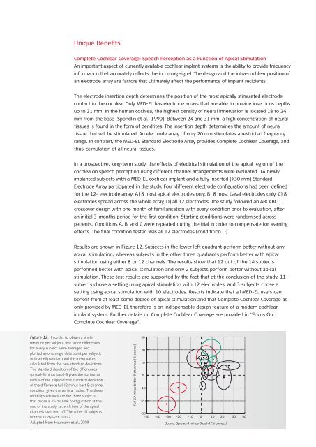
Figure 12: In order to obtain a single<br />
measure per subject, test score differences<br />
for every subject were averaged and<br />
plotted as one single data point per subject,<br />
with an ellipsoid around the mean value,<br />
calculated from the two standard deviations.<br />
The standard deviation of the differences<br />
spread-8 minus basal-8 gives the horizontal<br />
radius of the ellipsoid; the standard deviation<br />
of the difference full-12 minus best 8-channel<br />
condition gives the vertical radius. The three<br />
red ellipsoids indicate the three subjects<br />
that chose a 10-channel configuration at the<br />
end of the study, i.e. with two of the apical<br />
channels switched off. The other 11 subjects<br />
left the study with full-12.<br />
Adapted from Haumann et al., 2009.<br />
8<br />
Complete <strong>Cochlear</strong> Coverage: Speech Perception as a Function of Apical Stimulation<br />
An important aspect of currently available cochlear implant systems is the ability to provide frequency<br />
information that accurately reflects the incoming signal. The design and the intra‑cochlear position of<br />
an electrode array are factors that ultimately affect the performance of implant recipients.<br />
The electrode insertion depth determines the position of the most apically stimulated electrode<br />
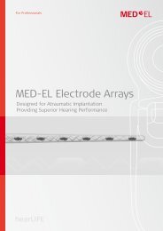
contact in the cochlea. Only <strong>MED</strong>‑<strong>EL</strong> has electrode arrays that are able to provide insertions depths<br />
up to 31 mm. In the human cochlea, the highest density of neural innervation is located 18 to 24<br />
mm from the base (Spöndlin et al., 1990). Between 24 and 31 mm, a high concentration of neural<br />
tissues is found in the form of dendrites. The insertion depth determines the amount of neural<br />
tissue that will be stimulated. An electrode array of only 20 mm stimulates a restricted frequency<br />
range. In contrast, the <strong>MED</strong>‑<strong>EL</strong> Standard Electrode Array provides Complete <strong>Cochlear</strong> Coverage, and<br />
thus, stimulation of all neural tissues.<br />
In a prospective, long‑term study, the effects of electrical stimulation of the apical region of the<br />
cochlea on speech perception using different channel arrangements were evaluated. 14 newly<br />
implanted subjects with a <strong>MED</strong>‑<strong>EL</strong> cochlear implant and a fully inserted (>30 mm) Standard<br />
Electrode Array participated in the study. Four different electrode configurations had been defined<br />
for the 12‑ electrode array: A) 8 most apical electrodes only, B) 8 most basal electrodes only, C) 8<br />
electrodes spread across the whole array, D) all 12 electrodes. The study followed an ABCABCD<br />
crossover design with one month of familiarisation with every condition prior to evaluation, after<br />
an initial 3‑months period for the first condition. Starting conditions were randomised across<br />
patients. Conditions A, B, and C were repeated during the trial in order to compensate for learning<br />
effects. The final condition tested was all 12 electrodes (conditition D).<br />
Results are shown in Figure 12. Subjects in the lower left quadrant perform better without any<br />
apical stimulation, whereas subjects in the other three quadrants perform better with apical<br />
stimulation using either 8 or 12 channels. The results show that 12 out of the 14 subjects<br />
performed better with apical stimulation and only 2 subjects perform better without apical<br />
stimulation. These test results are supported by the fact that at the conclusion of the study, 11<br />
subjects chose a setting using apical stimulation with 12 electrodes, and 3 subjects chose a<br />
setting using apical stimulation with 10 electrodes. Results indicate that all <strong>MED</strong>‑<strong>EL</strong> users can<br />
benefit from at least some degree of apical stimulation and that Complete <strong>Cochlear</strong> Coverage as<br />
only provided by <strong>MED</strong>‑<strong>EL</strong> therefore is an indispensable design feature of a modern cochlear<br />
implant system. Further details on Complete <strong>Cochlear</strong> Coverage are provided in “Focus On:<br />
Complete <strong>Cochlear</strong> Coverage”.<br />
Full-12 minus better 8-channels [% correct]<br />
30<br />
20<br />
10<br />
0<br />
-10<br />
-20<br />
-30<br />
-50 -40 -30 -20 -10 0 10 20 30 40<br />
Scores: Spread-8 minus Basal-8 [% correct]