Models of venous admixture
Models of venous admixture
Models of venous admixture

You also want an ePaper? Increase the reach of your titles
YUMPU automatically turns print PDFs into web optimized ePapers that Google loves.
<strong>Models</strong> <strong>of</strong> <strong>venous</strong> <strong>admixture</strong>Paul E. BigeleisenAdvan Physiol Educ 25:159-166, 2001.You might find this additional information useful...Medline items on this article's topics can be found at http://highwire.stanford.edu/lists/artbytopic.dtlon the following topics:Medicine .. Medical StudentsUpdated information and services including high-resolution figures, can be found at:http://ajpadvan.physiology.org/cgi/content/full/25/3/159Additional material and information about Advances in Physiology Education can be found at:http://www.the-aps.org/publications/advanThis information is current as <strong>of</strong> December 7, 2006 .Downloaded from ajpadvan.physiology.org on December 7, 2006Advances in Physiology Education is dedicated to the improvement <strong>of</strong> teaching and learning physiology, both in specializedcourses and in the broader context <strong>of</strong> general biology education. It is published four times a year in March, June, September andDecember by the American Physiological Society, 9650 Rockville Pike, Bethesda MD 20814-3991. Copyright © 2005 by theAmerican Physiological Society. ISSN: 1043-4046, ESSN: 1522-1229. Visit our website at http://www.the-aps.org/.
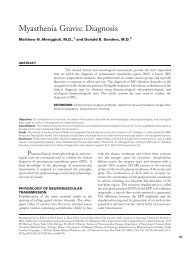
I N N O V A T I O N S A N D I D E A SMODELS OF VENOUS ADMIXTUREPaul E. BigeleisenDepartment <strong>of</strong> Anesthesiology, Strong Memorial Hospital,Rochester, New York 14642Medical students, residents, and allied health pr<strong>of</strong>essionals <strong>of</strong>ten have difficultyquantitating ventilation-perfusion mismatch in ill patients. Thismanuscript quantitates ventilation-perfusion mismatch using the underlyingphysiological concepts and equations that describe mismatch. In addition, clinicalproblems with diagrams and worked-out solutions are supplied to help studentsmaster these equations as well as their practical limitations.ADV PHYSIOL EDUC 25: 159–166, 2001.Key words: ventilationWhen patients have hypoxemia due to ventilationperfusionmismatch, clinicians need quantitativemethods to measure the severity <strong>of</strong> illness as afunction <strong>of</strong> time. Medical students, residents, andallied health pr<strong>of</strong>essionals usually have some familiaritywith the virtual shunt equation, shunt nomogram,alveolar-arterial gradient, and arterial/alveolarratio. These techniques are all commonly used toassess respiratory failure. Nonetheless, students <strong>of</strong>tenhave difficulty quantitating these measures anddo not understand when these methods fail. Standardtextbooks may provide the theoretical backgroundfor the material above, but they rarely provideclinical examples to solidify these concepts (1,3, 4). This manuscript presents the derivation <strong>of</strong>equations describing ventilation-perfusion mismatch.In addition, clinical examples with workedsolutions are provided to help students learn tomanipulate the formulas necessary to assess respiratoryfailure. The practical limitations <strong>of</strong> theseformulas are also discussed.MODELThe formal method <strong>of</strong> modeling pulmonary dysfunctionin a hypoxemic patient is the patient’s virtualshunt through his lungs. Figures 1 and 2 and Equations1-3 explain how this is done.Figure 1A shows a healthy alveolus and pulmonarycapillary. In this part <strong>of</strong> the lung, ventilation andperfusion are matched, and there is no <strong>venous</strong> <strong>admixture</strong>.Figure 1B shows a collapsed alveolus and ahealthy capillary. Here, all <strong>of</strong> the mixed <strong>venous</strong> bloodis shunted past the functional part <strong>of</strong> the lung. This iscalled <strong>venous</strong> <strong>admixture</strong>. Figure 1C shows a healthyalveolus and collapsed pulmonary capillary. This isthe definition <strong>of</strong> dead space. It is included here toavoid confusion, although it is not part <strong>of</strong> the model.Figure 2 shows how <strong>venous</strong> <strong>admixture</strong> to the systemicarterial blood can occur.Conservation <strong>of</strong> blood flow dictates that the pulmonarycapillary blood flow and shunted blood flow must equalthe total cardiac output through the lungs (Eq. 1)Q˙ T Q˙ C Q˙ S (1)where Q˙ T, Q˙ C, and Q˙ S are cardiac output, pulmonarycapillary blood flow, and shunted pulmonary capillaryblood flow, respectively.Conservation <strong>of</strong> mass dictates that the transport <strong>of</strong>oxygen through the lung is also conserved (Eq. 2)Q˙ TCa O2 Q˙ cC c O 2 Q˙ sC v O 2 (2)Downloaded from ajpadvan.physiology.org on December 7, 20061043 - 4046 / 01 – $5.00 – COPYRIGHT © 2001 THE AMERICAN PHYSIOLOGICAL SOCIETYVOLUME 25 : NUMBER 3 – ADVANCES IN PHYSIOLOGY EDUCATION – SEPTEMBER 2001159
I N N O V A T I O N S A N D I D E A SFIG. 1.Oxygen and blood flow in the lung. Cap, pulmonary capillary.Downloaded from ajpadvan.physiology.org on December 7, 2006FIG. 2.Virtual shunt model.where Ca O2is the O 2 content in the systemicarterial blood (pulmonary vein), C c O 2 is theO 2 content in the pulmonary capillary, and C v O 2is the O 2 content in the shunted or mixed<strong>venous</strong> blood. Substituting Eq. 1 into Eq. 2 andrearranging yields the familiar virtual shunt equation.Q˙ SQ˙ T C cO 2 C a O 2C c O 2 C v O 2(3)VOLUME 25 : NUMBER 3 – ADVANCES IN PHYSIOLOGY EDUCATION – SEPTEMBER 2001160
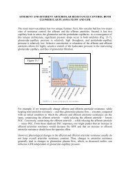
I N N O V A T I O N S A N D I D E A SFIG. 3.Pa O2 , arterial oxygen pressure in Torr; PA O2 , alveolar oxygen pressure in Torr; isoshunt,isoshunt lines in %.To calculate shunt (Eq. 3), one must measure thepartial pressure <strong>of</strong> O 2 in the mixed <strong>venous</strong> and systemicarterial blood. In addition, one must estimatethe partial pressure <strong>of</strong> O 2 in the pulmonary capillaryby calculating the partial pressure <strong>of</strong> oxygen deliveredto the alveolus (PA O2; Eq. 3A)PA O2 PB PH 2 O FI O2 Pa CO 2R(3A)where PB is atmospheric pressure, and PH 2 Oisthepressure <strong>of</strong> water vapor in the lung. Pa CO2 is thepartial pressure <strong>of</strong> CO 2 in the systemic arterial blood,and R is the respiratory quotient, which is assumed tobe 1 in this model. The use <strong>of</strong> Eq. 3 is impractical inroutine clinical care because the mixed <strong>venous</strong> bloodis rarely sampled.This nomogram allows one to estimate the patient’svirtual shunt and follow his pulmonary dysfunction ashis FI O2and ventillatory therapy are adjusted by measuringhis Pa O2. Despite their simplicity, the isoshuntlines did not achieve popularity because pulmonarycritical care involves treating more than a single parameterand because it was impractical to carry thenomogram around.A number <strong>of</strong> years later, interest was renewed in Eq.3 with the advent <strong>of</strong> pulse oximeters and oxymetricpulmonary artery catheters. Manipulation <strong>of</strong> Eq. 3shows why. The oxygen content <strong>of</strong> blood is given byEq. 4CO 2 1.34 Hgb saturation 0.003 PO 2 (4)Downloaded from ajpadvan.physiology.org on December 7, 2006To get around this, Eq. 3 was recast in nomogramform. Nunn assumed that the C v O 2 had a constantvalue and that the hemoglobin and Pa CO2 were withincommon ranges (2). Using these assumptions, he createdthe isoshunt lines shown in Fig. 3.where PO 2 is the partial pressure <strong>of</strong> oxygen in theblood. The term (0.003 PO 2 ) represents dissolved O 2in the plasma and will be ignored here. If we assumethat the amount <strong>of</strong> dissolved oxygen is negligible,then we can substitute Eq. 4 into Eq. 3 to yield.VOLUME 25 : NUMBER 3 – ADVANCES IN PHYSIOLOGY EDUCATION – SEPTEMBER 2001161
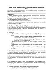
I N N O V A T I O N S A N D I D E A SQ˙ SQ T1.34 Hgb end-capillary sat arterial sat1.34 Hgb end-capillary sat mixed <strong>venous</strong> sat(5)With oximetry, both the arterial saturation and mixed<strong>venous</strong> saturation could be measured continuously togive a good estimate <strong>of</strong> shunt. Unfortunately, Eq. 5still involved a cumbersome calculation and the costand morbidity <strong>of</strong> an oxymetric pulmonary artery catheter.For these reasons, Eq. 5 never achieved commonusage either. Eq. 5, however, can be simplified tomake it useful for clinicians.In patients with stable cardiac outputs who are administereda high concentration <strong>of</strong> O 2 (FI O2), the differencebetween end-capillary saturation and mixed<strong>venous</strong> saturation is 0.25. Thus Eq. 5 becomesQ˙ SQ˙ Tend-capillary sat arterial sat0.25(6)Equation 6 is made usable by relating O 2 saturation toPa O2. Reference to the oxyhemoglobin dissociationcurve shows how this is done (Fig. 4).AtaPa O2100 Torr, the graph is nearly a flat line.Thus O 2 saturation and Pa O2are related by the formulafor a straight line (Eq. 7)O 2 saturation m Pa O2 b (7)where m is the slope <strong>of</strong> the line, and b is the intercepton the saturation axis. Substituting Eq. 7 into Eq. 6yieldsQ˙ SQ˙ T mPA O 2 Pa O2 0.25(8)where we have assumed that the PA O2is the same asPO 2 in the pulmonary capillary. Using the slope (m 1.4 10 4 saturation/Torr) on the flattest part <strong>of</strong> Fig.4 allows Eq. 8 to be recast asQ˙ SQ˙ T 1.4 104 PA O2 Pa O2 0.25(9)FIG. 4.Oxyhemoglobin dissociation curve.If we desire to use shunt fraction rather than shunt,then Eq. 9 becomesShunt fraction Q˙ SQ˙ T 100 PAO 2 PaO 218(10)Equation 10 shows that the commonly used alveolararterialgradient divided by a constant yields an easyestimate <strong>of</strong> shunt when the patient’s cardiac outputand hemoglobin are stable and when the PA O2andPa O2lie along the flat part <strong>of</strong> the oxyhemoglobindissociation curve. Using a similar approach, theshunt fraction can be estimated when the PA O2100 Torr and the Pa O2lies between 50 and 100 Torr.In this case, Eq. 10 becomesShunt fraction PAO 2 PaO 22(10A)Equations 10 and 10A are only estimates <strong>of</strong> shuntfraction and are insensitive to larger gradients in thedenominator <strong>of</strong> Eq. 5. Larger gradients in the denominator<strong>of</strong> Eq. 5 may occur in some patients withdecreased cardiac output or significant shunt. Nonetheless,as long as cardiac output is stable, Eqs. 10 and10A will track changes in shunt fraction as the patient’spulmonary function improves or declines. Ifone wishes to account for larger gradients in Eq. 5,then the slope in Eq. 7 must be adjusted to fit Eqs. 10and 10A to clinical data.The following clinical cases below are illustrative:An 18-yr-old male with a history <strong>of</strong> asthma presents tothe emergency department in respiratory distress. Anasal cannula supplies oxygen at 28%. His blood gasshows the following values: 7.32/31/74/23, O 2 saturation95%. In this case, his alveolar O 2 (PA O2) is givenDownloaded from ajpadvan.physiology.org on December 7, 2006VOLUME 25 : NUMBER 3 – ADVANCES IN PHYSIOLOGY EDUCATION – SEPTEMBER 2001162
I N N O V A T I O N S A N D I D E A STABLE 1FI O2 PA O2 Pa O2 A-a Gradient Virtual Shunt Estimated ShuntPatient 1 (asthma) 0.28 168 74 94 15%–20% 6%Patient 2 (surgery) 0.5 322 115 207 10%–15% 12%by Eq. 3A. Thus PA O2 (760 47) 0.28 31 168Torr. The patients’ alveolar-arterial gradient is givenby PA O2 [Pa O2] 168 74 94 Torr. Reference toFig. 3 shows the patient has a virtual shunt <strong>of</strong> 15–20%.His estimated shunt using the PA O2-Pa O2gradient (Eq.10) is6%.Now consider a very different patient. A 74-yr-oldmale with a 50-pack/yr history <strong>of</strong> cigarettes undergoesa Whipple procedure. At the end <strong>of</strong> the surgery,the patient is intubated and ventilated with an FI O2<strong>of</strong>50%. His blood gas shows the following values: 7.32/34/115/27, O 2 saturation 98%. Using the same analysisas above, we find that this patient has PA O2-Pa O2gradient <strong>of</strong> 207 Torr, a virtual shunt <strong>of</strong> 10–15%, andestimated shunt using the PA O2-Pa O2gradient (Eq. 10)<strong>of</strong> 12%. This is summarized in Table 1.Notice that patient 1 (asthma) is sicker (virtualshunt 15–20%) even though his PA O2-Pa O2gradientis smaller than patient 2. This shows the value<strong>of</strong> the virtual shunt method, i.e., virtual shunt remainsconstant regardless <strong>of</strong> the FI O2administered.The PA O2-Pa O2gradient, however, widens as the FI O2is increased, even in the absence <strong>of</strong> increased pulmonarydysfunction. The estimated shunt <strong>of</strong> patient1 (Eq. 10) is 6%, which compares poorly withhis virtual shunt (Eq. 3). This is because his Pa O2liesalong the steep part <strong>of</strong> the oxyhemoglobin dissociationcurve. In this situation, Eq. 10 is not a goodmeasure <strong>of</strong> <strong>venous</strong> <strong>admixture</strong>. Conversely, the estimatedshunt <strong>of</strong> patient 2 (12%) is similar to hisvirtual shunt. This is because both his PA O2and Pa O2lie along the flat part <strong>of</strong> the oxyhemoglobin dissociationcurve (Fig. 4).Figure 5 shows the relationship between virtualshunt and estimated shunt. Notice that when thePa O2is greater than 100 Torr, the virtual shunt linesand estimated shunt lines have slopes and absolutevalues that are similar. Below 100 Torr, the estimatedshunt (PA O2 Pa O2) reproduces the virtualshunt poorly.Now let’s look at the problem in a different way.Suppose we consider the right heart and lungs to bean engine whose job is to convert mixed <strong>venous</strong>blood into oxygenated blood. If the engine is ideal(Fig. 6A), then the blood will be fully saturated withoxygen when it leaves the engine.If the engine is less than ideal (Fig. 6B), then theblood will be only partially oxygenated when it leavesthe heart-lung engine. We can calculate an efficiencyfor this real engine by comparing it with the idealengine (Eq. 11).Efficiency O 2 content <strong>of</strong> partially oxygenated bloodO 2 content <strong>of</strong> fully oxygenated bloodSubstituting Eq. 4 into Eq. 11 yields1.34 Hgb arterial satEfficiency 1.34 Hgb end-capillary sat(11)(12)Now, recall that on the flat part <strong>of</strong> the oxyhemoglobindissociation curve (Fig. 4), the O 2 saturation m PO 2 b (Eq. 7). Substituting Eq. 7 into Eq. 11 yieldsEfficiency PaO 2PAO 2(13)This is commonly called the a/A ratio. Our derivationshows that the a/A ratio is only a good measure <strong>of</strong>pulmonary dysfunction (<strong>venous</strong> <strong>admixture</strong>) when thePa O2and PA O2both lie along the flat part <strong>of</strong> the oxyhemoglobindissociation curve.Figure 7 shows the graphical relationship <strong>of</strong> Pa O2/PA O2to the isoshunt lines. When the Pa O2-PA O2ratio is near1, isoshunt and Pa O2/PA O2correlate well. As the ratioDownloaded from ajpadvan.physiology.org on December 7, 2006VOLUME 25 : NUMBER 3 – ADVANCES IN PHYSIOLOGY EDUCATION – SEPTEMBER 2001163
I N N O V A T I O N S A N D I D E A SFIG. 5.A-a gradient, estimated shunt fraction; isoshunt, isoshunt lines in percent.falls, the correlation becomes poorer because thePa O2no longer lies along the flat part <strong>of</strong> the oxyhemoglobindissociation curve.As an example, consider patients 1 and 2 again. Theefficiency <strong>of</strong> patient 1 (Pa O2/PA O2) is 0.44, whereasthat <strong>of</strong> patient 2 is 0.36. By comparing a/A ratios,patient 1 appears healthier than patient 2, althoughhis virtual shunt is greater. This is because his Pa O2liesalong the steep part <strong>of</strong> the oxyhemoglobin dissociationcurve, and the a/A ratio is less accurate in thiscase. This is summarized in Table 2.Downloaded from ajpadvan.physiology.org on December 7, 2006FIG. 6.Heart-lung engine model.VOLUME 25 : NUMBER 3 – ADVANCES IN PHYSIOLOGY EDUCATION – SEPTEMBER 2001164
I N N O V A T I O N S A N D I D E A SSUMMARYVirtual shunt remains the best way <strong>of</strong> quantitating<strong>venous</strong> <strong>admixture</strong> in patients with pulmonary pathology.It is a cumbersome technique that requirespulmonary artery catheterization and data from theoxyhemoglobin dissociation curve. The isoshuntlines approximate virtual shunt when the mixed<strong>venous</strong> saturation, hemoglobin, and Pa CO2 arewithin common ranges and are stable. Most cliniciansdo not carry this nomogram with them. TheA-a gradient and a/A ratio both give a good estimate<strong>of</strong> <strong>venous</strong> <strong>admixture</strong> when the conditions for theisoshunt lines are met and when both Pa O2and PA O2FIG. 7.a-A ratio, arterial-to-alveolar oxygen pressure ratio.lie along the flat part <strong>of</strong> the oxyhemoglobin dissociationcurve. Pa O2is easily measured, and PA O2iseasily calculated. For these reasons, they have becomethe standards in clinical care.PRACTICE PROBLEMAn 18-mo-old child presents to the emergency departmentwith respiratory distress and a chestX-ray consistent with pneumonia. A facemask suppliesoxygen at 35%. His arterial blood gas showsthe following values: 7.31/29/67/23, O 2 saturation93%.Downloaded from ajpadvan.physiology.org on December 7, 2006TABLE 2FI O2 PA O2 Pa O2 A-a Gradient Virtual Shunt Estimated Shunt a/APatient 1 (asthma) 0.28 168 74 94 15%–20% 6% 0.44Patient 2 (surgery) 0.5 322 115 207 10%–15% 12% 0.36VOLUME 25 : NUMBER 3 – ADVANCES IN PHYSIOLOGY EDUCATION – SEPTEMBER 2001165
I N N O V A T I O N S A N D I D E A S1) Use Eq. 3A to calculate the patient’s alveolar O 2(PAO 2 ). Answer, 220 Torr.2) Calculate the patient’s alveolar arterial gradient,i.e., PAO 2 Pa O2. Answer, 153 Torr.3) Use Fig. 3 to estimate the patient’s virtual shunt.Answer, 25%.4) Now use Eq. 10 to estimate the patient’s shunt.Answer, 9%. Can you explain why Eq. 10 gives sucha poor estimate <strong>of</strong> virtual shunt? Hint, see the discussion<strong>of</strong> patient 1.5) Now use Eq. 19 to calculate the patient’s arterialalveolarratio. Answer, 0.3. Does Eq. 13 overestimateor underestimate the patient’s illness and why?Hint, see the discussion <strong>of</strong> patient 1.Address for reprint requests and other correspondence: P. E. Bigeleisen,Dept. <strong>of</strong> Anesthesiology, Box 604, Strong Memorial Hospital,601 Elmwood Ave., Rochester, NY 14642 (E-mail: Paul_Bigeleisen@urmc.rochester.edu).Received 12 November 2000; accepted in final form 4 May 2001REFERENCES1. Levitzky MG. Pulmonary Physiology. New York: McGraw-Hill,1999, p. 113–128.2. Lumb AB. Nunn’s Applied Respiratory Physiology. Oxford:Butterworth Heinemann, 2000, p. 249–298.3. Shapiro BA, Harrison A, Kacmarek RM, and Cane RD.Clinical Applications <strong>of</strong> Respiratory Care. Chicago, IL: YearBook, 1985, p. 52–61.4. West JB. Respiratory Physiology. Baltimore, MD: Williams &Wilkins, 1985, p. 49–68.Downloaded from ajpadvan.physiology.org on December 7, 2006VOLUME 25 : NUMBER 3 – ADVANCES IN PHYSIOLOGY EDUCATION – SEPTEMBER 2001166