A default mode of brain function: A brief history of an evolving idea
A default mode of brain function: A brief history of an evolving idea
A default mode of brain function: A brief history of an evolving idea

You also want an ePaper? Increase the reach of your titles
YUMPU automatically turns print PDFs into web optimized ePapers that Google loves.
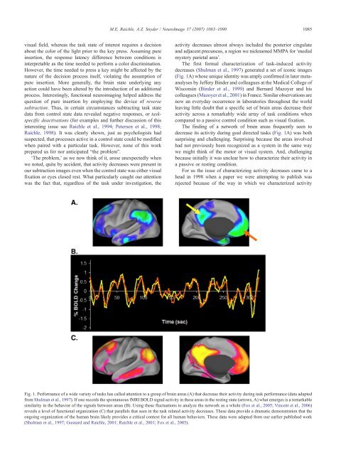
M.E. Raichle, A.Z. Snyder / NeuroImage 37 (2007) 1083–10901085visual field, whereas the task state <strong>of</strong> interest requires a decisionabout the color <strong>of</strong> the light prior to the key press. Assuming pureinsertion, the response latency difference between conditions isinterpretable as the time needed to perform a color discrimination.However, the time needed to press a key might be affected by thenature <strong>of</strong> the decision process itself, violating the assumption <strong>of</strong>pure insertion. More generally, the <strong>brain</strong> state underlying <strong>an</strong>yaction could have been altered by the introduction <strong>of</strong> <strong>an</strong> additionalprocess. Interestingly, <strong>function</strong>al neuroimaging helped address thequestion <strong>of</strong> pure insertion by employing the device <strong>of</strong> reversesubtraction. Thus, in certain circumst<strong>an</strong>ces subtracting task statedata from control state data revealed negative responses, or taskspecificdeactivations (for examples <strong>an</strong>d further discussion <strong>of</strong> thisinteresting issue see Raichle et al., 1994; Petersen et al., 1998;Raichle, 1998). It was clearly shown, just as psychologists hadsuspected, that processes active in a control state could be modifiedwhen paired with a particular task. However, none <strong>of</strong> this workprepared us for nor <strong>an</strong>ticipated “the problem”.‘The problem,’ as we now think <strong>of</strong> it, arose unexpectedly whenwe noted, quite by accident, that activity decreases were present inour subtraction images even when the control state was either visualfixation or eyes closed rest. What particularly caught our attentionwas the fact that, regardless <strong>of</strong> the task under investigation, theactivity decreases almost always included the posterior cingulate<strong>an</strong>d adjacent precuneus, a region we nicknamed MMPA for ‘medialmystery parietal area’.The first formal characterization <strong>of</strong> task-induced activitydecreases (Shulm<strong>an</strong> et al., 1997) generated a set <strong>of</strong> iconic images(Fig. 1A) whose unique identity was amply confirmed in later meta<strong>an</strong>alysesby Jeffery Binder <strong>an</strong>d colleagues at the Medical College <strong>of</strong>Wisconsin (Binder et al., 1999) <strong>an</strong>d Bernard Mazoyer <strong>an</strong>d hiscolleagues (Mazoyer et al., 2001) in Fr<strong>an</strong>ce. Similar observations arenow <strong>an</strong> everyday occurrence in laboratories throughout the worldleaving little doubt that a specific set <strong>of</strong> <strong>brain</strong> areas decrease theiractivity across a remarkably wide array <strong>of</strong> task conditions whencompared to a passive control condition such as visual fixation.The finding <strong>of</strong> a network <strong>of</strong> <strong>brain</strong> areas frequently seen todecrease its activity during goal directed tasks (Fig. 1A) was bothsurprising <strong>an</strong>d challenging. Surprising because the areas involvedhad not previously been recognized as a system in the same waywe might think <strong>of</strong> the motor or visual system. And, challengingbecause initially it was unclear how to characterize their activity ina passive or resting condition.For us the issue <strong>of</strong> characterizing activity decreases came to ahead in 1998 when a paper we were attempting to publish wasrejected because <strong>of</strong> the way in which we characterized activityFig. 1. Perform<strong>an</strong>ce <strong>of</strong> a wide variety <strong>of</strong> tasks has called attention to a group <strong>of</strong> <strong>brain</strong> areas (A) that decrease their activity during task perform<strong>an</strong>ce (data adaptedfrom Shulm<strong>an</strong> et al., 1997). If one records the spont<strong>an</strong>eous fMRI BOLD signal activity in these areas in the resting state (arrows, A) what emerges is a remarkablesimilarity in the behavior <strong>of</strong> the signals between areas (B). Using these fluctuations to <strong>an</strong>alyze the network as a whole (Fox et al., 2005; Vincent et al., 2006)reveals a level <strong>of</strong> <strong>function</strong>al org<strong>an</strong>ization (C) that parallels that seen in the task related activity decreases. These data provide a dramatic demonstration that theongoing org<strong>an</strong>ization <strong>of</strong> the hum<strong>an</strong> <strong>brain</strong> likely provides a critical context for all hum<strong>an</strong> behaviors. These data were adapted from our earlier published work(Shulm<strong>an</strong> et al., 1997; Gusnard <strong>an</strong>d Raichle, 2001; Raichle et al., 2001; Fox et al., 2005).