July 2005 - The Hong Kong College of Anaesthesiologists
July 2005 - The Hong Kong College of Anaesthesiologists
July 2005 - The Hong Kong College of Anaesthesiologists
Create successful ePaper yourself
Turn your PDF publications into a flip-book with our unique Google optimized e-Paper software.
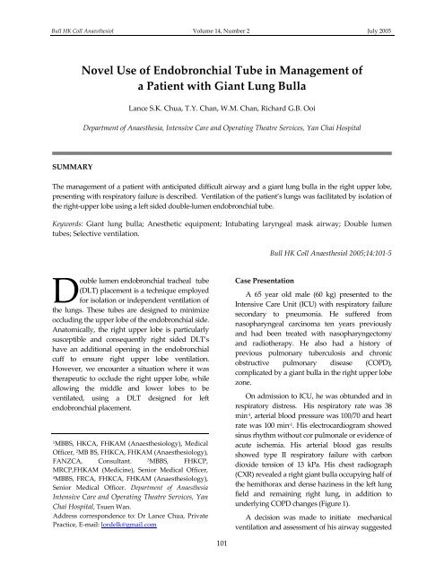
Bull HK Coll Anaesthesiol Volume 14, Number 2 <strong>July</strong> <strong>2005</strong>Novel Use <strong>of</strong> Endobronchial Tube in Management <strong>of</strong>a Patient with Giant Lung BullaLance S.K. Chua, T.Y. Chan, W.M. Chan, Richard G.B. OoiDepartment <strong>of</strong> Anaesthesia, Intensive Care and Operating <strong>The</strong>atre Services, Yan Chai HospitalSUMMARY<strong>The</strong> management <strong>of</strong> a patient with anticipated difficult airway and a giant lung bulla in the right upper lobe,presenting with respiratory failure is described. Ventilation <strong>of</strong> the patient’s lungs was facilitated by isolation <strong>of</strong>the right‐upper lobe using a left sided double‐lumen endobronchial tube.Keywords: Giant lung bulla; Anesthetic equipment; Intubating laryngeal mask airway; Double lumentubes; Selective ventilation.Bull HK Coll Anaesthesiol <strong>2005</strong>;14:101‐5Double lumen endobronchial tracheal tube(DLT) placement is a technique employedfor isolation or independent ventilation <strong>of</strong>the lungs. <strong>The</strong>se tubes are designed to minimizeoccluding the upper lobe <strong>of</strong> the endobronchial side.Anatomically, the right upper lobe is particularlysusceptible and consequently right sided DLT’shave an additional opening in the endobronchialcuff to ensure right upper lobe ventilation.However, we encounter a situation where it wastherapeutic to occlude the right upper lobe, whileallowing the middle and lower lobes to beventilated, using a DLT designed for leftendobronchial placement.1MBBS, HKCA, FHKAM (Anaesthesiology), MedicalOfficer, 2 MB BS, FHKCA, FHKAM (Anaesthesiology),FANZCA, Consultant. 3MBBS, FHKCP,MRCP,FHKAM (Medicine), Senior Medical Officer,4MBBS, FRCA, FHKCA, FHKAM (Anaesthesiology),Senior Medical Officer. Department <strong>of</strong> AnaesthesiaIntensive Care and Operating <strong>The</strong>atre Services, YanChai Hospital, Tsuen Wan.Address correspondence to: Dr Lance Chua, PrivatePractice, E‐mail: lordelk@gmail.comCase PresentationA 65 year old male (60 kg) presented to theIntensive Care Unit (ICU) with respiratory failuresecondary to pneumonia. He suffered fromnasopharyngeal carcinoma ten years previouslyand had been treated with nasopharyngectomyand radiotherapy. He also had a history <strong>of</strong>previous pulmonary tuberculosis and chronicobstructive pulmonary disease (COPD),complicated by a giant bulla in the right upper lobezone.On admission to ICU, he was obtunded and inrespiratory distress. His respiratory rate was 38min ‐1 , arterial blood pressure was 100/70 and heartrate was 100 min ‐1 . His electrocardiogram showedsinus rhythm without cor pulmonale or evidence <strong>of</strong>acute ischemia. His arterial blood gas resultsshowed type II respiratory failure with carbondioxide tension <strong>of</strong> 13 kPa. His chest radiograph(CXR) revealed a right giant bulla occupying half <strong>of</strong>the hemithorax and dense haziness in the left lungfield and remaining right lung, in addition tounderlying COPD changes (Figure 1).A decision was made to initiate mechanicalventilation and assessment <strong>of</strong> his airway suggested101


