Diagnostic Testing - The Chester County Hospital
Diagnostic Testing - The Chester County Hospital
Diagnostic Testing - The Chester County Hospital

You also want an ePaper? Increase the reach of your titles
YUMPU automatically turns print PDFs into web optimized ePapers that Google loves.
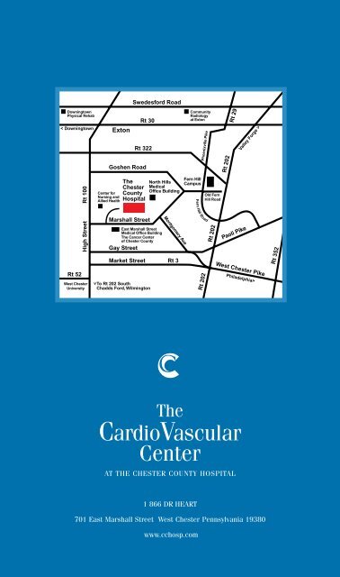
Swedesford RoadDowningtownPhysical Rehab< DowningtownRt 30ExtonRt 322Goshen RoadCommunityRadiologyat ExtonPhoenixville PikeRt 202Rt 29Valley Forge >High Street Rt 100Center forNursing andAllied Health<strong>The</strong><strong>Chester</strong><strong>County</strong><strong>Hospital</strong>Marshall StreetEast Marshall StreetMedical Office Building<strong>The</strong> Cancer Centerof <strong>Chester</strong> <strong>County</strong>Gay StreetNorth HillsMedicalOffice BuildingMontgomery AveMarket Street Rt 3Fern HillCampusFern Hill RoadOld FernHill RoadRt 202Paoli PikeRt 352Rt 52West <strong>Chester</strong>UniversityTo Rt 202 SouthChadds Ford, Wilmington>Rt 202West <strong>Chester</strong> PikePhiladelphia><strong>The</strong>CardioVascularCenterAT THE CHESTER COUNTY HOSPITAL1 866 DR HEART701 East Marshall Street West <strong>Chester</strong> Pennsylvania 19380www.cchosp.com
P A T I E N T G U I D EDIAGNOSTIC CARDIOLOGY TESTING<strong>The</strong>CardioVascular CenterAT THE CHESTER COUNTY HOSPITAL
<strong>Diagnostic</strong> Cardiology <strong>Testing</strong>WELCOME 1ADULT AND PEDIATRIC ECHOCARDIOGRAM 1STRESS ECHOCARDIOGRAM 1DOBUTAMINE STRESS ECHOCARDIOGRAM 2HOLTER MONITORING 3TRANSESOPHAGEAL ECHOCARDIOGRAM (TEE) 3NUCLEAR CARDIOLOGY STRESS TEST 4PERSANTINE STRESS TEST 5WELCOMEThank you for choosing <strong>The</strong> CardioVascular Center at <strong>The</strong> <strong>Chester</strong> <strong>County</strong><strong>Hospital</strong> for your cardiology care. At <strong>The</strong> CardioVascular Center, our philosophyfocuses on patients and incorporates a new paradigm in the treatment ofcardiovascular disease. Our goal is the prevention and treatment of heartdisease as the patient experiences it, avoiding the segmentation anddiscontinuity of the traditional distinctions between medical and surgicaltherapies.We seek to assist our patients, their families and their referring physicians inthe continuum of cardiovascular care – from primary prevention to earlydiagnosis, timely intervention, aggressive rehabilitation and secondaryprevention.Your physician has recommended that you undergo diagnostic cardiologytesting to better evaluate the condition of your heart and vascular system.<strong>The</strong> following information is intended to explain what you can expect duringyour procedure.
ADULT AND PEDIATRIC ECHOCARDIOGRAMAt <strong>The</strong> CardioVascular Center at <strong>The</strong> <strong>Chester</strong> <strong>County</strong> <strong>Hospital</strong> in the friendly atmosphereof the Echo Lab you will experience a painless procedure known as an echocardiogram.During the test, a small microphone-like device called a transducer will send ultrasoundwaves that echo off various parts of your heart. <strong>The</strong> echoes are converted into a movingimage of the heart. This image is displayed on a screen for a cardiologist to review.<strong>The</strong> echocardiogram provides your physician with important information such as:> Size of the heart chambers> Heart valve function> <strong>The</strong> pumping action and blood flow of the heart<strong>The</strong>re are three parts to an echocardiogram, which include M-mode, 2-D, and colorDoppler. M-mode produces a tracing like image used for measuring heart chambers. 2-D shows the actual shape and motion of the heart. Color Doppler shows blood directionby color changes seen on the monitor. You will also hear swishing noises during the test,which is the sound of blood flowing through the heart.This test takes approximately 40 minutes and no special instructions prior to the test arenecessary. You will be asked to take everything off from the waist up and put on ahospital gown opening in the front. <strong>The</strong> echocardiography technologist will apply threeelectrodes to your chest and ask you to lie on your left side. A gel similar to Vaseline willbe applied to the transducer, and then the transducer is placed onto your chest. <strong>The</strong>technologist will apply some pressure on the transducer to obtain the heart images,which may result in minimal discomfort.><strong>The</strong> Echocardiography Laboratory (Echo Lab) is located on the second floor of the<strong>Hospital</strong>. A <strong>Chester</strong> <strong>County</strong> Cardiologist reads all echocardiograms. <strong>The</strong> results will bereported to your physician within three working days.Preparation Instructions: This test takes approximately 40 minutes and no specialinstructions prior to the test are necessary.EXERCISE STRESS ECHOCARDIOGRAMAn exercise stress echocardiogram is a diagnostic tool made up of two components:a stress test and an echocardiogram. During the stress test component of the test, youwill be walking on a treadmill. <strong>The</strong> treadmill will increase in speed and elevation atpredetermined times. During the echocardiogram portion of the test, you will be lying ona stretcher while ultrasound images of your heart are recorded.Prior to walking on the treadmill, the echocardiography technologist will obtain restingEKG, blood pressures and echocardiogram images. When the Cardiologist arrives in theEcho Lab, you will be asked to walk on the treadmill starting at a slow speed with a >DIAGNOSTIC CARDIOLOGY TESTING| 1
small hill. You must be able to walk on the treadmill long enough to increase you heartrate considerably. We will continue to monitor your progress with EKG and bloodpressures as you walk on the treadmill and increase your heart rate. At your peak heartrate, we will obtain an EKG and more echocardiography images. <strong>The</strong>re is only a 60-second window to obtain the peak heart rate echocardiogram images because yourheart rate starts to drop immediately. As soon as the treadmill stops you should movequickly to the stretcher. You may be asked to hold your breath on inhalation or exhalationwhen your echocardiogram images are obtained.><strong>The</strong> Echocardiography Laboratory is located on the second floor of the <strong>Hospital</strong>. You willbe informed if there is any abnormality following the test and the final report will be sentto your doctor within three working days.Preparation Instructions: Please wear comfortable walking shoes and a two piece outfitbecause you will be asked to wear a hospital gown during the test. No caffeine ornicotine 12 hours prior to the start of the test.DOBUTAMINE STRESS ECHOCARDIOGRAM<strong>The</strong> dobutamine stress echocardiogram is very similar to the exercise stressechocardiogram except an intravenous (IV) medication called Dobutamine is used toelevate your heart rate instead of you walking on the treadmill. You will be asked tochange into a hospital gown and preliminary echocardiogram images will be taken. <strong>The</strong>registered nurse in the Cardiology Department will assess you and start an intravenousline (IV). During this procedure, you will be placed on a cardiac monitor and your bloodpressure will be checked frequently. A small non-invasive clip that will be placed on yourfinger will monitor your oxygen saturation. Once the Cardiologist is in attendance, the IVDobutamine will be started. Echocardiogram images are obtained with every change inDobutamine dosage and also in the recovery phase. You will be monitored closely forany adverse symptoms during the procedure.><strong>The</strong> Echocardiography Laboratory is located on the second floor of the <strong>Hospital</strong>. You willbe informed if there is any abnormality following the test and the final report will be sentto your doctor within three working days.Preparation Instructions: To prepare for this test do not eat or drink anything for 4 hoursprior to the procedure. If you have been on a beta-blocker medication, please notify thestaff when your last dose was taken.2
HOLTER MONITORINGA Holter monitor is a test, which diagnoses abnormal heart rhythms and records yourEKG for 24 hours while you perform your normal daily activities. <strong>The</strong> Holter monitor isa small, portable recorder that is attached with 5 electrodes onto your chest. Prior toelectrode placement, your chest will be prepped with an abrasive alcohol pad removingthe skin oils to ensure adequate contact. If necessary, men’s chests will be shaved toapply the electrodes securely. <strong>The</strong> monitor is placed in a pouch that can be worn aroundthe waist or over the arm. You will be given a diary to document the times of any abnormalsymptoms that you experience while wearing the monitor. While wearing the monitor beaware of the electrode connections and reattach them if needed. Do not get any part ofthe monitor wet and please avoid electric blankets, metal detectors, magnets, and areasof high voltage as this may affect the results of your test. Following the 24 hour timeperiod, do not turn the monitor off before returning it to the Cardiology Department.><strong>The</strong> Holter monitor is applied in the Cardiology Department located at <strong>The</strong>CardioVascular Center on the second floor of the <strong>Hospital</strong>. Your Holter monitor tape isscanned by a trained cardiology staff member and reviewed by a Cardiologist. <strong>The</strong> finalresults are available in 3 to 5 working days.Preparation Instructions: <strong>The</strong> Holter monitor must be worn for 24 hours continuously soplease bathe or shower prior to arriving at the <strong>Hospital</strong>.TRANSESOPHAGEAL ECHOCARDIOGRAM (TEE)A transesophageal echocardiogram (TEE) is indicated if a normal transthoracicechocardiogram does not provide adequate images to obtain necessary information ofa patient’s cardiac anatomy. <strong>The</strong> TEE allows the cardiologist to view the heart posteriorlyin order to give more definition to the heart chambers and valves. This is done by insertinga long flexible tube into the mouth and down the esophagus. This tube or probe has atransducer at the end that sends ultrasound waves that echo off of the heart’s structures.As an outpatient, you will be admitted to the Ambulatory Care Center (ACC) prior to thetest. <strong>The</strong> ACC is located on the ground floor of the <strong>Hospital</strong>. In ACC, you will be assessedby a registered nurse and an intravenous line (IV) will be started. A member of theCardiology Department will arrive to transport you to the Echo Lab. Prior to the test, aCardiology registered nurse will place you on the monitoring equipment necessary for theprocedure. Electrodes will be placed on your chest to monitor your heart and a bloodpressure cuff will be placed on your arm. A small clip will be placed on your finger tomonitor the amount of oxygen in your blood and intravenous fluids will be started. <strong>The</strong>Cardiologist performing the test will obtain the consent for the test and then spray atopical anesthetic to the back of your throat to numb the area. <strong>The</strong> registered nurse willplace you on nasal oxygen and administer a sedative in the IV to help your relax. Onceyou are comfortable, the Cardiologist will insert the probe into your mouth. You will beasked to swallow and the probe will be directed into your esophagus. You will feel thetube moving but you should not be in pain. During this time, your vital signs will be >DIAGNOSTIC CARDIOLOGY TESTING| 3
monitored closely and your mouth will be suctioned.<strong>The</strong> TEE procedure takes about 30 minutes to complete. After the sedative is administeredyou will be monitored for approximately 30 minutes before returning to ACC.>This test is performed in the Echocardiography Laboratory on the second floor of the<strong>Hospital</strong> in the Cardiology Department. <strong>The</strong> Cardiologist performing the test will give youthe preliminary results before you leave the <strong>Hospital</strong>.Preparation Instructions: To prepare for the TEE do not eat or drink anything for 6 hoursbefore the procedure. Plan on having someone drive you home if you are having this testas an outpatient. If you have any difficulty swallowing, please notify your physician.NUCLEAR CARDIOLOGY STRESS TESTThis test is a screening tool for coronary artery disease. It involves the insertion of anintravenous (IV) catheter and an injection of a radioactive isotope before and during thetest. <strong>The</strong> isotope is a chemical element that when injected moves into the heart muscle.Prior to the stress or exercise portion of the test, you will be placed under the nuclearmedicine camera where resting images of your heart are obtained. At this time, you willbe laying flat and the camera will come in close to view your heart. <strong>The</strong>n you will be takento the stress test area. Your skin will be prepped with an abrasive alcohol pad and thenten EKG electrodes will be placed on your chest. If necessary, men’s chests will beshaved to ensure good electrode contact.For the stress portion of the test, the cardiac technician will take an initial EKG and bloodpressure. We will then ask you to move to the treadmill. A Cardiologist will be presentduring the test to observe your EKG. <strong>The</strong> treadmill will increase in speed and inclineevery three minutes and additional blood pressures will be taken. <strong>The</strong> staff will bemonitoring your symptoms continuously. It is necessary to notify the staff when you canonly walk one more minute because at that time you will again be injected with theradioactive isotope. This additional minute of exercise allows the isotope to circulate toyour heart muscle for an accurate test. After the stress test there is a short recoveryphase and then you will be asked to have a rest in the waiting room. To complete thestudy, you will be placed under the nuclear medicine camera for more cardiac imaging.><strong>The</strong> Nuclear Cardiology Stress Laboratory is located in the Nuclear Medicine Departmentadjacent to Radiology on the first floor of the <strong>Hospital</strong>. If there is any gross abnormalityin your test you will be notified following the stress portion. A Cardiologist following thesecond set of images will read the final results.Preparation Instructions: <strong>The</strong> nuclear cardiology stress test takes approximately threehours to perform. It is necessary to avoid caffeine for 12 hours prior to the test. Pleaseeat a light breakfast such as cereal with milk or a bagel and juice. You may bring a snackto eat following the test. Women, please wear a supportive bra. Remember to wearcomfortable walking shoes and a two piece outfit.4
PERSANTINE STRESS TESTThis is an alternative test for patients with potential coronary artery disease, who cannot exercise on a treadmill. This includes patients who have surgical, orthopedic, orpulmonary problems.When you arrive for your test, an intravenous (IV) will be started in your arm and you willbe given an injection of a radioactive isotope. <strong>The</strong> isotope is a chemical that wheninjected moves into the heart muscle. You will be asked to fill out a questionnaire and aconsent form for the procedure. <strong>The</strong>re will be images of your heart taken at rest by thegamma camera in the Nuclear Medicine Department. Following these images, you will beescorted to the Nuclear Stress Laboratory. Here your chest will be prepped with anabrasive alcohol pad for the placement of ten electrodes. A baseline EKG and bloodpressure will be obtained before the IV Persantine is given. <strong>The</strong> Persantine is injectedvery slowly while the cardiologist is observing your EKG. Persantine causes the coronaryarteries to dilate and therefore causes increased blood flow to the heart muscle. Whilethis medication is given you may experience a flushed feeling, chest pressure, shortnessof breath, nausea, headache or dizziness. <strong>The</strong>se side effects are normal but please letthe staff know how you are feeling because an additional medication can be given toreverse these effects. <strong>The</strong> radioactive isotope is again injected. Following this portion ofthe test, you will again be placed under the gamma camera for the "stress" images.><strong>The</strong> Nuclear Cardiology Stress Laboratory is located in the Nuclear Medicine Departmentadjacent to Radiology on the first floor of the <strong>Hospital</strong>. If there is any gross abnormalityin your test, you will be notified following the stress portion. A Cardiologist following thesecond set of images will read the final results.Preparation Instructions: <strong>The</strong> persantine stress test takes about 3 hours to perform.Please do not eat or drink for 4 hours prior to the test. Avoid caffeine for 24 hours beforethe test and do not take any medication containing <strong>The</strong>ophylline for 48 hours prior to thetest. If you have a history of asthma, emphysema or lung disease, please notify the staffbecause the test could cause difficulty with breathing.For more information, please contact:<strong>The</strong> CardioVascular Centerat <strong>The</strong> <strong>Chester</strong> <strong>County</strong> <strong>Hospital</strong> by calling1 866 DR HEARTDIAGNOSTIC CARDIOLOGY TESTING| 5