Genome-Wide Gene Expression Analysis ... - Academia Sinica
Genome-Wide Gene Expression Analysis ... - Academia Sinica
Genome-Wide Gene Expression Analysis ... - Academia Sinica
Create successful ePaper yourself
Turn your PDF publications into a flip-book with our unique Google optimized e-Paper software.
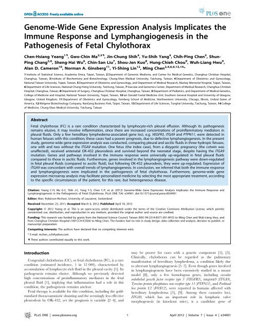
<strong>Genome</strong>-<strong>Wide</strong> <strong>Gene</strong> <strong>Expression</strong> <strong>Analysis</strong> Implicates theImmune Response and Lymphangiogenesis in thePathogenesis of Fetal ChylothoraxChen-Hsiang Yeang 1. , Gwo-Chin Ma 2,3. , Jin-Chung Shih 4 , Yu-Shih Yang 4 , Chih-Ping Chen 5 , Shun-Ping Chang 2,6 , Sheng-Hai Wu 6 , Chin-San Liu 7 , Shou-Jen Kuo 8 , Hung-Chieh Chou 9 , Wuh-Liang Hwu 9 ,Alan D. Cameron 10 , Norman A. Ginsberg 11 , Yi-Shing Lin 12 , Ming Chen 2,4,6,9,13,14 *1 Institute of Statistical Science, <strong>Academia</strong> <strong>Sinica</strong>, Taipei, Taiwan, 2 Department of Genomic Medicine, and Center for Medical <strong>Gene</strong>tics, Changhua Christian Hospital,Changhua, Taiwan, 3 Institute of Biochemistry and Biotechnology, Chung-Shan Medical University, Taichung, Taiwan, 4 Department of Obstetrics and Gynecology,National Taiwan University, Taipei, Taiwan, 5 Department of Obstetrics and Gynecology, and Department of Medical Research, Mackay Memorial Hospital, Taipei, Taiwan,6 Department of Life Sciences, National Chung-Hsing University, Taichung, Taiwan, 7 Vascular and Genomics Center, Department of Medical Research, Changhua ChristianHospital, Changhua, Taiwan, 8 Department of Surgery, Changhua Christian Hospital, Changhua, Taiwan, 9 Department of Pediatrics, and Department of Medical <strong>Gene</strong>tics,College of Medicine and Hospital, National Taiwan University, Taipei, Taiwan, 10 Ian Donald Foetal Medicine Unit, Southern <strong>Gene</strong>ral Hospital and University of Glasgow,Glasgow, United Kingdom, 11 Department of Obstetrics and Gynecology, Feinberg School of Medicine, Northwestern University, Chicago, Illinois, United States ofAmerica, 12 Welgene Biotechnology Company, NanGang Business Park, Taipei, Taiwan, 13 Department of Life Sciences, Tunghai University, Taichung, Taiwan, 14 Collegeof Medicine, Chung-Shan Medical University, Taichung, TaiwanAbstractFetal chylothorax (FC) is a rare condition characterized by lymphocyte-rich pleural effusion. Although its pathogenesisremains elusive, it may involve inflammation, since there are increased concentrations of proinflammatory mediators inpleural fluids. Only a few hereditary lymphedema-associated gene loci, e.g. VEGFR3, ITGA9 and PTPN11, were detected inhuman fetuses with this condition; these cases had a poorer prognosis, due to defective lymphangiogenesis. In the presentstudy, genome-wide gene expression analysis was conducted, comparing pleural and ascitic fluids in three hydropic fetuses,one with and two without the ITGA9 mutation. One fetus (the index case), from a dizygotic pregnancy (the cotwin wasunaffected), received antenatal OK-432 pleurodesis and survived beyond the neonatal stage, despite having the ITGA9mutation. <strong>Gene</strong>s and pathways involved in the immune response were universally up-regulated in fetal pleural fluidscompared to those in ascitic fluids. Furthermore, genes involved in the lymphangiogenesis pathway were down-regulatedin fetal pleural fluids (compared to ascitic fluid), but following OK-432 pleurodesis, they were up-regulated. <strong>Expression</strong> ofITGA9 was concordant with overall trends of lymphangiogenesis. In conclusion, we inferred that both the immune responseand lymphangiogenesis were implicated in the pathogenesis of fetal chylothorax. Furthermore, genome-wide geneexpression microarray analysis may facilitate personalized medicine by selecting the most appropriate treatment, accordingto the specific circumstances of the patient, for this rare, but heterogeneous disease.Citation: Yeang C-H, Ma G-C, Shih J-C, Yang Y-S, Chen C-P, et al. (2012) <strong>Genome</strong>-<strong>Wide</strong> <strong>Gene</strong> <strong>Expression</strong> <strong>Analysis</strong> Implicates the Immune Response andLymphangiogenesis in the Pathogenesis of Fetal Chylothorax. PLoS ONE 7(4): e34901. doi:10.1371/journal.pone.0034901Editor: Marc Robinson-Rechavi, University of Lausanne, SwitzerlandReceived November 23, 2011; Accepted March 6, 2012; Published April 18, 2012Copyright: ß 2012 Yeang et al. This is an open-access article distributed under the terms of the Creative Commons Attribution License, which permitsunrestricted use, distribution, and reproduction in any medium, provided the original author and source are credited.Funding: This research was funded by grants from the National Science Council, Taiwan (NSC-99-2314-B371-001-MY2) to Ming Chen and Wuh-Liang Hwu, andfrom Changhua Christian Hospital (100-CCH-ICO04) to Ming Chen. The funders had no role in study design, data collection and analysis, decision to publish, ormanuscript preparation.Competing Interests: The authors have declared that no competing interests exist.* E-mail: mchen_cch@yahoo.com. These authors contributed equally to this work.IntroductionCongenital chylothorax (CC), or fetal chylothorax (FC), is a rarecondition (estimated incidence, 1 in 12 000), characterized byaccumulation of lymphocyte-rich fluid in the pleural cavity [1]. Itspathogenesis remains elusive. Although we previously detectedhigh concentrations of proinflammatory mediators in the fetalpleural fluid [1], implying that inflammation had a role in thiscondition, the pathogenesis remains unclear.Fetal therapy is available for this condition, including the goldstandardthoracoamniotic shunting and the seemingly less effectivepleurodesis by OK-432, yet the prognosis is variable [2–4], andmay be poorer for cases with a genetic component [1], [5].Clinically, chylothorax can be regarded as the pulmonarymanifestation of hereditary lymphedema, a condition likely dueto aberrant lymphangiogenesis [5–7]. Even though genes involvedin lymphangiogenesis have been extensively studied in a mousemodel [8], only a few homologous genes, including vascularendothelial growth factor receptor type 3 (VEGFR3), integrina9 (ITGA9),Tyrosine-protein phosphatase non-receptor type 11 (PTPN11), and Forkheadbox protein C2 (FOXC2), were reported in humans affected withhereditary lymphedema [5], [9]. Among these causative loci,ITGA9, which has an important role in lymphatic valvemorphogenesis (in knockout mice), is a candidate gene ofPLoS ONE | www.plosone.org 1 April 2012 | Volume 7 | Issue 4 | e34901
<strong>Genome</strong>-<strong>Wide</strong> <strong>Gene</strong> <strong>Expression</strong>s in Fetal ChylothoraxFigure 1. Fetal treatment of bilateral fetal pleural effusion (Ind case). The fetal chylothorax (FC) and hydrops had lessened from (A) bilateralpleural effusion and hydrops to (B) unilateral pleural effusion only (arrow) after OK-432 pleurodesis.doi:10.1371/journal.pone.0034901.g001implicated in the pathogenesis of FC by a genome-wide geneexpression analysis. Using GSEA to compare genome-wide geneexpression levels in fetal pleural fluid versus fetal ascitic fluids inthe hydropic fetus yielded valuable insights regarding thepathogenesis of FC. It is noteworthy that GO categories/pathwaysinvolving immune or stress responses were universally upregulatedin the two FC samples before treatment (Ind-B andFC-r). It was unlikely that this phenomenon was caused bydifferential expression between pleural fluid and ascites, since asimilar trend was not observed in the reference case without FC(NFC-r). Therefore, the merit of this genome-wide approach wasto elucidate overall trends, instead of only a few known loci, offunctional groups of genes and pathways among the tens ofthousands of genes included in the microarray [12–14]. Thepresent findings supported previous assertions that inflammation isone etiology of chylothorax [1], [15], [16].Additionally, genes involved in the lymphangiogenesis pathwaywere down-regulated in the pleural fluid when compared to ascitesin the two FC fetuses before treatment (Ind-B and FC-r), consistentwith the conclusion that FC can result from defects in thelymphatic drainage system, including aberrant lymphangiogenesis[2], [5–7], [9–11]. Since genes involved in lymphangiogenesisPLoS ONE | www.plosone.org 3 April 2012 | Volume 7 | Issue 4 | e34901
<strong>Genome</strong>-<strong>Wide</strong> <strong>Gene</strong> <strong>Expression</strong>s in Fetal ChylothoraxFigure 2. Mutation detection by aCGH. (A) Two microduplications in the 59-region of ITGA9, arr 3p21.3 (37,493,992–37,494,056)63 and arr 3p21.3(37,494,150–37,494,255)63, were detected. The former (64 bp) located over the exon 1-intron 1 boundary and the latter (105 bp) located within theintron 1. (B) The two microduplications were confirmed with a dye swap test. The aCGH was performed with the Agilent customer array, ITGA9 Tilingchip (designed by Welgene Biotechnology Company and Changhua Christian Hospital, Taiwan; Appendix S1).doi:10.1371/journal.pone.0034901.g002Figure 3. Real-time PCR analysis of ITGA9. Representative RT-PCR plot resulting from the amplification of the ITGA9 gene using primer pairs ofq1F/q1R (duplicated tests; red and green lines), qIVS1F/qIVS1R (duplicated tests; yellow and blue lines) and q15F/q15R (duplicated tests; violet andpurple lines; PATIENTS AND METHODS). No evidence for the existence of duplicated segments in the 59 -end of ITGA9 indicated a false-positive resultof aCGH, as shown in Figure 2.doi:10.1371/journal.pone.0034901.g003PLoS ONE | www.plosone.org 4 April 2012 | Volume 7 | Issue 4 | e34901
<strong>Genome</strong>-<strong>Wide</strong> <strong>Gene</strong> <strong>Expression</strong>s in Fetal ChylothoraxTable 1. Functional <strong>Gene</strong> Ontology (GO) categories and pathways enriched with differentially expressed genes between pleuraland ascitic fluids.Direction Type <strong>Gene</strong> set Ind-A Ind-B FC-r NFC-rup GO MHC class II receptor activity 1 (1.0000e204) 8 (6.7000e203) - 2 (4.0000e204)up GO protein kinase C activity - 7 (1.3800e202) 12 (2.4000e202) -up GO response to UV 13 (2.8700e202) 13 (7.2000e203) - -up GO poly(A) binding 4 (1.9300e202) 16 (1.6800e202) - -up GO positive regulation of interleukin-2 biosynthetic process 14 (5.7100e202) 20 (2.8700e202) - 3 (7.0000e203)up pathway FasL/CD95L signaling 4 (9.3000e203) 2 (3.3000e203) 19 (1.1850e201) -up pathway Canonical NF kappa B Pathway 2 (1.7000e203) 1 (0.0000e+00) - -up pathway cd40l signaling pathway - 6 (1.0000e204) 1 (3.0000e204) -up pathway ifn gamma signaling pathway 7 (4.8500e202) - 3 (2.7100e202) -up pathway MyD88 cascade 11 (5.2200e202) 3 (1.3000e203) - -up pathway Effects of Botulinum Toxin 10 (1.9000e202) - 14 (2.6300e202) -up pathway spliceosomal assembly - 17 (1.8400e202) 10 (3.2400e202) 4 (1.5600e202)down GO hemoglobin complex 6 (1.0000e204) 4 (0.0000e+00) 5 (1.0000e204) 3 (0.0000e+00)down GO spindle organization and biogenesis 1 (0.0000e+00) 3 (0.0000e+00) - 4 (0.0000e+00)down GO cadmium ion binding 4 (6.0000e204) 1 (0.0000e+00) - 6 (9.0000e204)down GO cysteine metabolic process - 7 (4.0000e204) 4 (1.0000e203) -down GO cellular copper ion homeostasis 12 (1.2200e202) 9 (3.0000e204) - -down GO cholesterol transport 3 (1.0000e203) 16 (2.3000e203) - -down GO regulation of angiogenesis - 19 (1.6000e203) 12 (4.3000e203) 16 (2.9000e203)down pathway Heme biosynthesis - 1 (0.0000e+00) 1 (1.5000e203) -down pathway hemoglobin’s chaperone - 4 (0.0000e+00) 2 (0.0000e+00) -down pathway lectin induced complement pathway - 14 (0.0000e+00) 11 (1.0000e204) -down pathway Retinoic acid receptors mediated signaling 2 (1.7000e203) - 15 (3.1300e202) -down pathway Formation of ATP by chemiosmotic coupling 18 (2.1300e202) 11 (5.0000e204) - -down pathway btg family proteins and cell cycle regulation 19 (8.3000e203) - 18 (1.9500e-02) -down pathway classical complement pathway - 19 (0.0000e+00) 5 (0.0000e+00) -Functional <strong>Gene</strong> Ontology (GO) categories and pathways enriched with differentially expressed genes between pleural and ascitic fluids in the index hydropic case withan ITGA9 p.G404S mutation before (Ind-B) and after (Ind-A) OK-432 treatment, and in hydropic cases with and without FC (FC-r and NFC-r, respectively) but withoutmutations in the ITGA9, FOXC2, PTPN11, andVEGFR3 genes. Columns indicate the following information: direction of differential expressions (Up or Down), type of geneset (GO category or pathway), gene set name and ranks and permutation P-values (in parentheses) of the gene set enrichment scores in each sample (among the 4822GO categories or 789 pathways).doi:10.1371/journal.pone.0034901.t001Table 2. <strong>Expression</strong> responses of the lymphangiogenesispathway in chylothorax fetuses.Ind-A Ind-B FC-r NFC-rEnrichment direction up down down upEnrichment rank 154 165 30 212Enrichment score 0.1937 0.2544 0.4216 0.1831Enrichment P-value 0.3248 0.1550 0.0071 0.3766ITGA9 log ratios 1.2466 21.1786 20.2068 1.2513ITGA9 P-values 0.2573 0.0194 0.3169 ,0.0001Ind-B and Ind-A, pleural versus ascitic fluids in the index hydropic case with anITGA9 p.G404S mutation before (Ind-B) and after (Ind-A) OK-432 treatment,respectively. FC-r and NFC-r, pleural versus ascitic fluids in hydropic cases withand without FC, respectively; both cases lacked mutations in the ITGA9, FOXC2,PTPN11, andVEGFR3 genes.doi:10.1371/journal.pone.0034901.t002changed their status from down-regulated to up-regulated afterantenatal OK-432 pleurodesis treatment (Ind-A versus Ind-B), weinferred that OK-432 pleurodesis may regulate lymphangiogenesis.This mechanism should be further studied by assessinglymphangiogenesis before and after the OK-432 treatment, eitherwith animal models of lymphedema [8], or postnatal human casesreceiving OK-432 for cystic lymphangioma or congenitalchylothorax [5], [15].The therapeutic effect of OK-432 in sclerosing pleurodesis inthe prenatal and postnatal cases (including malignant pleuraleffusion and chylothorax) was believed to include induction of asclerosing adhesion between the parietal and visceral thoracicpleurae, thereby alleviating pleural effusion [16], [17]. However,the effects were also thought to be both cellular- and cytokinemediated[15]. In that regard, the present study provided novelinsight regarding a potential therapeutic mechanism of OK-432,namely up-regulation of local lymphangiogenesis.Among the three hydropic cases we enrolled (Ind, FC-r andNFC-r), two were complicated with FC (Ind and FC-r). The Indcase had the ITGA9 mutation. Although the FC-r case had noPLoS ONE | www.plosone.org 5 April 2012 | Volume 7 | Issue 4 | e34901
<strong>Genome</strong>-<strong>Wide</strong> <strong>Gene</strong> <strong>Expression</strong>s in Fetal ChylothoraxITGA9 mutation, it was included due to similar severity of FC andfetal hydrops. Both cases were subjected to diagnostic thoracocentesis,although the FC-r case died immediately after birth,despite vigorous resuscitation efforts. In contrast, the Ind casesurvived beyond the age of 6 months. This case was apparently thefirst reported to be successfully rescued, despite having a putativedeleterious ITGA9 allele (p.G404S) [4]. The ITGA9 is a locus withautosomal recessive inheritance in mice [7], [10]; it was associatedwith a poor prognosis in human fetuses with a specific deleteriousallele (p.G404S) [11]. In the Ind case, the expression arrayrevealed that both ITGA9 expression and the overall trend of genesinvolving the lymphangiogenesis pathway were down regulatedbefore fetal treatment, but were subsequently up regulated afterfetal treatment. Based on aCGH, the fetus probably had twomicroduplications at the 59-end of the ITGA9 gene, in addition tothe confirmed p.G404S deleterious mutation. However, neitherthe ITGA9 cDNA assay nor real-time PCR provided evidence tosupport the existence of the two proposed microduplications inITGA9. Therefore, we concluded that the microduplicationsegments detected in the aCGH may have arisen frompseudogenes or duplicated gene segments located elsewhere inthe genome [18]. Since the normal cotwin of the Ind case washealthy at birth, we inferred that these two fetuses shared afavorable maternal uterine compartment in utero.Contrary to previous reports of a poor prognosis for patientswith ITGA9 mutations, the Ind case was successfully rescued byOK-432 pleurodesis, despite carrying a putatively deleteriousallele (p.G404S). Since expressions of both ITGA9 and genes in thepathway of lymphangiogenesis were up-regulated after treatment,the response of the lymphangiogenesis pathway to OK-432pleurodesis may be a better prognostic indicator than simply thepresence of the ITGA9 mutation per se. Therefore, it would be ofinterest to determine the association between treatment-inducedresponses of lymphangiogenesis pathway activities and patientoutcomes.The clinical progression of fetal pleural effusion, includingchylothorax, was highly variable, with an inconsistent prognosisamong case series and systematic reviews [2], [19], [20].Nevertheless, the rationale and efficacy of fetal therapy need tobe carefully examined in subsets with distinct etiologies. In thatregard, a better understanding of pathogenesis should guidedevelopment of more effective treatments. Currently availablemodalities for fetal therapy in chylothorax include thoracoamnioticshunting, repeated thoracocentesis, and OK-432 pleurodesis.Thoracoamniotic shunting is regarded as the gold standard [2],[4], despite some very rare complications [21]. However, OK-432seemed promising in a small prenatal Danish series [3], and it isgaining acceptance for postnatal use [17], [22]. The finding thatOK-432 treatment changed lymphangiogenesis from downregulatedto up-regulated in this study may offer an explanationof the therapeutic effect of this agent. Although there were safetyconcerns regarding prenatal use (based on studies in fetal sheep[23], [24]) and an outcome inferior to thoracoamniotic shunting ina large series [4], OK-432 treatment may be of value in selectedcases, with appropriate case selection and patient counseling.In conclusion, based on the current study, we inferred that bothlymphangiogenesis and the immune response were implicated inthe pathogenesis of FC. Future studies involving more cases for thethree groups of fetuses (fetuses with FC and hydrops and carryingmutations in hereditary lymphedema associated loci, those withFC and hydrops but without mutations in relevant loci, and fetuseswith hydrops but without FC and not-carrying mutations inrelevant loci) are warranted to validate the insights obtained fromthis study. <strong>Genome</strong>-wide gene expression analysis may provide amore accurate and objective prognosis and facilitate personalizedmedicine by selecting the most appropriate treatment, accordingto the specific circumstances of the patient, for this rare, butheterogeneous disease.Patients and MethodsClinical Courses of Enrolled CasesCase 1 (Ind): a hydropic fetus with FC and an ITGA9mutation (p.G404S). A 38-year-old, gravid 3, spontaneousabortion 1, para 1, Taiwanese woman without consanguineousfamily history was referred to Changhua Christian Hospital forfurther management at GA 20 weeks, due to bilateral massivepleural effusion, polyhydramnios, and fetal ascites in the femalehydropic cotwin (the index case, Ind) of her dizygotic twinpregnancy. Amniocentesis at GA 17 weeks yielded 46,XY and46,XX. Following comprehensive, non-directive geneticcounseling, diagnostic thoracocentesis, palliative paracentesis,and amnioreduction were performed due to persistence of fetalhydrothorax, despite modification of the maternal diet (mediumchain triglyceride oil and nutrients) for 1 week. The aspiratedpleural fluid was a lymphocyte-rich (.99% of the cells) pleuraleffusion, a feature that is diagnostic for fetal chylothorax (FC). Thepleural and aspirated fluids were sent for genetic investigation(sample Ind-B; Tables 1 and 2). Since FC persisted in the femalecotwin 1 week after thoracocentesis, the couple chose fetal OK-432 pleurodesis of the affected female cotwin at GA 22 weeks. Adetailed explanation of the risk and possible harm to the normalmale cotwin was given. The dosage of the pleurodesis treatment(0.1 Klinische Einheit per side) followed our previous report [4].Paracentesis was also performed. Both pleural and aspirated fluidswere sent for genetic investigation (sample Ind-A; Tables 1 and 2).Two weeks later, the fetal hydrothorax had improved from severehydrops (Figure 1A) to the much milder unilateral pleural effusion(Figure 1B), and the condition was much improved. Consequently,at GA 26 weeks, the woman was referred to the National TaiwanUniversity Hospital for further perinatal care. Due topolyhydramnios affecting the female cotwin, 2 000 mL ofamniotic fluid was drained. A scheduled cesarean section (GA36 weeks) was uneventful. The unaffected male baby weighed2608 gm, with Apgar scores of 89 (1 minute) and 99 (5 minutes).The male baby was normal and discharged with the mother.The affected female baby weighed 2752 grams, with Apgarscores of 79 (1 minute) and 89 (5 minutes). At birth, this baby hadneonatal respiratory distress syndrome, along with pulmonaryhypertension. She received neonatal intensive care. A chest tubewas placed in her left pleural cavity within a few hours after birth.Intensive respiratory supports, including high frequency oscillatoryventilation (HFOV), inhaled nitric oxide (iNO), and surfactant,were applied. The baby responded well to treatment, was healthyat discharge (42 days) and is now 6 months of age (at manuscriptresubmission), with normal growth and development.Case 2 (FC-r): a hydropic fetus with FC but without ITGA9mutation. The FC-r case (please refer to Table 1, 2) is a casewith hydrops fetalis (pleural effusion, ascites, and skin edema) butdid not have the ITGA9 mutation. The samples of pleural fluid andascites from this case were obtained before OK-432 pleurodesis.This case did not survive after birth (immediate neonatal death)despite repeated OK-432 pleurodesis and thoracoamnioticshunting.Case 3 (NFC-r): a hydropic fetus without FC and ITGA9mutation. The NFC-r case (Tables 1 and 2) is a hydropic casecaused by transient severe fetal anemia due to Parvovirus B19infection. This fetus only received prenatal aspirations; thePLoS ONE | www.plosone.org 6 April 2012 | Volume 7 | Issue 4 | e34901
<strong>Genome</strong>-<strong>Wide</strong> <strong>Gene</strong> <strong>Expression</strong>s in Fetal Chylothoraxcondition subsided 2 months after initial manifestation and thebaby was born alive at term.Genotyping of ITGA9, VEGFR3, FOXC2 and PTPN11 genesGenomic DNAs were isolated using the PUREGENEH DNAPurification Kit (Gentra Systems, Minneapolis, MN, USA), fromperipheral blood lymphocytes or from cultured amniocytes ofaffected fetuses. Mutation screening of all coding sequences andexon-intron boundaries were performed for the four candidategenes, namely ITGA9, VEGFR3, FOXC2, and PTPN11, aspreviously described [6], [11].In brief, each 20 mL PCR reaction mixture contained 5 ngDNA, 16 PCR buffer, 1.25 mmol/L MgCl 2 , 0.35 mmol/L ofeach dNTP, 0.5 mmol/L of each primer, 16 GC-RICH solution,and 1 U Faststart Taq DNA polymerase (Roche MolecularBiochemicals, Mannheim, Germany). The cycling condition were95uC for 5 minutes, followed by 35 cycles of 95uC for 45 seconds,55uC for 45 seconds, and 71uC for 1 minute, and a final extensioncycle at 71uC for 90 seconds. Amplified products were subjected tobi-directional sequencing, using amplification primers from theBig-Dye Terminator v3.1 Cycle Sequencing Kit and an ABI Prism3100 genetic analyzer (Applied Biosystems, Foster City, CA,USA). Sequence data were compared with the available referencesequences in the National Center for Biotechnology Information(NCBI; accession numbers: NT 022517, NT 023133, NT 010498,and NT 009775 for ITGA9, PTPN11, VEGFR3, FOXC2, andPTPN11, respectively) and 96 reference individuals. The DNAmutation nomenclature followed the guidelines of the Human<strong>Genome</strong> Variation Society (HGVS; http://www.HGVS.org/mutnomen), with cDNA numbering using the A of the ATGtranslation initiation codon as nucleotide +1. Descriptions of themutations, at the protein level, were based on protein sequencesNP 002198 for ITGA9, NP 002825 for PTPN11, NP 891555 forVEGFR3, and NP 005242 for FOXC2. The initiation codon wascodon 1.Array Comparative Genomic Hybridization (aCGH)DNA extraction. Extraction of DNA from the culturedamniotic cells was done using PUREGENEH DNA PurificationKits (Gentra Systems). Purity, quality, and concentration of DNAwere assessed by ultraviolet spectrophotometry (260 and 280 nm).Genomic DNA fragmentation. The DNA wasintermittently sonicated using Branson digital sonifier (model450, Branson Ultrasonics Corp., Danbury, CT, USA) for 5, 30,90, or 120 seconds. The DNA fragments were then run on a 1.2%agarose gel to determine fragment-size distributionsDNA labeling and hybridization. Agilent’s Genomic DNALabeling Kit PLUS (Agilent part number 5188–5309) was used tolabel the sonicated DNA with either Cyanine 3 (Cy 3) or Cyanine5 (Cy 5). As recommend by the manufacturer, 0.5 mg of genomicDNA was used as the input starting material for each labelingreaction. Samples labeled with Cy 3 and Cy5 (CyDye,PerkinElmer, Norwalk, CT, USA) were mixed and hybridized tothe ITGA9 Tiling chip (Agilent Customer Array, ChanghuaChristian Hospital, Changhua, Taiwan) following themanufacturer’s standard processing recommendations. TheITGA9 Tiling chip consisted of a probe backbone of AgilentSurePrint G3 Human CGH Microarray 8660 K (Agilent designID: 021924), with two customer-designed ITGA9 45-mer probegroups: 1)10,441 tiling probes with a space of 20 bp; and 2) 4,201interval probes with space of 50 bp (Appendix S1). Thecustomized probes spanned across chromosome 3 region of37,483,813–37,861,279, which were designed based on thehuman reference genome NCBI Build 37.2 and using the tool ofAgilent eArray (https://earray.chem.agilent.com/earray/). A dyeswap test (modified from [25]) was also performed to confirm theresults.Microarray scanning and data analysis. Scanning andimage analysis were conducted according to Agilent’sOligonucleotide Array-based CGH for genomic DNA analysisProtocol (Version 4.0). Microarrays were scanned using an AgilentDNA Microarray Scanner (G2565BA). Agilent’s FeatureExtraction software (Version 9.1.3) was used to extract datafrom raw microarray image files in preparation for analysis.Agilent CGH Analytics software (Version 3.4) was used tovisualize, detect, and analyze aberration patterns from CGHmicroarray profiles. The chromosomal coordinates for fragmentalrearrangements and genes included were based on NCBI Build 37.<strong>Analysis</strong> of Complementary DNA (cDNA)RNA extraction and reverse transcription-polymerasechain reaction (RT-PCR). Total RNA was extracted from1.5 mL of whole blood or from 3610 6 cultured cells, using theQIAamp RNA Blood Mini Kit (Qiagen, Hilden, Germany). Then,cDNA was synthesized using the Quantitect ReverseTranscription kit (Qiagen), according to the manufacturer’sinstructions.cDNA cloning of ITGA9. To confirm whether the twovariants were on the same alleles, a specific oligonucleotide primerset was used to amplify (by PCR) and clone the partial sequence(from exon 1 to exon 12) of the ITGA9 gene. The sense primer c1F(59-CCCGCTGACTCGTTCTTC-39) and antisense primer c12R(59-GGATAGCCATTTCCATCCAT-39) were designed to becomplementary to the coding sequences of exons 1 and 12,respectively, of the ITGA9 gene. The PCR reaction mixture andcycling conditions were as described above, but with an 1199-bpproduct. Preparation of cDNA and subcloning of plasmids were alldone as described [26]. After subcloning and transformation, thehost was screened by blue-white selection on a medium containinga selective antibiotic (ampicillin). Each individual colony wascollected and inoculated into 2 mL of LB broth (containing theantibiotic) and incubated overnight at 37uC with shaking. PlasmidDNA was isolated with <strong>Gene</strong>-Spin MiniPrep Purification Kit(Protech Technology, Taipei, Taiwan); the presence of pureplasmids was verified by cleavage with several restriction enzymes.Finally, each pure plasmid DNA was analyzed by sequencing.Real-time PCR for ITGA9Real-time PCR was performed to verify the two probablyduplicated regions detected in aCGH analysis. A total of six pairsof primers were used, two for targeted sequence and four forreference exons of the same gene. Primers q1F: 59-GGAG-CATTTCCACGACAACAC-39/q1R: 59-CGTGGAAGAGAC-CGGAAAGG-39 are specific for the 39 -region of exon 1, andqIVS1F: 59-GGCGATTTAAATGTCTCCGTTG-39/qIVS1R:59-GGCGGAGGAGACAACTCTAGC-39 are specific for theIVS1 region; the amplified fragments were 121 and 185 bp,respectively. The four reference primer sets, specific for exon 4, 6,15 and 16, respectively, were designed and selected based on theclosest Tm value and highest amplification efficiency (nearly100%), which were q4F: 59-CTGCTACATCATCCCCTC-CAAC-39/q4R: 59-ATGTTCAGCTGGTGGTGCATAG-39 forthe exon 4 (150 bp), q6F: 59-GCTGAACCTTACGGACAA-CACC-39/q6R: 59-TCACTCCCATTTCCCCTTTCTT-39 forthe exon 6 (121 bp), q15F: 59-ACTTTGTGCTGCTGGGAGA-GAC-39/q15R: 59-AAGGAGGAGAGGACTGACCTTCA-39for the exon 15 (116 bp), and q16F: 59-TGTGACTGGAGAG-GAGGAGAGG-39/q16R: 59-CCACCTTGGGTGCTGAGT-PLoS ONE | www.plosone.org 7 April 2012 | Volume 7 | Issue 4 | e34901
<strong>Genome</strong>-<strong>Wide</strong> <strong>Gene</strong> <strong>Expression</strong>s in Fetal ChylothoraxATG-39 for the exon 16 (126 bp). The qPCR was performed onthe 7700 ABI Prism Sequence Detector (Applied Biosystems) in20 mL reaction volume, which consisted of 0.5 ng DNA,0.5 mmol/L of each primer, 16 ROX, and 16 SYBR GreenPCR Master Mix (Finnzymes, Espoo, Finland). Each reaction wasdone in duplicate. The cycling conditions were: 95uC for15 minutes, followed by 45 cycles at 95uC for 10 seconds, 58uCfor 10 seconds, and 71uC for 10 seconds.<strong>Genome</strong>-wide <strong>Expression</strong> ArraySamples. Four pleural fluid versus ascetic fluid sample-pairswere enrolled. Two sample-pairs were from the index case (Ind-Band Ind-A for samples collected before and after fetal therapy withOK-432 pleurodesis, respectively). The remaining two samplepairswere from two additional hydropic cases, one complicatedwith FC (FC-r) and one without FC (NFC-r); both cases lackedmutations in the ITGA9, FOXC2, PTPN11, and VEGFR3 genes.Total RNA purification. Cell lines were lyzed by TrizolHReagent (Invitrogen, Carlsbad, CA, USA), followed by processingwith an RNeasy Mini Kit (Qiagen). The isolated RNA wasquantified at OD 260 nm with a ND-1000 spectrophotometer(Nanodrop Technology, Wilmington, DE, USA) andcharacterized with a Bioanalyzer 2100 (Agilent Technologies,Santa Clara, CA, USA) with RNA 6000 nano labchip kit (AgilentTechnologies).Experiments of <strong>Expression</strong> Array. The RNA from pleuraland from ascitic fluids were labeled with Cy5 and Cy3,respectively. For this, 0.2 mg of total RNA was amplified by aLow Input Quick-Amp Labeling kit (Agilent Technologies) andlabeled with Cy5 or Cy3 (PerkinElmer) during the in vitrotranscription process. Then, 0.3 mg of Cy-labled cRNA wasfragmented (average size of ,50 to 100 nucleotides) by incubationwith fragmentation buffer at 60uC for 30 min. Correspondinglyfragmented labeled cRNA was then pooled and hybridized to aSurePrint G3 Human GE 8660 K oligo microarray (AgilentTechnologies) at 65uC for 17 h. After washing and drying (blowingwith a nitrogen gun), microarrays were scanned with an Agilentmicroarray scanner (Agilent Technologies) at 535 and 625 nm forCy3 and Cy5, respectively. Scanned images were analyzed byFeature extraction 10.5.1.1 software (Agilent Technologies), andimage analysis and normalization software was used to quantifysignal and background intensity for each feature. This softwaresubstantially normalized the background-subtracted signal byrank-consistency-filtering, using the LOWESS method. The logratio, the log 2 Cy5/Cy3 ratio, of a specific locus is applied tonormalize the differential expression of dyes. To determine thedifferentially expressed genes, the P-values of the log rations arecalculated by Student’s t-test. The P-value is a measure of theconfidence (viewed as a probability) that the feature is notdifferentially expressed. In that regard, a very small P-value(typically, ,0.01), indicates to a 99% confidence level that thegene is differentially expressed.Finally, all data were MIAME compliant and raw andprocessed/normalized data were deposited in the <strong>Gene</strong> <strong>Expression</strong>Omnibus (GEO) database (accession number GSE33872; AppendixS2).<strong>Gene</strong> Set Enrichment <strong>Analysis</strong> on differentially expressedgenesThe <strong>Gene</strong> Ontology (GO) annotations of human genes weredownloaded from http://www.geneontology.org/. They consistedof 4822 categories with overlapped member genes. Pathwayinformation was extracted from three sources: Reactome [14],NCI pathway interaction database [13], and BioCarta (http://www.biocarta.com/). In total, there were 889 pathways withoverlapped member genes. For each sample, <strong>Gene</strong> Set Enrichment<strong>Analysis</strong> (GSEA) was applied to identify the GO categoriesand pathways enriched with differentially expressed genes [12]. Inbrief, for each sample, log-ratios of gene expressions betweenpleural and ascitic fluids were sorted by a decreasing order.Denote L ={g 1 ,…, g N } the sorted genes. To evaluate theenrichment of up-regulated genes in a gene set S (a GO categoryor pathway) with N S members, define a function f over thenumbers 1,…, N. For each rank 1#i#N, f (i) evaluates the relativefrequency of finding members in the gene set S minus the relativefrequency of finding non-members of S among genes above orequal to rank i:f (i)~Xjƒi,g j [S1{N SXjƒi,g j =[S1:N{N SThe enrichment score ES(S) is the maximum of f (i) over1#i#N: ES(S) = max 1#i#N f (i). f can be viewed as a random walkalong the sorted genes, with an up increment 1/N S when a genebelongs to S and a down increment 1/N2N S otherwise. Intuitively,if the majority of members of S have high rankings in L, then fencounters more up increments and thus yields a high maximum.The enrichment score of down-regulated genes can be evaluatedreciprocally by reversing the order of genes in L.To assess the significance of an enrichment score, gene orderswere randomly permuted in L10 000 times and enrichment scoreswere evaluated on S of the permuted genes. The P-value was thefraction of random permutations that yielded higher enrichmentscores than the empirical value from the unpermuted L.Ethics StatementThis study was approved by the Ethical Review Board ofChanghua Christian Hospital (IRB ID: CCH-IRB-091209). Allpatients and/or legal guardians gave written informed consent toparticipate in this study and for publication of clinical pictures anddetails in scientific journals.Supporting InformationAppendix S1 The probe list of the customer ITGA9Tiling chip (Agilent Customer Array, Changhua ChristianHospital, Taiwan) used in this study.(PDF)Appendix S2 The results of genome-wide expressionarray submitted to <strong>Gene</strong> <strong>Expression</strong> Omnibus (GEO)(http://www.ncbi.nlm.nih.gov/geo/).(RAR)Appendix S3 The ITGA9 genotypings for the mutationc.1210G.A, p.G404S in the familial members of theindex case (Ind).(PDF)AcknowledgmentsThe authors appreciate the advice of Dr. John Kastelic (Lethbridge,Canada) during manuscript preparation. Mr. Dong-Jay Lee and Mr. Wen-Hsiang Lin provided technical assistance. We also express our appreciationto Dr. Dick Oepkes (Leiden, The Netherlands) and Dr. Marc Dommergues(Paris, France) for their stimulating discussions when this area of researchwas proposed and planned.PLoS ONE | www.plosone.org 8 April 2012 | Volume 7 | Issue 4 | e34901
<strong>Genome</strong>-<strong>Wide</strong> <strong>Gene</strong> <strong>Expression</strong>s in Fetal ChylothoraxAuthor ContributionsConceived and designed the experiments: SJK CSL MC. Performed theexperiments: GCM SPC SHW MC. Analyzed the data: CHY YSL GCMReferences1. Chen M, Hsieh CY, Shih JC, Chou CH, Ma GC, et al. (2007) Proinflammatorymacrophage migratory inhibitory factor and IL-6 are concentrated in pleuraleffusion of human fetuses with prenatal chylothorax. Prenat Diagn 27: 435–441.2. Deurloo KL, Devlieger R, Loproire E, Klumper FJ, Oepkes D (2007) Isolatedfetal hydrothorax with hydrops: a systematic review of prenatal treatmentoptions. Prenat Diagn 270: 893–899.3. Nygaard U, Sundberg K, Nielsen HS, Hertel S, Jørgensen C (2007) Newtreatment of early fetal chylothorax. Obstet Gynecol 105: 1088–1092.4. Yang YS, Ma GC, Shih JC, Chen CP, Chou CH, et al. (2012) Experimentaltreatment of bilateral fetal chylothorax using in utero pleurodesis. UltrasoundObstet Gynecol 39: 56–62.5. Chen CH, Chen TH, Kuo SJ, Chen CP, Lee DJ, et al. (2009) <strong>Gene</strong>ticevaluation and management of fetal chylothorax: review and insights from a caseof Noonan syndrome. Lymphology 42: 134–138.6. Tartaglia M, Kalidas K, Shaw A, Song X, Musat DL, et al. (2002) PTPN11mutations in Noonan syndrome: molecular spectrum, genotype-phenotypecorrelation, and phenotypic heterogeneity. Am J Hum <strong>Gene</strong>t 70: 1555–1563.7. Bazigou E, Xie S, Chen C, Weston A, Miura N, et al. (2009) Integrin-a9 isrequired for fibronectin matrix assembly during lymphatic valve morphogenesis.Dev Cell 17: 175–186.8. Saharinen P, Tammela T, Kaikkainen MJ, Alitalo K (2004) Lymphaticvasculature: development, molecular regulation, and role in tumor metastasisand inflammation. Trends Immunol 25: 387–385.9. Tammela T, Alitalo K (2010) Lymphangiogenesis: molecular mechanisms andfuture promise. Cell 140: 460–476.10. Huang XZ, Wu JF, Ferrando R, Lee JH, Wang YL, et al. (2000) Fetal bilateralchylothorax in mice lacking the integrin a9b1. Mol Cell Biol 20: 5208–5215.11. Ma GC, Liu CS, Chang SP, Yeh KT, Ke YY, et al. (2008) A recurrent ITGA9missense mutation in human fetuses with severe chylothorax: possiblecorrelation with poor response to fetal therapy. Prenat Diagn 28: 1057–1063.12. Subramanian A, Tamayo P, Mootha VK, Mukherjee S, Ebert BL, et al. (2005)<strong>Gene</strong> set enrichment analysis: A knowledge-based approach for interpretinggenome-wide expression profiles. Proc Natl Acad USA 102: 15545–15550.13. Schaefer CF, Anthony K, Krupa S, Buchoff J, Day M, et al. (2009) PID: thePathway Interaction Database. Nucl Acids Res 37: D674–679.14. Joshi-Tope G, Gillespie M, Vastrik I, D’Eustachio P, Schmidt E, et al. (2005)Reactome: a knowledgebase of biological pathways. Nucl Acids Res 33:D428–432.CPC. Contributed reagents/materials/analysis tools: JCS YSY CPC HCCWLH MC. Wrote the paper: CHY GCM MC. Initiated the idea for thisresearch: WLH ADC NAG MC.15. Samuel M, McCarthy L, Broddy SA (2000) Efficacy and safety of OK-432sclerotherapy for giant cystic hygroma in a newborn. Fetal Diagn Ther 15:93–96.16. Chen M, Shih JC, Wang BT, Chen CP, Yu CL (2005) Fetal OK-432pleurodesis: complete or incomplete? Ultrasound Obstet Gynecol 26: 791–793.17. Matsukuma E, Aoki Y, Sakai M, Kawamoto N, Watanabe H, et al. (2009)Treatment of OK-432 for persistent congenital chylothorax in newborn infantsresistant to octreotide. J Pediatr Surg 44: e37–39.18. Zheng D, Frankish A, Baertsch R, Kapranov P, Reymond A, et al. (2007)Pseudogenes in the ENCODE regions: Consensus annotation, analysis oftranscription, and evolution. <strong>Genome</strong> Res 17: 839–851.19. Yinon Y, Kelly E, Ryan G (2008) Fetal pleural effusions. Best Prac Res ClinObstet Gynecol 22: 77–96.20. Ruano R, Ramalho AS, Cardoso AK, Moise K, Jr., Zugaib M (2011) Prenataldiagnosis and natural history of fetuses presenting with pleural effusion. PrenatDiagn 31: 496–499.21. Chao AS, Chao A, Chang YL, Wang TH, Lien R, et al. (2010) Chest walldeformities in a newborn infant after in utero thoracoamniotic shunting formassive pleural effusion. Eur J Obstet Gynecol Reprod Biol 151: 112–113.22. Ono S, Iwai N, Chiba F, Furukawa T, Fumino S (2010) OK-432 therapy forchylous pleural effusion or ascites associated with lymphatic malformations.J Pediatr Surg 45: e7–10.23. Cowie RV, Stone PR, Parry E, Jensen EC, Gunn AJ, et al. (2009) Acutebehavioral effects of intrapleural OK-432(Picibanil) administration in pretermfetal sheep. Fetal Diagn Ther 25: 304–313.24. Bennett L, Cowie RV, Stone PR, Barrett R, Naylor AS, et al. (2010) The neuraland vascular effects of killed Su-Streptococcus pyogenes (OK-432) in pretermfetal sheep. Am J Physiol Regul Integr Comp Physiol 299: R664–672.25. Ma GC, Ke YY, Lee ML, Tsao LY, Yang CW, et al. (2010) De novo triplesegmental aneuploid of 1p, 1q, and 4q in a girl with hypertrophiccardiomyopathy, muscle hypotonia, and multiple congenital anomalies.Am J Med <strong>Gene</strong>t A 152A: 784–788.26. Pemble SE, Taylor JB (1992) An evolutionary perspective on glutathionetransferases inferred from class-theta glutathione transferase cDNA sequences.Biochem J 287: 957–963.PLoS ONE | www.plosone.org 9 April 2012 | Volume 7 | Issue 4 | e34901


