Targeted Drug Delivery Systems Mediated by a ... - Academia Sinica
Targeted Drug Delivery Systems Mediated by a ... - Academia Sinica
Targeted Drug Delivery Systems Mediated by a ... - Academia Sinica

Create successful ePaper yourself
Turn your PDF publications into a flip-book with our unique Google optimized e-Paper software.
<strong>Targeted</strong> <strong>Drug</strong> <strong>Delivery</strong> <strong>Systems</strong> <strong>Mediated</strong> <strong>by</strong> a NovelPeptide in Breast Cancer Therapy and ImagingRuei-Min Lu 1. , Min-Shan Chen 1,2. , De-Kuan Chang 1. , Chien-Yu Chiu 1 , Wei-Chuan Lin 1 , Shin-Long Yan 1 ,Yi-Ping Wang 1 , Yuan-Sung Kuo 3 , Chen-Yun Yeh 1 , Albert Lo 1 , Han-Chung Wu 1,2 *1 Institute of Cellular and Organismic Biology, <strong>Academia</strong> <strong>Sinica</strong>, Taipei, Taiwan, 2 Graduate Institute of Oral Biology, College of Medicine, National Taiwan University,Taipei, Taiwan, 3 Department of Surgery, National Taiwan University Hospital, National Taiwan University, Taipei, TaiwanAbstract<strong>Targeted</strong> delivery of drugs to tumors represents a significant advance in cancer diagnosis and therapy. Therefore,development of novel tumor-specific ligands or pharmaceutical nanocarriers is highly desirable. In this study, we utilizedphage display to identify a new targeting peptide, SP90, which specifically binds to breast cancer cells, and recognizestumor tissues from breast cancer patients. We used confocal and electron microscopy to reveal that conjugation of SP90with liposomes enables efficient delivery of drugs into cancer cells through endocytosis. Furthermore, in vivo fluorescentimaging demonstrated that SP90-conjugated quantum dots possess tumor-targeting properties. In tumor xenograft andorthotopic models, SP90-conjugated liposomal doxorubicin was found to improve the therapeutic index of thechemotherapeutic drug <strong>by</strong> selectively increasing its accumulation in tumors. We conclude that the targeting peptide SP90has significant potential in improving the clinical benefits of chemotherapy in the treatment and the diagnosis of breastcancer.Citation: Lu R-M, Chen M-S, Chang D-K, Chiu C-Y, Lin W-C, et al. (2013) <strong>Targeted</strong> <strong>Drug</strong> <strong>Delivery</strong> <strong>Systems</strong> <strong>Mediated</strong> <strong>by</strong> a Novel Peptide in Breast Cancer Therapyand Imaging. PLoS ONE 8(6): e66128. doi:10.1371/journal.pone.0066128Editor: Keith L. Black, Cedars-Sinai Medical Center, United States of AmericaReceived February 12, 2013; Accepted May 1, 2013; Published June 11, 2013Copyright: ß 2013 Lu et al. This is an open-access article distributed under the terms of the Creative Commons Attribution License, which permits unrestricteduse, distribution, and reproduction in any medium, provided the original author and source are credited.Funding: Funding provided <strong>by</strong> <strong>Academia</strong> <strong>Sinica</strong> and National Science Council (NSC-100-2325-B-001-011 and NSC-100-2321-B-001-038), Taiwan (HCW). Thefunders had no role in study design, data collection and analysis, decision to publish, or preparation of the manuscript.Competing Interests: The authors have declared that no competing interests exist.* E-mail: hcw0928@gate.sinica.edu.tw. These authors contributed equally to this work.IntroductionBreast cancer is the most common malignancy among women,and the second leading cause of cancer deaths in the United States[1]. The 5-year survival rate for women diagnosed with metastaticbreast cancer is only about 27.4% [2]. Although adjuvantchemotherapy, as well as radiation, hormonal, and targetedtherapy, have all been used to treat different stages of breastcancer, chemotherapy continues to be the major therapeuticoption for patients with metastatic breast cancer [3]. While manycytotoxic drugs (including doxorubicin, vinorelbine, gemcitabine,nab-paclitaxel, pemetrexed, platinum salts, etoposide, and irinotecan)have been developed for the treatment of metastatic breastcancer, the response rates for these chemotherapeutic agents areusually poor, and the frequency at which patients develop drugresistanceremains high [4].One major problem with chemotherapy for the treatment ofcancer is the lack of selective toxicity, which results in a narrowtherapeutic index, and there<strong>by</strong> compromises clinical prognosis. Inorder to reduce damage to normal tissues, sub-optimal doses ofanticancer chemotherapeutics are often administered. Additionally,the high interstitial fluid pressure (IFP) of solid tumors results inpoor biodistribution and penetration of drugs [5,6]. It has beenshown that the amount of drug accumulated in normal viscera is,10- to 20-fold higher than that in the same weight of tumor site[7,8], and that many anticancer drugs are not able to penetratemore than 40–50 mm (equivalent to the combined diameter of 3–5cells) from the vasculature [9,10]. These defects often lead toincomplete tumor response, multiple drug resistance, andultimately, therapeutic failure [11].Nanotechnology holds significant promise for circumventingthese challenges, <strong>by</strong> enabling large amounts of therapeutic drugs tobe encapsulated into nanoparticles. This simultaneously increasesthe half-life and reduces toxic adverse effects of drugs, improvingtheir pharmacokinetic profile and therapeutic efficacy [12–14].Such therapeutic nanoparticles have been broadly employed incancer drug delivery, and have been found to result in higher drugaccumulation in tumors through passive targeting, or theenhanced permeability and retention (EPR) effect, which arisesas a consequence of leaky vasculature and inefficient lymphaticdrainage in the tumor [15,16]. There are more than twentytherapeutic nanomaterials that have been approved for clinical use[17]. Of these, lipid-based formulations, such as liposomaldoxorubicin (Doxil/Caelyx) and liposomal daunorubicin (DaunoXome),are considered to be a major class of nanocarriers foruse in cancer therapy [18]. More recently, albumin has begun toemerge as an ideal protein vehicle for drug delivery. From aphysiological perspective, albumin’s long serum half-life (19 dayson average) and its ability to carry various protein andhydrophobic molecules (such as vitamins and hormones) throughoutthe body, make it a suitable candidate for nanoparticle carrierdesign [19]. One such nanoparticle, albumin-bound paclitaxel(Abraxane), has a diameter comparable to that of liposomal drugs(130 nm), and was approved <strong>by</strong> the FDA for the treatment ofPLOS ONE | www.plosone.org 1 June 2013 | Volume 8 | Issue 6 | e66128
<strong>Targeted</strong> <strong>Drug</strong> <strong>Delivery</strong> to Breast Cancermetastatic breast cancer in 2005 [20]. However, not all solidtumors exhibit the EPR effect [15,16], and nanoparticles are nomore efficacious than free drugs in the passive targeting of bloodcancers [21].Therapeutic efficacy and the regulation of drug release can beimproved <strong>by</strong> using selective targeting drug delivery systems. Thisapproach utilizes ligands that specifically interact with receptorsexpressed on cancer cells, in order to enhance binding andinternalization of nanoparticle-based drugs [22–25]. Nanoparticleswith targeting moieties on their external surface exhibit muchhigher affinity for cancer cells [26–28]. To date, studies ontargeted drug delivery for breast cancer have largely centered onpre-clinical studies using anti-HER2 antibody-linked liposomaldrugs [29,30]. However, few reports have studied the use ofpeptides for targeted drug delivery in breast cancer treatment[31,32].Phage display is an efficient tool for rapid selection of ligands forvarious molecular targets [33]. Using this method, we havepreviously identified several peptides and antibodies which targettumors and tumor vasculature [8,34,35]. In the current study, weuse phage display to identify a new peptide that binds to breastcancer cells, as a means of improving drug delivery for therapyand early diagnosis. Furthermore, we outline the proof-of-conceptstudies in murine animal models to investigate the therapeutic anddiagnostic potential of nanoparticles linked to the novel peptide.Materials and MethodsCell Lines and CulturesBreast cancer cell lines, BT483, MDA-MB-231, MD1-MB-361,MCF-7 and SK-BR-3, were obtained from the American TypeCulture Collection. BT483 cells were grown in DMEM (Invitrogen)containing 20 mM L-glutamine. MDA-MB-231, MD1-MB-361 and SK-BR-3 cells were cultured in DMEM/F12 (Invitrogen),Leibovitz’s L-15 medium (Invitrogen) and McCoy’s 5A medium(Sigma-Aldrich), respectively. The human normal nasal mucosalepithelial (NNM) cells were a primary culture derived from a nasalpolyp [36] and were grown in DMEM. Normal mammaryepithelial cells (HMEpiC) were purchased from ScienCellResearch Laboratories and were cultured in Mammary EpithelialCell Medium (ScienCell Research Laboratories). All cell lines werecultured in a condition containing 10% fetal bovine serum(Invitrogen) and 100 mg/ml penicillin/streptomycin in a humidifiedincubator with 5% CO 2 at 37uC.Phage Display Biopanning ProceduresBreast cancer cell line BT483 cells were incubated with UVtreatedinactive control helper phage (insertless phage). Thephage-displayed peptide library (New England BioLabs), whichinitially contained 5 6 10 10 plaque-forming units (pfu), was thenadded. After washing, the bound phages were eluted with a lysisbuffer (150 mM NaCl, 50 mM Tris-HCl, 1 mM EDTA, 1% NP-40, 0.5% sodium deoxycholate, 0.1% SDS, pH 7.4) on ice. Thiseluted phage pool was amplified and titered in an Escherichia coliER2738 culture (New England BioLabs). Recovered phages wereused as input for the next round of panning as described previously[36].Identification of Phage Clones using Cellular EnzymelinkedImmunosorbent Assay (ELISA)Ninety-six-well ELISA plates were seeded with either cancer orhuman normal nasal mucosal epithelial (NNM) control cells.Individual phage particles were added to the cell-coated plates andwere incubated with horseradish peroxidase (HRP)-conjugatedmouse anti-M13 monoclonal antibody (GE Healthcare), followed<strong>by</strong> incubating with the peroxidase substrate o-phenylenediaminedihydrochloride (Sigma-Aldrich). The reaction was stopped andabsorbance was measured at 490 nm using an ELISA reader.Sequencing of Recovered PhagesThe selected phage clones were further analyzed using DNAsequencing with the primer 59-CCCTCATAGTTAGCG-TAACG-39 corresponding to the pIII gene sequence. The phageclones with higher BT483-binding activities, PC34, PC65, PC73,PC82 and PC90, displayed QNIYAGVPMISF,EATNSHGSRTMG, TVSWSTTGRIPL, QLEFYTQLAHLIand SMDPFLFQLLQL peptide sequence, respectively. Thepeptide sequences of PC34, PC65, PC73, PC82 and PC90 havesubmitted to GenBank and the accession numbers are KC802225,KC802226, KC802227, KC802228 and KC802229, respectively.Flow Cytometry AnalysisThe breast cancer cell lines or control cells were collected usingPBS containing 10 mM EDTA, and then were incubated with1610 10 pfu/mL PC90 phages or insert-less control phages at 4uCfor 1 hour. After washing, the phage-bound cells were incubatedwith anti-M13 mAbs (GE Healthcare) at 4uC for 1 hour and thentreated with PE-conjugated goat anti-mouse IgG antibody at 4uCfor 30 minutes. Cells were washed and analyzed <strong>by</strong> flow cytometer(Becton Dickinson).Peptide SynthesisThe synthetic targeting peptide SP90 (SMDPFLFQLLQL) andcontrol peptide (MP5-2, TDSILRSYDGGG) [8] were synthesizedand purified using reverse-phase high-performance liquid chromatographyto 95% purity <strong>by</strong> <strong>Academia</strong> <strong>Sinica</strong> (Taipei, Taiwan).In vivo Homing Experiments and Tissue Distribution ofPhagesSCID mice were injected subcutaneously in the dorsolateralflank with 1 6 10 7 BT483 cells. The mice bearing size-matchedbreast cancer xenografts (approximately 300 mm 3 ) were intravenouslyadministered with 10 9 pfu of the targeting phage or controlphage. After eight minutes of phage circulation, the mice weresacrificed and perfused with 50 ml PBS to wash out unboundphage. Subsequently, xenograft tumors and mouse organs weredissected and homogenized. The phages bound to each tissuesample were recovered <strong>by</strong> adding ER2738 bacteria and titered onIPTG/X-Gal agar plates. In the peptide competitive inhibitionexperiments, the phages were injected along with 100 mg synthetictargeting peptide. The organs and tumor masses were fixed inBouin’s solution (Sigma-Aldrich). After fixation, the samples wereembedded in paraffin blocks. The paraffin sections were deparaffinized,rehydrated, and subjected to immunostaining using themouse anti- M13 mAb.Immunohistochemistry Staining for Human SurgicalSpecimensA total of twenty cases of frozen tissue blocks consisted ofinfiltrating ducal carcinoma of the breast were obtained fromtissue bank of National Taiwan University Hospital (NTUH) withapproval from the Institutional Review Board in NTUH(IRB9461702021). Each tissue section was cut at 4 mm and fixedin 1% paraformadehyde. For localization of phages binding to thebreast cancer tissues, the tissues were incubated with PC90 orcontrol phages. For the peptide competitive inhibition assay, thephages were mixed with 100 ng/ml synthetic SP90 or controlPLOS ONE | www.plosone.org 2 June 2013 | Volume 8 | Issue 6 | e66128
<strong>Targeted</strong> <strong>Drug</strong> <strong>Delivery</strong> to Breast Cancerpeptides. After washing in PBS with 0.1% Tween 20 (PBST0.1),sections were treated with anti-M13 mouse mAb (GE Healthcare)for 1 hour at room temperature. Following washing in PBST0.1, abiotin-free super sensitive polymer-HRP detection system (Biogenex)was used to detect immunoreactivity. The slides werelightly counterstained with hematoxylin, mounted with Aquatex(Merck) and examined <strong>by</strong> light microscopy. The percentage ofpositive staining cells was quantified and labeling index (LI) ofeach section was judged <strong>by</strong> the medical pathologist of NTUH.Synthesis of SP90-PEG-DSPE Conjugates8.5 mg of NHS-PEG-DSPE [N-hydroxysuccinimido-carboxylpolyethyleneglycol (MW, 3400)-derived distearoylphosphatidylethanolamine] (NOF Corporation) dissolved in 0.25 ml ofdichloromethane (Sigma-Aldrich) was added to 0.25 ml of DMSO(Sigma-Aldrich) containing 3.1 mg of SP90 peptide. This was thenmixed with 0.011 ml of triethylamine (Sigma-Aldrich) to catalyzethe reaction. The stoichiometric molar ratio of SP90 and NHS-PEG-DSPE was 1.1:1. The reaction was carried out for 72 hoursat room temperature. The SP90-PEG-DSPE conjugates werepurified <strong>by</strong> dialysis with a 2 kDa cut-off membrane (Spectrum),and were then dried through lyophilization.To determine the percentage of conjugation between PEG andSP90, equal concentrations of SP90, NHS-PEG-DSPE and SP90-PEG-DSPE conjugates prior to purification were subjected toelectrophoresis on a Tricine-SDS polyacrylamide gel (20%acrylamide; 6% crosslinker, relative to the total concentration),as modified from a previous paper [37]. Precision Plus ProteinDual Xtra Standards (Bio-Rad) were used as protein markers.Coomassie blue and barium iodide staining were used to detectpeptide and PEG molecules, respectively. After Coomassie bluestaining, gels were rinsed with ddH 2 O, and immersed in 5% (w/v)barium chloride solution (Sigma-Aldrich) for 10 min. After asecond rinse with ddH 2 O, the gel was placed in 0.05 M iodinesolution (Sigma-Aldrich) for color development. The conjugatewas also analyzed <strong>by</strong> MALDI-TOF-MS (BRUKER microflex)operated in the linear mode. The matrix used was a-cyano-4-hydroxycinnamic acid (HCCA).Preparation of SP90-conjugated Liposomal NanoparticlesA lipid film hydration method was used to prepare PEGylatedliposomes composed of distearoylphosphatidylcholine, cholesterol,and PEG-DSPE, which were then used to encapsulate doxorubicin(Sigma-Aldrich) or to incorporate sulforhodamine B-DSPE(Avanti) [25,36]. After preparation, the liposomes contained 110to 130 mg doxorubicin per mmol phospholipid and had a particlesize ranging from 65 to 75 nm in diameter. SP90-PEG-DSPE wassubsequently incorporated into pre-formed liposomes <strong>by</strong> coincubationat 60uC, the transition temperature of the lipid bilayer,for 1 hour with gentle shaking. After incubation, the surface ofeach liposome displayed about 500 peptide molecules. Sepharose4B (GE Healthcare) gel filtration chromatography was used toremove released free drug, unconjugated peptides, and unincorporatedconjugates. Doxorubicin concentrations in the fractions ofeluent were determined <strong>by</strong> measuring fluorescence at lEx/Em = 485/590 nm using a spectrofluorometer (Spectra Max M5,Molecular Devices).MTT Cell Proliferation AssayBT483 cells were seeded in 96-well plates (5000 cells/well) inDMEM media and were incubated overnight. After 24 hours,supernatants were removed and cells were treated with differentconcentration of drug in 2% FBS/DMEM for 72 hours. At theend of time point, 50 ml of MTT (Thiazolyl Blue TetrazoliumBromide; Sigma-Aldrich) was added to each well of the plate andwas incubated for 3.5 hours. After incubation, 150 ml of Dimethylsulfoxide (DMSO; Mallinckrodt Baker) was added to each well for10 min, and the absorbance was determined with microplatereader (SpectraMax M5, Molecular Devices) at 540 nm.Synthesis of SP90-conjugated Quantum Dots (SP90-QD)Qdot 800 ITK amino PEG quantum dots (Invitrogen) wereused in this study. The procedures for synthesis of ligandconjugatedQDs were described in a previous study [38]. Briefly,QDs were conjugated to sulfo-SMCC (Sulfosuccinimidyl-4-(Nmaleimidomethyl)cyclohexane-1-carboxylate; Thermo Scientific)to generate a maleimide-activated surface on the QD, and freesulfo-SMCC was then removed using a NAP-5 desalting column(GE HealthCare). SP90 peptide were thiolated (addition ofsulfhydryl) using Traut’s reagent (Thermo Scientific), and thenpurified with HPLC. Subsequently, the maleimide-functionalizedQDs were incubated with SP90-SH at 4uC overnight. SP90-conjugated QDs were purified using a NAP-5 desalting column toremove any free peptides. The concentration of quantum dots wasexamined using a spectrofluorometer, and calculated throughinterpolation using a standard curve.Uptake of SP90-LD <strong>by</strong> Human Breast Cancer CellsTumor cells were grown on a 24-well plate to 90% confluency,and 2.5 mg/ml SP90-LD or LD in complete culture medium wasadded. The cells were incubated at 37uC, for the following periodsof time: 1, 2, 4, 8, 16, 32 and 48 hours. At the indicated time point,cells were washed with PBS, and SP90-LD or LD on the cellsurface was removed <strong>by</strong> adding 0.1 M Glycine, pH 2.8 for10 min. Cells were then lysed with 200 ml 1% Triton X-100. Forextraction of doxorubicin, 300 ml IPA (0.75 N HCl in isopropanol)was added to the lysate and shaken for 30 min. After the lysate wascentrifuged at 12,000 rpm for 5 min, total doxorubicin wasdetermined <strong>by</strong> measuring fluorescence at l Ex/Em = 485/590 nmusing a spectrofluorometer (SpectraMax M5, Molecular Devices).The concentration of doxorubicin was calculated <strong>by</strong> interpolationusing a standard curve.Animal Model for the Study of Ligand-targeted TherapyAnimal care was carried out according to guidelines established<strong>by</strong> <strong>Academia</strong> <strong>Sinica</strong>, Taiwan. The protocol was approved <strong>by</strong> theCommittee on the Ethics of Animal Experiments of <strong>Academia</strong><strong>Sinica</strong> (Permit Number: MMi-ZOOWH2009102). Mice 4–6weeks old were injected subcutaneously in the dorsolateral flankwith human breast cancer cells. Mice with size-matched tumors(approximately 75 mm 3 ) were then randomly assigned to differenttreatment groups, and were injected with either liposomaldoxorubicin (LD), SP90-conjugated liposomal doxorubicin(SP90-LD), free doxorubicin (FD) or equivalent volumes of salinethrough the tail vein. The dosage of doxorubicin was 3 mg/kginjected once a week for three weeks (on days 0, 7 and 14). Micebearing large tumors (approximately 500 mm 3 ) were treated withSP90-LD, LD or FD through tail vein injection, at a dose of 3 mg/kg twice a week, for a total dose of 9 mg/kg. Mouse body weightand tumor size were measured twice a week. At the end of theexperiment, tumor tissue and the visceral organs of each mousewere removed and fixed with 3% formaldehyde, and OCT wasembedded for further histopathological examination.For the breast cancer orthotopic model, 1610 7 MCF-7-Luc(clones stably expressing luciferase) cells mixed with matrigel (BD)were orthotopically injected into the right inguinal mammary fatpad of each female SCID mouse. Once tumor size reached 500mm 3 , the mice were treated with either SP90-LD, FD or LD <strong>by</strong>PLOS ONE | www.plosone.org 3 June 2013 | Volume 8 | Issue 6 | e66128
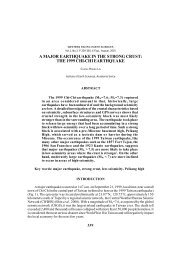
<strong>Targeted</strong> <strong>Drug</strong> <strong>Delivery</strong> to Breast Cancerintravenous injection, with a total doxorubicin dosage of 9 mg/kg(3 mg/kg; twice a week for three injections). For measurement oftumor luciferase activity, the mice were injected intraperitoneallywith luciferin (Caliper Life Sciences) and imaged using a XenogenIVIS200 imaging system. Tumor size was monitored using avernier caliper, and the tumor volume was calculated using theformula: length 6 (width) 2 6 0.52.Endocytosis of SP90-conjugated Liposomes <strong>by</strong> BT483CellsBT483 cells were plated and grown to ,80% confluence oncoverslips. The cells were then incubated with either liposomalSRB (LS), SP90-Lipo-SRB (SP90-LS) or control peptide-Lipo-SRB (CP-LS) at 37uC in complete medium. After 1 hour ofincubation, the cells were washed with PBS, stained with DAPI,and then examined using a Leica confocal microscope (TCS-SP5-AOBS). Images were processed <strong>by</strong> Leica Application SuiteAdvanced Fluorescence software.To assay the liposomes in endosomes of BT483 cells, the cellswere incubated with SP90-conjugated liposomal doxorubicin(SP90-LD) or control peptide-conjugated LD (CP-LD) at 37uC.After 5 min of incubation, the cells were washed with PBS, fixedusing a high pressure freezer (EM PACT2, Leica), and thenanalyzed <strong>by</strong> transmission electron microscopy (CM100, Philips).Total White Blood Cell (WBC) CountThree days after final treatment (on day 17), blood wasextracted from the submaxillary vein and mixed gently with 15%EDTA solution to prevent coagulation. Red blood cell lysis buffercontaining 2% acetic acid and 1% of Gentian violet (Sigma-Aldrich) was then added and incubated at room temperature. Thetotal WBC was calculated using a hemacytometer.Terminal Deoxynucleotidyl Transferase–mediated dUTPNick End Labeling (TUNEL) StainingThe frozen tumor tissue sections were incubated with terminaldeoxynucleotidyl transferase-mediated dUTP nick end labelingreaction mixture (Roche Diagnostics) at 37uC for one hour. Theslides were counterstained with mounting medium with DAPI(Vector Laboratories). The slides were then visualized under afluorescent microscope and areas of TUNEL positive cells werequantified <strong>by</strong> pixel area count normalize with DAPI usingMetaMorph software (Molecular Devices).Animal Model for Studying Tumor Localization ofLiposomal DoxorubicinSCID mice bearing breast cancer xenografts (,300 mm 3 ) wereinjected in the tail vein with either free drug doxorubicin (FD),targeting (SP90-LD) or non-targeting (CP-LD, and LD) liposomaldoxorubicin, at a dose of 2 mg/kg. At 24 hours post-injection,three mice in each group were anaesthetized and sacrificed. Themice were perfused with 50 ml PBS to wash out doxorubicin inblood, and xenograft tumors were then removed and homogenized.Total doxorubicin was quantified <strong>by</strong> measuring fluorescenceat l Ex/Em = 485/590 nm using a spectrofluorometer (SpectraMaxM5, Molecular Devices).To determine the presence of the drug in tumor tissues, frozentumor sections were fixed and stained with DAPI. The fluorescencesignal of doxorubicin was detected using an invertedfluorescence microscope (Axiovert, Zeiss) with a 546 nm excitationand 590 nm emission filter set.In vivo Imaging AnalysisSix 6-week old SCID female mice were subcutaneouslyimplanted with 5610 6 BT483 cells. Mice with size-matchedtumors (approximately 300 mm 3 ) were then randomly divided intotwo groups and intravenously injected with 200 pmole of SP90-QD or QD. The mice were anesthetized using isofluoran, and thefluorescence images were captured using a Xenogen IVIS 200imaging system (Excitation: 520/50 nm; Emission: 832/65 nm) atthe indicated times. To quantitatively compare tumor accumulationof SP90-QD versus QD, the fluorescence intensity wascalculated with background subtraction, using Living imagesoftware (Xenogen).Statistical AnalysisStudent’s t-test (unpaired and two-sided) was used to calculate Pvalues. P , 0.05 was considered significant for all analyses. Allvalues are represented as mean 6 standard deviation (s.d.).ResultsIdentification of Novel Peptides that Bind to BreastCancer CellsIn this study, we used a phage-displayed random peptide libraryto isolate phages that were able to bind to BT483 breast cancercells. After four rounds of affinity selection (biopanning), the titerof bound phage increased <strong>by</strong> up to 60-fold (Fig. 1a). ThroughELISA screening and DNA sequencing, we identified five phageclones (PC34, PC65, PC73, PC82 and PC90) with unique peptidesequences that bind to BT483, but not to control normal nasalmucosal (NNM) epithelial cells (Fig. 1b). The phage clones withhigher BT483-binding activities; PC34, PC65, PC73, PC82 andPC90, displayed QNIYAGVPMISF, EATNSHGSRTMG,TVSWSTTGRIPL, QLEFYTQLAHLI and SMDPFLFQLLQLpeptide sequence, respectively.To verify whether these five phage clones would bind to targetmolecules expressed on the surface of breast cancer cells, thesurface binding activity of each individual phage clone wasanalyzed <strong>by</strong> flow cytometry (Fig. S1a). The five phage clonesexhibited prominent binding to BT483 cells, with PC90 displayingthe best reactivity (Fig. S1a). We further demonstrated that PC90did not bind to normal human mammary epithelial cells(HMEpiC) using flow cytometry analysis (Fig. S1b). Based onthese findings, we chose to focus on PC90 for the rest of the study.To investigate whether PC90 may be effective against a broadspectrum of cancer cells, we used flow cytometry to analyze thebinding ability of PC90 to five breast cancer cell lines (Fig. 1c). Wefound markedly positive shifts in fluorescence in BT483, MDA-MB-231, MCF-7 and MDA-MB-361, and a small shift in SK-BR-3 cells. These results indicate that the PC90 phage specificallybinds to several breast cancer cell lines.To investigate the targeting ability of the selected phage clonesin vivo, we intravenously injected each clone into mice bearingBT483-derived tumor xenografts. After perfusion, we measuredthe phage titers in the tumor and normal organs [34,39]. Theresults showed that the five phage clones (PC34, PC65, PC73,PC82 and PC90) but not control helper phages had tumor-homingability (Fig.1d and Fig. S1c). Of these, PC90 targeted to tumorsmost efficiently, and was identified in tumor mass at concentrations§ 61-fold higher than that in the control organs. Wesubsequently synthesized the peptide displayed <strong>by</strong> the PC90phage, SP90, which has the amino acid sequenceSMDPFLFQLLQ. We found that co-administration of SP90and PC90 into mice reduced recovery of PC90 from tumor masses<strong>by</strong> nearly 97%, suggesting that SP90 is able to competitivelyPLOS ONE | www.plosone.org 4 June 2013 | Volume 8 | Issue 6 | e66128
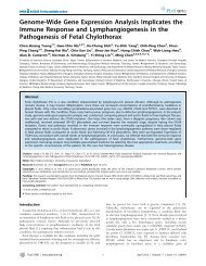
<strong>Targeted</strong> <strong>Drug</strong> <strong>Delivery</strong> to Breast CancerFigure 1. Identification of breast cancer cell-targeting peptides using in vitro phage display. a, A phage-display random peptide librarywas used to identify peptides that bind to the breast cancer cell line, BT483. After four rounds of biopanning, the titer of phage eluted from thebreast cancer cells had increased 60-fold relative to the first round of selection. PFU, plaque-forming units. b, Binding activity and specificity betweenindividual phage clones and breast cancer cells were tested <strong>by</strong> cellular ELISA. Bar, mean; Error bar, s.d.; n = 4; OD490 nm, optical density at 490 nm. c,The binding activity of the PC90 phage to five breast cancer cell lines was analyzed <strong>by</strong> flow cytometry. Con-P, control phage. d, Verification of tumorhomingability of phages in vivo. SCID mice bearing human breast cancer xenografts received intravenous injections of PC90 and control phage. Afterperfusion with PBS buffer, xenograft tumor masses were removed and phage titers were measured. e, The tumor-homing ability of the PC90 phagewas competitively inhibited <strong>by</strong> its cognate peptide SP90. f, Immunohistochemical localization of PC90 in figure 1d. Scale bar: 50 mm. g,Immunohistochemical staining of human surgical specimens of breast infiltrating ductal carcinoma using PC90 phage. Tumor sections of surgicalspecimens incubated with PC90 or control phage. The phage signal was detected using HRP-conjugated anti-M13 phage antibody. Scale bar: 20 mm.doi:10.1371/journal.pone.0066128.g001PLOS ONE | www.plosone.org 5 June 2013 | Volume 8 | Issue 6 | e66128
<strong>Targeted</strong> <strong>Drug</strong> <strong>Delivery</strong> to Breast Cancerinhibit the binding of PC90 to breast cancer cells (Fig. 1e). We alsoexamined the tissue distribution of PC90, <strong>by</strong> using anti-phageantibody to immunostain the tissue sections derived from homingexperiments. PC90 was found to be selectively localized in tumortissues rather than in normal tissues, whereas no immunoreactiveproduct was found through control phage staining (Fig. 1f and Fig.S2). Moreover, when PC90 was co-injected with the syntheticpeptide SP90, no immunoreactivity was found in the tumor tissue(Fig. 1f).To determine whether this targeting ligand had affinity forhuman breast cancer surgical specimens, we performed immunohistochemistrystaining with PC90 on breast infiltrating ductalcarcinoma tissue sections. We found that PC90 could recognizethe tumor cells of breast cancer surgical specimens, and that cotreatmentwith SP90 could competitively inhibit PC90 binding(Fig. 1g). Of the 20 breast cancer specimens from differentpatients, 90% (18/20) stained positive for PC90 (Table S1). Thesedata indicate that PC90 can recognize unidentified moleculesexpressed on breast cancer cell lines, as well as on cells from thesurgical specimens of breast cancer.SP90-conjugated Liposomes Exhibit Enhanced <strong>Drug</strong>Intracellular <strong>Delivery</strong> and CytotoxicityTo investigate whether SP90 could promote liposomal drugdelivery in human breast cancer cells, SP90 was conjugated toNHS-PEG-DSPE. Upon insertion of the phospholipid DSPE, thePEGylated SP90 conjugates became coupled to the externalsurface of liposomal nanoparticles. These nanoparticles containedsulforhodamine B (SRB; fluorescence reagent) or doxorubicin(Fig. 2a). The PEGylation efficiency of SP90 was validated <strong>by</strong>Tricine-SDS-PAGE and MALDI-TOF MS (Fig. 2b and c).The internalization ability of the targeting ligand is an essentialproperty for successful tumor-targeted liposomal drug delivery. Assuch, we examined internalization of SP90-conjugated liposomalSRB (SP90-LS) in tumor cells using confocal microscopy. Weobserved a large amount of SRB in the cytoplasm of BT483 cellsincubated with SP90-LS at 37uC, whereas little SRB fluorescencewas detectable in cells incubated with non-targeting liposomalSRB (LS) or control peptide-conjugated liposomal SRB (CP-LS)(Fig. 2d). This indicates that conjugation of the liposome withSP90 resulted in effective internalization of SRB. Furthermore, wefound that SP90 markedly enhanced intracellular SRB uptake <strong>by</strong>cancer cells at each time point examined (Fig. 2e).To verify internalization of SP90-conjugated liposomes throughreceptor-mediated endocytosis, we used transmission electronmicroscopy (TEM) to analyze the endosomes of tumor cells treatedwith either SP90-conjugated liposomal doxorubicin (SP90-LD) orcontrol peptide-conjugated liposomal doxorubicin (CP-LD) at37uC for 5 minutes. As shown in Figure 3a, SP90-LD accumulatedin the endosomes of cancer cells to a much greater extent than CP-LD. Endocytosed liposomes were observed in 90% of cells treatedwith SP90-LD, but only in 51% of cells treated with CP-LD(Fig. 3b). The average number of liposomes observed in eachendosome was 2.4 fold higher in cells treated with SP90-LD ascompared to cells treated with CP-LD (Fig. 3c).Doxorubicin is a small-molecule chemotherapeutic agent that ishistorically important in the treatment of breast cancer. Toidentify whether SP90-conjugated liposomes may enhance deliveryof doxorubicin, we treated BT483 cells with equal concentrationsof LD or SP90-LD. The uptake of doxorubicin wasquantitatively measured, based on fluorescent intensities at severaltime points (Fig. 3d). Cellular uptake of doxorubicin was elevatedin BT483 cells through treatment with SP90-LD. The area underthe concentration-time curve (AUC 0–48 hour) was 2.36-fold largerfor SP90-LD as compared to LD in breast cancer cells (Table S2).To assess whether SP90-conjugation enhanced the therapeuticpotential of LD, we performed in vitro cytotoxicity assays for SP90-LD in BT483 cells. Compared with LD, SP90-LD significantlyreduced the viability of cancer cells, and promoted a 4.9-folddecrease of the half maximal inhibitory concentration (IC 50 )inBT483 cells (Fig. 3e and Table S2). However, enhancement of thecytotoxic effect of SP90-LD was subject to competitive inhibitionupon co-treatment with free SP90 peptides, in a dose-dependentmanner (Fig. S3a). The cell viability of BT483 cells was notaffected <strong>by</strong> treatment with SP90 peptides alone (Fig. S3b).Therapeutic Efficacy of SP90-mediated <strong>Drug</strong> <strong>Delivery</strong>System in Mouse ModelsTo evaluate the potential of SP90 in improving the efficacy ofanticancer chemotherapy in vivo, we formulated a targeted drugdelivery system <strong>by</strong> coupling SP90 with PEGylated liposomaldoxorubicin (SP90-LD). SCID mice bearing BT483-derivedxenografts (,75 mm 3 ) were treated with SP90-LD, liposomaldoxorubicin (LD), free doxorubicin (FD) or equivalent volumes ofPBS. All formulations were injected intravenously at a totaldoxorubicin dosage of 9 mg/kg (3 mg/kg at weekly intervals).Anticancer efficacy was evaluated <strong>by</strong> determining the averagetumor volume throughout the 32 days treatment period. Thetumors in mice administered with SP90-LD were found to be 2.3-fold smaller in volume than those administered with LD alone onday 32 (Fig. 4a). To evaluate the side-effects caused <strong>by</strong> the systemicdelivery of chemotherapeutic drugs, we measured total whiteblood cell (WBC) count and body weight. The average total WBCcount of the SP90-LD group (4.8610 3 /mm 3 ) was similar to that ofthe PBS (5.0610 3 /mm 3 ) and FD (5.6610 3 /mm 3 ) groups, buthigher than that of the LD (2.6610 3 /mm 3 ) group (Fig. S4a).Administration of SP90-LD did not cause an appreciablereduction in body weight as compared to the LD group (Fig.S4b and c).To verify whether SP90-LD treatment would be effectiveagainst human breast tumor xenografts with a greater volume,mice bearing large BT483-derived xenografts (500 mm 3 ) wereintravenously administered with one of three drug formulations ata total doxorubicin dosage of 9 mg/kg (3 mg/kg twice a week). Bythe cessation of treatment, tumors treated with SP90-LD weresignificantly smaller than those treated with non-targeting LD(Fig. 4b). The histopathology of tumor tissues in each treatmentgroup was subsequently examined <strong>by</strong> H&E staining. Markedlydisseminated necrotic/apoptotic areas were presented throughoutthe whole section of SP90-LD-treated xenografts, while fewernecrotic/apoptotic areas were present in the tumors of the LDtreatedgroup (Fig. S5). We further carried out TUNEL assay toexamine apoptotic cells in tumor region. The number of apoptoticcells in the SP90-LD-treated group was significantly greater (2.5-fold) than in the LD-treated group (Fig. S6).Orthotopic tumor models are more pertinent with respect toboth host-tumor interactions and response to therapy. Therefore,we established an orthotopic mouse model of breast tumor <strong>by</strong>implanting MCF-7 cells stably expressing luciferase (MCF-7-Luc)into murine mammary pads, enabling us to further elucidate thetherapeutic response of SP90-targeting liposomes. Once tumorsize reached 500 mm 3 , mice were treated with either SP90-LD,LD or FD. Tumor growth was monitored using luminescentimaging and a vernier caliper twice a week. By the cessation oftreatment (day 10), the level of bioluminescence in the tumors ofSP90-LD-treated mice was lower than that of FD- and LD-treatedmice (<strong>by</strong> 8- and 2-fold, respectively) (Fig. 4c and S7a). Similarly,the average tumor volume and weight in the SP90-LD-treatedPLOS ONE | www.plosone.org 6 June 2013 | Volume 8 | Issue 6 | e66128
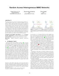
<strong>Targeted</strong> <strong>Drug</strong> <strong>Delivery</strong> to Breast CancerFigure 2. Generation of peptide-conjugated liposomal nanoparticles. a, Schematic illustration of the targeting liposome, depicting the lipidbilayer membrane encapsulating a large amount of doxorubicin or sulforhodamine B, and the breast cancer targeting ligands that can be displayedPLOS ONE | www.plosone.org 7 June 2013 | Volume 8 | Issue 6 | e66128
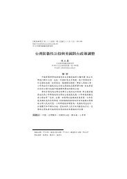
<strong>Targeted</strong> <strong>Drug</strong> <strong>Delivery</strong> to Breast Canceron the surface of the liposome. Targeting peptide SP90 was chemically conjugated to NHS-PEG-DSPE in a 1.1:1 molar ratio. There were about 500peptide molecules per liposome. b, The percentage of conjugation between SP90 and NHS-PEG-DSPE was confirmed <strong>by</strong> subjecting the reactionproduct to electrophoresis on a Tricine-SDS gel, staining with coomassie blue for peptide, followed <strong>by</strong> barium chloride for PEG (SP90:1.4 kDa, NHS-PEG-DSPE: 4.3 kDa, SP90-PEG-DSPE: 5.7 kDa). PEGylation efficiency for SP90 was 85% based on quantification of band intensity <strong>by</strong> densitometry. c,The SP90-PEG-DSPE conjugate was analyzed <strong>by</strong> MALDI-TOF mass spectrometry (bottom panel). A major peak appears at m/z 5702.8, which can beassigned as the PEGylated SP90 conjugate. Unconjugated SP90 (split peak at m/z 1452) and unreacted PEG (a broad peak around m/z 4250) were alsovisualized in the mass spectrum, which correspond to the peaks in the top and middle panels, respectively. d, Internalization of SP90-liposomal SRB(SP90-LS) and nontargeted LS (CP-LS and LS) <strong>by</strong> BT483 cells was studied <strong>by</strong> confocal microscopy (SRB, red). Nuclear staining was <strong>by</strong> DAPI (blue) (Scalebar: 10 mm). e, Time-course of SRB uptake <strong>by</strong> BT483 cells treated with SP90-LS, CP-LS and LS at the indicated times.doi:10.1371/journal.pone.0066128.g002group was significantly lower compared to that of the FD- andLD- treated groups (Fig. 4d and S7b).SP90-conjugated Liposomes Improved <strong>Drug</strong> <strong>Delivery</strong> invivoTo explore the mechanisms underlying the enhanced inhibitoryeffects using SP90-conjugated liposomal drugs, we examined drugaccumulation in tumor tissues <strong>by</strong> injecting SCID mice bearingBT483 xenografts with FD, LD, control peptide-conjugated LD(CP-LD) or SP90-LD. The mean intra-tumor doxorubicinconcentration in the SP90-LD group was 12.0-, 2.2- and 2.6-fold higher than that in the FD, LD and CP-LD groups,respectively (Fig. 5a). As it is possible that extensive perfusioncaused LD to be washed out of the tumor tissues, we repeated thisexperiment without PBS perfusion. Accumulation of SP90-LD intumors continued to be higher than that of FD (9.4-fold) and nonconjugatedLD (1.5-fold) (Fig. S8). To visualize the drug deliveryprofile of the three doxorubicin formulations, we examinedintracellular uptake of doxorubicin in tumor tissues <strong>by</strong> fluorescencemicroscopy. We found areas with detectable doxorubicin tobe larger in the nucleus of SP90-LD-treated tumors than in LDtreatedtumors, while no detectable doxorubicin was found in FDtreatedtumors (Fig. 5b). These experiments demonstrate thatSP90 can elevate delivery and penetration of anticancer drugs intothe tumor, resulting in accumulation of the drug at its intracellulartarget site, and there<strong>by</strong> enhancing its therapeutic effect.In vivo Imaging of SP90-conjugated NanoparticlesTo investigate whether SP90 could be used to enhance imagingof tumors, we constructed SP90-conjugated quantum dots (SP90-Figure 3. SP90-conjugated liposomes enhanced drug delivery and cytotoxicity towards cancer cells, through increasedendocytosis. a, Electron micrographs revealing liposomes in the endosomes of BT483 cells treated with SP90-LD (arrows) or CP-LD. Scale bar:100 nm. b, The percentage of breast cancer cells harboring endocytosed liposomes following treatment with SP90-LD or CP-LD (n = 50 in each group;*P,0.01). c, The average number of liposomes in each endosome following treatment with SP90-LD or CP-LD (n = 20 in each group; **P,0.01). d,Cells were incubated with either SP90-LD or LD at 37uC. Doxorubicin uptake <strong>by</strong> the cells was quantified at the indicated times, following the removalof surface-bound liposomal drugs. e, Cells were treated with SP90-LD and LD at varying concentrations. Cell viability was determined <strong>by</strong> MTT assay,and calculated as a percentage of living cells. Each point represents the mean of four experiments. Error bar, s.d.doi:10.1371/journal.pone.0066128.g003PLOS ONE | www.plosone.org 8 June 2013 | Volume 8 | Issue 6 | e66128
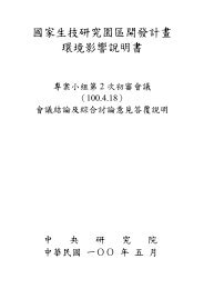
<strong>Targeted</strong> <strong>Drug</strong> <strong>Delivery</strong> to Breast CancerFigure 4. Treatment of SCID mice bearing human breast cancer xenografts with SP90-LD. a, Mice bearing BT483-derived breast cancerxenografts with average tumor size of ,75 mm 3 were treated with 3 mg/kg at weekly intervals (for a total of three injections) of either FD, LD, SP90-LD, or an equal volume of PBS <strong>by</strong> intravenous injection. n = 6 each group. Points, mean tumor volumes. b, Mice bearing size-matched BT483-derivedbreast cancer xenografts with tumor size of ,500 mm 3 were treated with SP90-LD, LD, or FD, for a total doxorubicin dosage of 9 mg/kg (3 mg/kg/injection, twice a week). n = 8. The average tumor volumes at cessation of treatment in the LD, FD and control PBS groups were 1.5, 6.2 and 8.4 timeslarger than that in the SP90-LD-treated group, respectively. c, Mice bearing orthotopic breast cancer tumors with average tumor size of ,500 mm 3 ,MCF-7-Luc, were injected with either SP90-LD, FD or LD, for a total doxorubicin dosage of 9 mg/kg (3 mg/kg/injection, twice a week). The tumorswere imaged and luminescence was quantified at the indicated days <strong>by</strong> IVIS200. n =5. d, Volume of tumors depicted in (c). Error bar, s.d.; *P,0.05;**P,0.01; ***P,0.001.doi:10.1371/journal.pone.0066128.g004QD; Fig. 6a), and used flow cytometry to confirm their tumorbindingactivity (Fig. S9). BT483-xenograft mice were thenintravenously injected with QD and SP90-QD. At 30 minutespost-injection, we detected a higher near-infrared (NIR) fluorescencesignal in the tumor sites of SP90-QD-treated mice thanthose in QD-treated mice (Fig. 6b). The greatest contrast betweenthe fluorescence intensity of tumors in SP90-QD versus QDoccurred at 2 hours post-injection, with a ratio of 22:1 (Fig. 6c).Kinetic evaluation of imaging over 24 hours further confirmedthat SP90-QD was both efficiently targeted to, and retained intumors (Fig. 6d). These results indicate that the SP90-mediateddrug delivery systems show great promise for their applications intumor-targeted drug delivery and imaging.DiscussionEarlier diagnosis and better targeted drug delivery wouldsignificantly improve the efficacy of cancer therapy. Identificationof tumor-targeting agents is instrumental in achieving theseobjectives. One promising approach to treating cancer is the useof ligand-conjugated liposome-encapsulated drugs that targettumor cells and blood vessels. Peptides that specifically bind totumor targets can be coupled to the PEG terminus of stericallystabilized liposomes to precisely deliver chemotherapeutic agentsto tumor cells. In this study, we searched for specific peptideligands that would target breast cancer specifically, in order todevelop a ligand-mediated target therapy capable of treating thisdisease.The peptide-mediated targeting liposomes designed in thisstudy offer several advantages over the use of free drugs intreatment of breast cancer. First, each peptide-conjugatedliposome can efficiently deliver 15,000 anticancer drug moleculesdirectly into endosome compartments with low pH; theliposomes then break down and release the encapsulated druginto the intracellular space of the target tumor cells [40],enabling controlled drug release and improving drug bioavail-PLOS ONE | www.plosone.org 9 June 2013 | Volume 8 | Issue 6 | e66128
<strong>Targeted</strong> <strong>Drug</strong> <strong>Delivery</strong> to Breast CancerFigure 5. SP90-conjugated liposomes enhanced drug delivery to tumor. a, BT483 breast cancer-bearing mice were treated with differentformulations of liposomal (LD, CP-LD and SP90-LD) and free doxorubicin (FD). After perfusion with 50 ml PBS, doxorubicin concentration wasdetermined in tumor tissue (n = 3 in each group; **P,0.01, ***P,0.005). b, Representative two-color images showing the distribution of doxorubicin(red) in relation to nuclei (blue) in tumor tissue sections. Accumulation of doxorubicin in tumor nuclei was examined at day 3 post-injection. Scale bar,50 mm. Bar, mean; Error bar, s.d.doi:10.1371/journal.pone.0066128.g005Figure 6. In vivo tumor targeting and imaging with SP90-conjugated quantum dots. a, SP90 was thiolated using Traut’s reagent togenerate thiol-modified SP90 (SP90-SH). SP90-SH was subsequently conjugated with sulfo-SMCC-activated QD to produce SP90-QD. Mal, Maleimide.b, In vivo fluorescence imaging of SCID mice bearing BT483-derived tumors was performed after intravenous injection of QD (left) or SP90-QD (right).n = 3. Red arrows indicate the tumor loci. The NIR fluorescence images were captured at the indicated time points. c, Fluorescence signals withintumors were quantified using IVIS200 software. n = 3; Error bar, s.d; ***P,0.001. d, Quantification and kinetics of in vivo targeting of SP90-QD.Fluorescence intensity was recorded as photons per second per square centimeter per steradian (p/s/cm 2 /sr). Representative images from threeindependent sets of studies all gave similar results. n =3.doi:10.1371/journal.pone.0066128.g006PLOS ONE | www.plosone.org 10 June 2013 | Volume 8 | Issue 6 | e66128
<strong>Targeted</strong> <strong>Drug</strong> <strong>Delivery</strong> to Breast Cancerability (Fig. 3 and 5). Second, targeted and sustained release ofthe drug molecules from encapsulated carriers can increasetherapeutic index of these chemotherapeutic agents against thetumor, while simultaneously reducing the toxicity of these drugson the normal tissues (Fig. 4); importantly, conjugation to SP90significantly reduced the toxicity of liposomal doxorubicin towhite blood cells (Fig. S2), one of the major adverse effects ofthis drug in the clinical setting [41]. Third, enhanced drugaccumulation in tumor tissues may help circumvent theproblems associated with delivering drugs to solid tumors withhigh interstitial fluid pressure (Fig. 5) [5,42,43]. Our resultsrevealed that SP90-LD more severely damaged blood vessels(data not shown) and cancer cells in tumor tissue as comparedto LD in therapeutic experiments (Fig. S5 and S6). However,we have no evidence to suggest that SP90 can bind vascularendothelial cells, and hence tumor vessels were most likely killedindirectly, as a result of the diffusion of small-molecule drugsfrom targeted cells to neighboring cells through the <strong>by</strong>standereffect.Although monoclonal antibodies have shown clinical benefits asanti-tumor agents, their potential for use as a drug delivery systemhas been limited due to a number of factors, including largemolecular size, poor tumor penetration, and high immunogenicitywhen used in immunoliposomes [44]. Additionally, antibodybaseddrug delivery may lead to higher than normal levels oftoxicity in liver and bone marrow, due to nonspecific antibodyuptake <strong>by</strong> Fc receptor-expressing normal cells [44]. Theselimitations can be overcome <strong>by</strong> using peptide ligands, which aresmaller, less immunogenic, and more cost-effective to produce andmanipulate [34,39]. Furthermore, multivalent peptide ligandshave only a moderate affinity to tumor antigens, which ispotentially advantageous for targeted drug delivery [26], sincethe strong affinity of antibodies may limit the penetration depth oftheir cargo into tumors [45,46].We found that most surgical specimens from breast cancerpatients could be detected <strong>by</strong> SP90-bearing phage, PC90 (Fig. 1and Table S1), further supporting the potential clinicalapplication of this novel peptide ligand. Conjugating pharmaceuticalnanocarriers or tumor imaging agents with SP90 mayimprove the effectiveness of current chemotherapeutic anddiagnostic options for human breast cancer, <strong>by</strong> increasing theirsensitivity and specificity. To explore whether the putativereceptor can recognize a peptide ligand with sequence of SP90,we performed Alignment Search for this 12-mer peptide usingBLASTP program [47]. No human protein sequences orconserved domains were found to be homologous to SP90based on our results. However, the targeted cell surfacemolecule recognized <strong>by</strong> SP90 needs to be identified, in orderto elucidate the mechanism of action of SP90 binding and toaddress safety concerns prior to clinical trials.In conclusion, we identified several novel peptides, includingSP90, capable of binding specifically to the cell surface of breastcancer cells both in vitro and in vivo. Linking SP90 to liposomescontaining doxorubicin increased the therapeutic efficacy in micewith human breast cancer xenografts, through enhanced tumorapoptosis and decreased tumor angiogenesis. Quantification andvisualization of doxorubicin levels also revealed increased drugconcentrations in tumor tissues targeted <strong>by</strong> the liposome,highlighting the enhancement in both delivery and penetrationof doxorubicin into the tumor. Our results indicate that the SP90peptide may be used to enable specific targeting of tumor cells inthe treatment of breast cancer, as well as to facilitate diagnosis ofthis malignancy.Supporting InformationFigure S1 Verification of binding and in vivo tumorhomingability of phages. a, The surface binding activity ofeach selected phage to breast cancer and NNM cells wasdetermined <strong>by</strong> flow cytometry. b, The binding activity of PC90phage to normal human mammary epithelial cells (HMEpiC) wasdetermined <strong>by</strong> flow cytometry. BT483 cells were used as positive.c, SCID mice bearing a BT483 xenograft tumor receivedintravenously injections of PC34, PC65, PC73, PC82, and controlhelper phage. After perfusion with PBS buffer, xenograft tumormasses and organs were removed and phage titers were measured.Phage titer in control organs are compared with tumor tissues, asindicated.(JPG)Figure S2 The low-magnification images of PC90 immunohistochemicalstaining in tumor-homing analysis(Fig. 1e). The PC90 phage was localized on tumor tissues and nolocalization was observed in normal organs such as the brain,heart, and lungs. Neither tumor cells nor normal organs werefound to have immunoreactivity with control phage.(JPG)Figure S3 Competition analysis of SP90-LD-inducedcytotoxic effect <strong>by</strong> free SP90 peptides. a, BT483 cells weretreated with various concentrations of LD or SP90-LD in thepresence of 10, 1, 0.1 or 0 mg/ml of SP90 peptides. b, BT483 cellswere incubated with free SP90 and control peptides at variousconcentrations. After incubation for three days, cell viability wasdetermined <strong>by</strong> MTT assay, and was calculated as a percentage ofliving cells. Each point represents the mean of three experiments.Error bar, s.d.(JPG)Figure S4 Response of SCID mice bearing BT483-derived xenografts to the administration of SP90-LD inFigure 3a. a, The effect of different treatments on white bloodcell (WBC) counts. SP90-LD reduced the WBC toxicity ofliposomal doxorubicin in the breast cancer xenograft model(n = 6 in each group; **P,0.001). b, The body weight of eachgroup. c, The effect of different treatments on change in bodyweight during the period from day 0 to day 20 (n = 6 in eachgroup).(JPG)Figure S5 Histopathological examination of SP90-LDtreatedbreast cancer xenografts. After cessation of treatment,PBS- and FD-treated tumors were removed on day 20,while LD- and SP90-LD-treated tumors were removed on day 32for histopathological examination. a, Tumor tissues were examinedafter staining with H&E. Markedly disseminated necrotic/apoptotic areas were observed throughout the entire section ofSP90-LD-treated xenografts. LD-treated xenografts presentedwith moderate necrotic/apoptotic areas, while normal breastcancer cells were observed in the FD- and PBS-treated groups.(Scale bar, 100 mm). b, The percentage areas of necrosis/apoptosis were determined (n=6) at low magnification. Theaverage percentage area of necrosis/apoptosis was markedlyincreased in the SP90-LD treated group as compared to theLD-, FD- or PBS-treated groups (n =6, **P,0.01).(JPG)Figure S6 SP90-conjugated targeting liposomes increasedtherapeutic efficacy through enhanced cancercell apoptosis. a, Sections were TUNEL-labeled to visualizeapoptotic tumor cells (green). TUNEL-positive tumor cells werePLOS ONE | www.plosone.org 11 June 2013 | Volume 8 | Issue 6 | e66128
<strong>Targeted</strong> <strong>Drug</strong> <strong>Delivery</strong> to Breast Cancerdistributed more extensively in the SP90-LD-treated groups thanin the LD, FD or PBS groups. b, Areas of TUNEL positive cellswere quantified <strong>by</strong> pixel area count, and normalized to DAPIusing MetaMorph Software. A significantly greater averageapoptotic area was observed for the xenografts of the SP90-LDtreatedgroup, as compared to those of the LD, FD or PBS treatedgroups. Scale bar, 85 mm.(JPG)Figure S7 Treatment of SCID mice with SP90-LD inorthotopic human breast models. a, Representative imagesused for the analysis described in Figure 4c. Luminescent radiancewas assessed <strong>by</strong> IVIS200 imaging on the indicated days. n =5. b,At the end of treatment, mice were sacrificed, and tumors weredissected and weighed. *P,0.05.(JPG)Figure S8 SP90-conjugated liposomes enhanced drugdelivery to tumor. Accumulation of doxorubicin in tumors ofbreast cancer-bearing mice treated with different formulations ofliposomal and free doxorubicin, without PBS perfusion (n =3 ineach group; *P,0.05).(JPG)References1. Siegel R, Naishadham D, Jemal A (2012) Cancer statistics, 2012. CA: A CancerJournal for Clinicians 62: 10–29.2. DeSantis C, Siegel R, Bandi P, Jemal A (2011) Breast cancer statistics, 2011.CA: A Cancer Journal for Clinicians 61: 408–418.3. O’Shaughnessy JA, Kaufmann M, Siedentopf F, Dalivoust P, Debled M, et al.(2012) Capecitabine Monotherapy: Review of Studies in First-Line HER-2-Negative Metastatic Breast Cancer. The Oncologist 17: 476–484.4. Cobleigh MA (2011) Other Options in the Treatment of Advanced BreastCancer. Seminars in Oncology 38, Supplement 2: S11–S16.5. Heldin CH, Rubin K, Pietras K, Ostman A (2004) High interstitial fluid pressure- an obstacle in cancer therapy. Nat Rev Cancer 4: 806–813.6. Provenzano PP, Cuevas C, Chang AE, Goel VK, Von Hoff DD, et al. (2012)Enzymatic targeting of the stroma ablates physical barriers to treatment ofpancreatic ductal adenocarcinoma. Cancer Cell 21: 418–429.7. Bosslet K, Straub R, Blumrich M, Czech J, Gerken M, et al. (1998) Elucidationof the mechanism enabling tumor selective prodrug monotherapy. Cancer Res58: 1195–1201.8. Chang DK, Lin CT, Wu CH, Wu HC (2009) A novel peptide enhancestherapeutic efficacy of liposomal anti-cancer drugs in mice models of humanlung cancer. PLoS One 4: e4171.9. Hambley TW, Hait WN (2009) Is anticancer drug development heading in theright direction? Cancer Res 69: 1259–1262.10. Minchinton AI, Tannock IF (2006) <strong>Drug</strong> penetration in solid tumours. Nat RevCancer 6: 583–592.11. Szakacs G, Paterson JK, Ludwig JA, Booth-Genthe C, Gottesman MM (2006)Targeting multidrug resistance in cancer. Nat Rev <strong>Drug</strong> Discov 5: 219–234.12. Davis ME, Chen Z, Shin DM (2008) Nanoparticle therapeutics: an emergingtreatment modality for cancer. Nat Rev <strong>Drug</strong> Discov 7: 771–782.13. Farokhzad OC, Langer R (2009) Impact of nanotechnology on drug delivery.ACS Nano 3: 16–20.14. Shi J, Votruba AR, Farokhzad OC, Langer R (2010) Nanotechnology in <strong>Drug</strong><strong>Delivery</strong> and Tissue Engineering: From Discovery to Applications. Nano Letters10: 3223–3230.15. Matsumura Y, Maeda H (1986) A new concept for macromolecular therapeuticsin cancer chemotherapy: mechanism of tumoritropic accumulation of proteinsand the antitumor agent smancs. Cancer Res 46: 6387–6392.16. Maeda H, Wu J, Sawa T, Matsumura Y, Hori K (2000) Tumor vascularpermeability and the EPR effect in macromolecular therapeutics: a review.Journal of Controlled Release 65: 271–284.17. Wagner V, Dullaart A, Bock A-K, Zweck A (2006) The emerging nanomedicinelandscape. Nat Biotech 24: 1211–1217.18. Slingerland M, Guchelaar H-J, Gelderblom H (2012) Liposomal drugformulations in cancer therapy: 15 years along the road. <strong>Drug</strong> DiscoveryToday 17: 160–166.19. Iber FL, Nassau K, Plough IC, Berger FM, Meroney WH, et al. (1958) The useof radioiodinated albumin in metabolic studies; the effects of the level of dietaryprotein and L-triiodothyroinine on the catabolism of radioiodinated humanserum albumin. J Clin Invest 37: 1442–1452.20. Kratz F (2008) Albumin as a drug carrier: Design of prodrugs, drug conjugatesand nanoparticles. Journal of Controlled Release 132: 171–183.Figure S9 Analysis of tumor binding activity of SP90-QDs in vitro. The binding activity of QD-labeled SP90 toBT483 cells was analyzed <strong>by</strong> flow cytometry.(JPG)Table S1 Detection of human breast cancer surgicalspecimens <strong>by</strong> PC90 phages using immunohistochemistry.(DOCX)Table S2 AUC and IC 50 of BT483 treated with SP90-LDand LD.( DOCX)AcknowledgmentsThe authors thank the Core Facility of the Institute of Cellular andOrganismic Biology, <strong>Academia</strong> <strong>Sinica</strong>, and Dr. Wann-Neng Jane for theirassistance in fluorescence and electron microscopy, and Dr. DuncanWright for reading the manuscript.Author ContributionsConceived and designed the experiments: HCW RML. Performed theexperiments: RML MSC DKC CYC WCL CYY YPW AL YSK.Analyzed the data: MSC DKC SLY. Wrote the paper: HCW RML.21. Kohlschütter J, Michelfelder S, Trepel M (2008) <strong>Drug</strong> delivery in acute myeloidleukemia. Expert Opinion on <strong>Drug</strong> <strong>Delivery</strong> 5: 653–663.22. Torchilin V (2008) Antibody-modified liposomes for cancer chemotherapy.Expert Opin <strong>Drug</strong> Deliv 5: 1003–1025.23. Wu HC, Chang DK (2010) Peptide-mediated liposomal drug delivery systemtargeting tumor blood vessels in anticancer therapy. J Oncol 2010: 723798.24. Svensen N, Walton JGA, Bradley M (2012) Peptides for cell-selective drugdelivery. Trends in pharmacological sciences 33: 186–192.25. Chang DK, Chiu CY, Kuo SY, Lin WC, Lo A, et al. (2009) Antiangiogenictargeting liposomes increase therapeutic efficacy for solid tumors. J Biol Chem284: 12905–12916.26. Ashley CE, Carnes EC, Phillips GK, Padilla D, Durfee PN, et al. (2011) Thetargeted delivery of multicomponent cargos to cancer cells <strong>by</strong> nanoporousparticle-supported lipid bilayers. Nat Mater 10: 389–397.27. Cho HJ, Yoon IS, Yoon HY, Koo H, Jin YJ, et al. (2012) Polyethylene glycolconjugatedhyaluronic acid-ceramide self-assembled nanoparticles for targeteddelivery of doxorubicin. Biomaterials 33: 1190–1200.28. Park J-H, von Maltzahn G, Zhang L, Derfus AM, Simberg D, et al. (2009)Systematic Surface Engineering of Magnetic Nanoworms for In vivo TumorTargeting. Small 5: 694–700.29. Park JW, Hong K, Kirpotin DB, Colbern G, Shala<strong>by</strong> R, et al. (2002) Anti-HER2 immunoliposomes: enhanced efficacy attributable to targeted delivery.Clin Cancer Res 8: 1172–1181.30. Nellis DF, Ekstrom DL, Kirpotin DB, Zhu J, Andersson R, et al. (2005)Preclinical Manufacture of an Anti-HER2 scFv-PEG-DSPE, Liposome-InsertingConjugate. 1. Gram-Scale Production and Purification. Biotechnology Progress21: 205–220.31. Wang T, D’Souza GGM, Bedi D, Fagbohun OA, Potturi LP, et al. (2010)Enhanced binding and killing of target tumor cells <strong>by</strong> drug-loaded liposomesmodified with tumor-specific phage fusion coat protein. Nanomedicine 5: 563–574.32. Shadidi M, Sioud M (2003) Identification of novel carrier peptides for thespecific delivery of therapeutics into cancer cells. FASEB J 17: 256–258.33. Lee CM, Iorno N, Sierro F, Christ D (2007) Selection of human antibodyfragments <strong>by</strong> phage display. Nat Protoc 2: 3001–3008.34. Lee TY, Lin CT, Kuo SY, Chang DK, Wu HC (2007) Peptide-mediatedtargeting to tumor blood vessels of lung cancer for drug delivery. Cancer Res 67:10958–10965.35. Lu R-M, Chang Y-L, Chen M-S, Wu H-C (2011) Single chain anti-c-Metantibody conjugated nanoparticles for in vivo tumor-targeted imaging and drugdelivery. Biomaterials 32: 3265–3274.36. Lee TY, Wu HC, Tseng YL, Lin CT (2004) A novel peptide specifically bindingto nasopharyngeal carcinoma for targeted drug delivery. Cancer Res 64: 8002–8008.37. Schagger H (2006) Tricine-SDS-PAGE. Nat Protoc 1: 16–22.38. Cai W, Chen X (2008) Preparation of peptide-conjugated quantum dots fortumor vasculature-targeted imaging. Nat Protoc 3: 89–96.39. Lo A, Lin CT, Wu HC (2008) Hepatocellular carcinoma cell-specific peptideligand for targeted drug delivery. Mol Cancer Ther 7: 579–589.PLOS ONE | www.plosone.org 12 June 2013 | Volume 8 | Issue 6 | e66128
<strong>Targeted</strong> <strong>Drug</strong> <strong>Delivery</strong> to Breast Cancer40. Barenholz Y (2012) Doxil(R)–the first FDA-approved nano-drug: lessonslearned. J Control Release 160: 117–134.41. O’Brien ME, Wigler N, Inbar M, Rosso R, Grischke E, et al. (2004) Reducedcardiotoxicity and comparable efficacy in a phase III trial of pegylated liposomaldoxorubicin HCl (CAELYX/Doxil) versus conventional doxorubicin for firstlinetreatment of metastatic breast cancer. Ann Oncol 15: 440–449.42. Wu H, Chang D, Huang C (2006) <strong>Targeted</strong>-therapy for cancer. J Cancer Mol 2:57–66.43. Carmeliet P, Jain RK (2011) Molecular mechanisms and clinical applications ofangiogenesis. Nature 473: 298–307.44. Cheng WW, Allen TM (2010) The use of single chain Fv as targeting agents forimmunoliposomes: an update on immunoliposomal drugs for cancer treatment.Expert Opin <strong>Drug</strong> Deliv 7: 461–478.45. Adams GP, Schier R, McCall AM, Simmons HH, Horak EM, et al. (2001) Highaffinity restricts the localization and tumor penetration of single-chain fvantibody molecules. Cancer Res 61: 4750–4755.46. Rudnick SI, Lou J, Shaller CC, Tang Y, Klein-Szanto AJP, et al. (2011)Influence of Affinity and Antigen Internalization on the Uptake and Penetrationof Anti-HER2 Antibodies in Solid Tumors. Cancer Research 71: 2250–2259.47. Altschul SF, Madden TL, Schaffer AA, Zhang J, Zhang Z, et al. (1997) GappedBLAST and PSI-BLAST: a new generation of protein database searchprograms. Nucleic Acids Res 25: 3389–3402.PLOS ONE | www.plosone.org 13 June 2013 | Volume 8 | Issue 6 | e66128