NPC Valorisation Voucher - Netherlands Proteomics Centre
NPC Valorisation Voucher - Netherlands Proteomics Centre
NPC Valorisation Voucher - Netherlands Proteomics Centre

You also want an ePaper? Increase the reach of your titles
YUMPU automatically turns print PDFs into web optimized ePapers that Google loves.
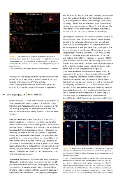
with TIC in y-axis) does not give much information on a sampleother than a vague indication of its magnitude and complexity.With the publicly available software MSight (www.expasy.org/MSight), LC-MS data are assembled into a more informativetwo-dimensional image (see Figure 2, for details see [3])which can be readily interpreted with respect to structuralfeatures (i.e. peptide PTMs) of interest to the biologist.Figure 2 | A. Orthogonal plot of LC-MS run of amphibian skin secretionsample. Each spot represents an isotope cluster of a peptide-like ion. Insert:zoom-in area of 2D LC-MS plot revealing amidated peptide ions, as slightlymore hydrophobic (increased RT on reversed phase HPLC) and exactly one Dalighter isotopes.B. Similar display of same sample after chemical treatment (i.c. reduction).investigation. This is the part of the peptide which fits in thebinding pocket of a receptor or within a groove of the catalyticsite of an enzyme modifying its activity.In this respect post-translational modifications (PTMs) arecritically important biochemical alterations of a peptide’s10 | <strong>NPC</strong> Highlights 16 | Peter Verhaertprimary structure, as these often dramatically affect (part of)their tertiary (3D) structure, making it fit its binder or not.Using high-end MS based peptide analytics (employing Q-TOFand orbitrap analysers), we developed methods with sufficientMS resolution to specifically screen for these distinctivefeatures.Easily spotted Some PTMs are visible in untreated samples byvirtue of the fact that they are not present on one hundredpercent of the molecules, which means that for each posttranslationally-modifiedpeptide, the unmodified version isalso often present in a sample. Depending on the type of PTM,these can be seen at specific mass differences and more orless predictable retention time shifts. A PTM easily spottedthis way is C-terminal amidation. Replacement of the freeacid carboxyl end (-COOH) of a peptide to its amide (-CONH 2 )yields a modified peptide which differs exactly one mass unitfrom its unmodified variant, whereas its retention only slightlyshifts (with the amidated version typically a bit more hydrophobicthan the free acid; see insert in Figure 2).Other PTMs only reveal themselves after specific chemicaltreatment of the sample. In these cases the differential 2Ddisplay comparing treated and untreated samples very elegantlyshows peptides with the targeted PTM (see Figure 3).Such treatment can be a very simple one, such as reduction ofthe sample by dithiothreitol (DTT) or an alternative reducingagent. In this way we have been able to identify and fullystructurally characterise novel peptides with both intra- aswell as intermolecular disulfide bridges in various frog andtoad species [3, 4]. Subsequent bioactivity studies thenshowed that several of these newly discovered peptides haveFrog skin secretions A good example of a rich source ofbioactive peptides of sometimes very limited supply is thedefensive secretion by the dorsal skin glands of exotic amphibians(see Figure 1). Nowadays, this material — not infrequentlyhundreds of different peptides per sample — is typically noninvasivelycollected in the wild. As a third of all amphibianspecies are threatened with extinction, it is guaranteed thatpeptide donors remain unharmed and are released back intotheir habitat immediately after ‘milking’ [2]. Depending onthe biological need or ecological niche of a certain amphibianspecies, bioactivities to be found in its skin secretions are verydiverse, and, not seldom, unexpected. Focusing on a limitednumber of bioactivities, therefore, risks missing many potentiallyinteresting compounds.2D mapping We have successfully utilised a two dimensional(2D) peptide display, being an image generated from the 2Dplot of retention times versus mass-to-charge ratios of LC-MS(peptide) ions, to identify peptides with predefined PTMs/structural features indicative of their potential bioactivity. Aconventional one-dimensional LC-MS display (chromatogramFigure 3 | A. MSight overlay of two subsequent LC-MS runs (Fig. 2A and B) ofuntreated and reduced sample of amphibian skin secretion. This generates atwo-dimensional PTM driven differential display, revealing peptides with andwithout cysteine bridges.B. Zoom-in of selected areas focusing on peptides (from left to right panels)without cysteine bridges (far left), with a single disulfide bridge, with twoand with three S-S bonds (far right). Peptides originally carrying a disulfidebond are easily distinguished from the others (non cystine containing peptidesremain at identical RT and m/z in both untreated and treated sample)by their shift in both retention time (vertical axis) and m/z value (horizontalaxis; i.e. increase of 2 Da per disulfide bridge broken in the reduced sample).