Orthopedic Trauma in Pregnancy - The Journal of Family Practice
Orthopedic Trauma in Pregnancy - The Journal of Family Practice
Orthopedic Trauma in Pregnancy - The Journal of Family Practice

Create successful ePaper yourself
Turn your PDF publications into a flip-book with our unique Google optimized e-Paper software.
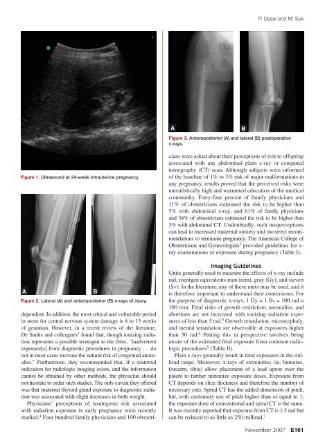
P. Desai and M. SukABFigure 3. Anteroposterior (A) and lateral (B) postoperativex-rays.Figure 1. Ultrasound at 24-week <strong>in</strong>trauter<strong>in</strong>e pregnancy.AFigure 2. Lateral (A) and anteroposterior (B) x-rays <strong>of</strong> <strong>in</strong>jury.Bdependent. In addition, the most critical and vulnerable period<strong>in</strong> utero for central nervous system damage is 8 to 15 weeks<strong>of</strong> gestation. However, <strong>in</strong> a recent review <strong>of</strong> the literature,De Santis and colleagues 3 found that, though ioniz<strong>in</strong>g radiationrepresents a possible teratogen to the fetus, “<strong>in</strong>advertentexposure[s] from diagnostic procedures <strong>in</strong> pregnancy … donot <strong>in</strong> most cases <strong>in</strong>crease the natural risk <strong>of</strong> congenital anomalies.”Furthermore, they recommended that, if a maternal<strong>in</strong>dication for radiologic imag<strong>in</strong>g exists, and the <strong>in</strong>formationcannot be obta<strong>in</strong>ed by other methods, the physician shouldnot hesitate to order such studies. <strong>The</strong> only caveat they <strong>of</strong>feredwas that maternal thyroid gland exposure to diagnostic radiationwas associated with slight decreases <strong>in</strong> birth weight.Physicians’ perceptions <strong>of</strong> teratogenic risk associatedwith radiation exposure <strong>in</strong> early pregnancy were recentlystudied. 4 Four hundred family physicians and 100 obstetricianswere asked about their perceptions <strong>of</strong> risk to <strong>of</strong>fspr<strong>in</strong>gassociated with any abdom<strong>in</strong>al pla<strong>in</strong> x-ray or computedtomography (CT) scan. Although subjects were <strong>in</strong>formed<strong>of</strong> the basel<strong>in</strong>e <strong>of</strong> 1% to 3% risk <strong>of</strong> major malformations <strong>in</strong>any pregnancy, results proved that the perceived risks wereunrealistically high and warranted education <strong>of</strong> the medicalcommunity. Forty-four percent <strong>of</strong> family physicians and11% <strong>of</strong> obstetricians estimated the risk to be higher than5% with abdom<strong>in</strong>al x-ray, and 61% <strong>of</strong> family physiciansand 34% <strong>of</strong> obstetricians estimated the risk to be higher than5% with abdom<strong>in</strong>al CT. Undoubtedly, such misperceptionscan lead to <strong>in</strong>creased maternal anxiety and <strong>in</strong>correct recommendationsto term<strong>in</strong>ate pregnancy. <strong>The</strong> American College <strong>of</strong>Obstetricians and Gynecologists 5 provided guidel<strong>in</strong>es for x-ray exam<strong>in</strong>ations or exposure dur<strong>in</strong>g pregnancy (Table I).Imag<strong>in</strong>g Guidel<strong>in</strong>esUnits generally used to measure the effects <strong>of</strong> x-ray <strong>in</strong>cluderad, roentgen equivalents man (rem), gray (Gy), and sievert(Sv). In the literature, any <strong>of</strong> these units may be used, and itis therefore important to understand their conversions. Forthe purpose <strong>of</strong> diagnostic x-rays, 1 Gy = 1 Sv = 100 rad =100 rem. Fetal risks <strong>of</strong> growth restriction, anomalies, andabortions are not <strong>in</strong>creased with ioniz<strong>in</strong>g radiation exposures<strong>of</strong> less than 5 rad. 5 Growth retardation, microcephaly,and mental retardation are observable at exposures higherthan 50 rad. 6 Putt<strong>in</strong>g this <strong>in</strong> perspective <strong>in</strong>volves be<strong>in</strong>gaware <strong>of</strong> the estimated fetal exposure from common radiologicprocedures 5 (Table II).Pla<strong>in</strong> x-rays generally result <strong>in</strong> fetal exposures <strong>in</strong> the milliradrange. Moreover, x-rays <strong>of</strong> extremities (ie, humerus,forearm, tibia) allow placement <strong>of</strong> a lead apron over thepatent to further m<strong>in</strong>imize exposure doses. Exposure fromCT depends on slice thickness and therefore the number <strong>of</strong>necessary cuts. Spiral CT has the added dimension <strong>of</strong> pitch,but, with customary use <strong>of</strong> pitch higher than or equal to 1,the exposure dose <strong>of</strong> conventional and spiral CT is the same.It was recently reported that exposure from CT is 1.5 rad butcan be reduced to as little as 250 millirad. 7November 2007 E161