The occipital The occipital lobes Occipital lobe - Mahidol University
The occipital The occipital lobes Occipital lobe - Mahidol University
The occipital The occipital lobes Occipital lobe - Mahidol University

You also want an ePaper? Increase the reach of your titles
YUMPU automatically turns print PDFs into web optimized ePapers that Google loves.
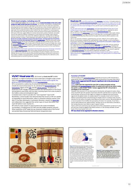
23/06/54Third visual complex, including area V3<strong>The</strong> term third visual complex refers to the region of cortex located immediately in front of V2, whichincludes the region named visual area V3 in humans. <strong>The</strong> "complex" nomenclature is justified by thefact that some controversy still exists regarding the exact extent of area V3, with some researchersproposing that the cortex located in front of V2 may include two or three functional subdivisions. Forexample, David Van Essen and others (1986) have proposed that the existence of a "dorsal V3" in theupper part of the cerebral hemisphere, which is distinct from the "ventral V3" (or ventral posteriorarea, VP) located in the lower part of the brain. Dorsal and ventral V3 have distinct connections withother parts of the brain, appear different in sections stained with a variety of methods, and containneurons that respond to different combinations of visual stimulus (for example, colour‐selectiveneurons are more common in the ventral V3). Additional subdivisions, including V3A and V3B havealso been reported in humans. <strong>The</strong>se subdivisions are located near dorsal V3, but do not adjoin V2.Dorsal V3 is normally considered to be part of the dorsal stream, receiving inputs from V2 and fromthe primary visual area and projecting to the posterior parietal cortex. It may be anatomically locatedin Brodmann area 19. Recent work with fMRI has suggested that area V3/V3A may play a role in theprocessing of global motion Other studies prefer to consider dorsal V3 as part of a larger area, namedthe dorsomedial area (DM), which contains a representation of the entire visual field. Neurons in areaDM respond to coherent motion of large patterns covering extensive portions of the visual field (Luiand collaborators, 2006).Ventral V3 (VP), has much weaker connections from the primary visual area, and stronger connectionswith the inferior temporal cortex. While earlier studies proposed that VP only contained arepresentation of the upper part of the visual field (above the point of fixation), more recent workindicates that this area is more extensive than previously appreciated, and like other visual areas itmay contain a complete visual representation. <strong>The</strong> revised, more extensive VP is referred to as theventrolateral posterior area (VLP) by Rosa and Tweedale.Visual area V4 is one of the visual areas in the extrastriate visual cortex. It is located anterior toV2 and posterior to posterior inferotemporal area (PIT). It comprises at least four regions (left and rightV4d, left and right V4v), and some groups report that it contains rostral and caudal subdivisions as well.It is unknown what the human homologue of V4 is, and this issue is currently the subject of muchscrutiny.V4 is the third cortical area in the ventral stream, receiving strong feedforward input from V2 andsending strong connections to the PIT. It also receives direct inputs from V1, especially for centralspace. In addition, it has weaker connections to V5 and dorsal prelunate gyrus (DP).V4 is the first area in the ventral stream to show strong attentional modulation. Most studies indicatethat selective attention can change firing rates in V4 by about 20%. A seminal paper by Moran andDesimone characterizing these effects was the first paper to find attention effects anywhere in thevisual cortex.Like V1, V4 is tuned for orientation, spatial frequency, and color. Unlike V1, V4 is tuned for objectfeatures of intermediate complexity, like simple geometric shapes, although no one has developed afull parametric description of the tuning space for V4. Visual area V4 is not tuned for complex objectssuch as faces, as areas in the inferotemporal cortex are.<strong>The</strong> firing properties of V4 were first described by Semir Zeki in the late 1970s, who also named thearea. Before that, V4 was known by its anatomical description, the prelunate gyrus. Originally, Zekiargued that the purpose of V4 was to process color information. Work in the early 1980s provedthat V4 was as directly involved in form recognition as earlier cortical areas. This research supportedthe Two Streams hypothesis, first presented by Ungerleider and Mishkin in 1982.Recent work has shown that V4 exhibits long‐term plasticity, encodes stimulus salience, is gated bysignals coming from the frontal eye fields and shows changes in the spatial profile of its receptive fieldswith attention.V5/MT Visual area V5, V also known as visual area MT (middletemporal), is a region of extrastriate visual cortex that is thought to play a majorrole in the perception of motion, the integration of local motion signals intoglobal percepts and the guidance of some eye movementsMT is connected to a wide array of cortical and subcortical brain areas. Its inputsinclude the visual cortical areas V1, V2, and dorsal V3 (dorsomedial area), thekoniocellular regions of the LGN, and the inferior pulvinar. <strong>The</strong> pattern ofprojections to MT changes somewhat between the representations of the fovealand peripheral visual fields, with the latter receiving inputs from areas located inthe midline ecortex and retrosplenial ospe regionA standard view is that V1 provides the "most important" input to MT.Nonetheless, several studies have demonstrated that neurons in MT are capableof responding to visual information, often in a direction‐selective manner, evenafter V1 has been destroyed or inactivated. Moreover, research by Semir Zekiand collaborators has suggested that certain types of visual information mayreach MT before it even reaches V1.MT sends its major outputs to areas located in the cortex immediatelysurrounding it, including areas FST, MST and V4t (middle temporal crescent).Other projections of MT target the eye movement‐related areas of the frontaland parietal <strong><strong>lobe</strong>s</strong> (frontal eye field and lateral intraparietal area).Function of V5/MT<strong>The</strong> first studies of the electrophysiological properties of neurons in MT showed that alarge portion of the cells were tuned to the speed and direction of moving visual stimuli<strong>The</strong>se results suggested that MT played a significant role in the processing ofvisual motion.Lesion studies have also supported the role of MT in motion perception and eyemovements and neuropsychological studies of a patient who could not see motion, seeingthe world in a series of static "frames" instead, suggested that MT in the primate ishomologous to V5 in the human.However, since neurons in V1 are also tuned to the direction and speed of motion, theseearly results left open the question of precisely what MT could do that V1 could not. Muchwork has been carried out on this region as it appears to integrate local visual motionsignals into the global motion of complex objects ] For example, lesion to the V5 lead todeficits in perceiving motion and processing of complex stimuli. It contains many neuronsselective for the motion of complex visual features (line ends, corners). Microstimulation ofa neuron located in the V5 affects the perception of motion. For example, if one finds aneuron with preference for upward motion, and then we use an electrode to stimulate it,the monkey becomes more likely to report 'upward' motion.<strong>The</strong>re is still much controversy over the exact form of the computations carried out in areaMT and some research suggests that feature motion is in fact already available at lowerlevels of the visual system such as V1MT was shown to be organized in direction columns.NEUROPSYCHIATRY 6010