Neurological Examination, clinical cases and neuropsychological ...
Neurological Examination, clinical cases and neuropsychological ...
Neurological Examination, clinical cases and neuropsychological ...

You also want an ePaper? Increase the reach of your titles
YUMPU automatically turns print PDFs into web optimized ePapers that Google loves.
23/07/54<br />
415703 Cognitive Neuropsychology<br />
Week 8:<br />
Review <strong>and</strong> Summary:<br />
<strong>Neurological</strong> <strong>Examination</strong>, <strong>clinical</strong> <strong>cases</strong><br />
<strong>and</strong> <strong>neuropsychological</strong><br />
interpretation<br />
etat Naiphinich Kotchabhakdi, Ph.D.<br />
Director, Salaya Stem Cell R & D Project,<br />
Research Center for Neuroscience,<br />
Institute of Molecular Biosciences,<br />
Mahidol University Salaya Campus,<br />
999 Phutthamonthol 4 Road, Salaya, Phutthamonthol,<br />
Nakornpathom 73170 Thail<strong>and</strong><br />
Email: scnkc@mahidol.ac.th or naiphinich@gmail.com<br />
Web: www.neuroscience.mahidol.ac.th<br />
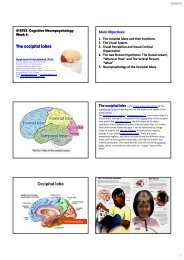
Main Objectives:<br />
1. The <strong>Neurological</strong> <strong>Examination</strong><br />
2. The Neuropsychological tests<br />
3. Clinical <strong>cases</strong> <strong>and</strong> <strong>neuropsychological</strong> interpretation<br />
4. Review <strong>and</strong> Summary<br />
4.1 Organization of the nervous system<br />
4.2 Functional Brain Imaging<br />
4.3 Sensory–Motor Systems <strong>and</strong> Cortical Functions<br />
4.4 Cerebral cortexes <strong>and</strong> lobe functions: Occipital,<br />
Parietal, Temporal <strong>and</strong> Frontal lobes<br />
A neurological examination is the<br />
assessment of sensory neuron <strong>and</strong> motor responses,<br />
especially reflexes, to determine whether the<br />
nervous system is impaired. It can be used both as a<br />
screening tool <strong>and</strong> as an investigative tool, the<br />
former of which when examining the patient when<br />
there is no expected neurological deficit <strong>and</strong> the<br />
latter of which when examining a patient where you<br />
do expect to find abnormalities. If a problem is found<br />
either in an investigative or screening process then<br />
further tests can be carried out to focus on a<br />
particular aspect of the nervous system (such as<br />
lumbar punctures <strong>and</strong> blood tests).<br />
Generally a neurological examination is focused<br />
towards finding out if there are lesions in the central<br />
<strong>and</strong> peripheral nervous systems or whether there is<br />
another diffuse process which is troubling the<br />
patient. Once the patient has been thoroughly<br />
tested, it is then the role of the physician to<br />
determine whether or not these findings combine to<br />
form a recognizable medical syndrome such as<br />
Parkinson's disease or motor neurone disease.<br />
Finally, it is the role of the physician to find the<br />
etiological reasons for why such a problem has<br />
occurred, for example finding if the problem was due<br />
to inflammation or congenital.<br />
Patient’s History<br />
A patient's history is the most important part of a neurological examination <strong>and</strong> must be<br />
performed before any other procedures unless impossible (i.e. the patient is unconscious).<br />
Certain aspects of a patients history will become more important depending upon the<br />
complaint issued. Important factors to be taken in the medical history include:<br />
1. Time of onset, duration <strong>and</strong> associated symptoms (e.g. is the complaint chronic or<br />
acute)<br />
2. Age, gender <strong>and</strong> occupation of the patient<br />
3. H<strong>and</strong>edness (right or left h<strong>and</strong>ed)<br />
4. Past medical history<br />
5. Drug history<br />
6. Family <strong>and</strong> social history<br />
H<strong>and</strong>edness is important in establishing the area of the brain important for language (as<br />
almost all right‐h<strong>and</strong>ed people have a left hemisphere which is responsible for language). As<br />
patients answer questions, it is important to gain an idea of the complaint thoroughly <strong>and</strong><br />
underst<strong>and</strong> its time course. Underst<strong>and</strong>ing the patient's neurological state at the time of<br />
questioning is important, <strong>and</strong> an idea should be obtained of how competent the patient is<br />
with various tasks <strong>and</strong> their level of impairment in carrying out these tasks. The interval of a<br />
complaint is important as it can help aid the diagnosis. For example, vascular disorders occur<br />
very frequently over minutes <strong>and</strong> hours, whereas congenital disorders occur over a matter of<br />
years.<br />
Carrying out a 'general' examination is just as important as the neurological exam as it may<br />
lead to clues to the etiology of the complaint. This is shown by <strong>cases</strong> of cerebral metastases<br />
where the initial complaint was of a mass in the breast.<br />
List of tests<br />
Specific tests in a neurological examination include:<br />
1. Mental Status <strong>Examination</strong><br />
2. Cranial Nerves <strong>Examination</strong><br />
3. Motor System <strong>Examination</strong><br />
4. Deep tendon Reflexes<br />
5. Sensory System <strong>Examination</strong><br />
6. Cerebellum <strong>Examination</strong> (Motor Coordination<br />
<strong>and</strong> Gaits)<br />
7. Higher Brain Functions<br />
1
23/07/54<br />
Interpretation<br />
The results of the examination are taken together to anatomically identify the lesion. This may<br />
be diffuse (e.g. neuromuscular diseases, encephalopathy) or highly specific (e.g. abnormal<br />
sensation in one dermatome due to compression of a specific spinal nerve by a tumor deposit).<br />
A differential diagnosis may then be constructed that takes into account the patient's<br />
background (e.g. previous cancer, autoimmune diathesis) <strong>and</strong> present findings to include the<br />
most likely causes. <strong>Examination</strong>s are aimed at ruling out the most <strong>clinical</strong>ly significant causes<br />
(even if relatively rare, e.g. brain tumor in a patient with subtle word finding abnormalities but<br />
no increased intracranial pressure) <strong>and</strong> ruling in the most likely causes<br />
Romberg's test or the Romberg maneuver is a<br />
test used by doctors in a neurological examination, <strong>and</strong> also as a test<br />
for drunken driving. The exam is based on the premise that a person<br />
requires at least two of the three following senses to maintain<br />
balanced while st<strong>and</strong>ing:<br />
Proprioception (the ability to know one's body in space); Vestibular<br />
function (the ability to know one's head position in space); <strong>and</strong><br />
Vision (which can be used to monitor [<strong>and</strong> adjust for] changes in<br />
body position).<br />
A patient who has a problem with proprioception can still maintain<br />
balance by using vestibular function <strong>and</strong> vision. In the Romberg test,<br />
the patient is stood up <strong>and</strong> asked to close his eyes. A loss of balance<br />
is interpreted as a positive Romberg sign.<br />
The Romberg test is a test of the body's sense of positioning<br />
(proprioception), which requires healthy functioning of the dorsal<br />
columns of the spinal cord, [1] .<br />
The Romberg test is used to investigate the cause of loss of motor<br />
coordination (ataxia). A positive Romberg test suggests that the<br />
ataxia is sensory in nature, that is, depending on loss of<br />
proprioception. If a patient is ataxic <strong>and</strong> Romberg's test is not<br />
positive, it suggests that ataxia is cerebellar in nature, that is,<br />
depending on localized cerebellar dysfunction instead.<br />
It is used as an indicator for possible alcohol or drug impaired<br />
driving <strong>and</strong> neurological decompression sickness. [2][3] When used to<br />
test impaired driving, the test is performed with the subject<br />
estimating 30 seconds in his head. This is used to gauge the subject's<br />
internal clock <strong>and</strong> can be an indicator of stimulant or depressant<br />
use. The test was named after the German neurologist Moritz<br />
Heinrich Romberg [1] (1795‐1873), who also gave his name to Parry‐<br />
Romberg syndrome <strong>and</strong> Howship‐Romberg sign.<br />
Procedure for Romberg's test or the Romberg maneuver<br />
Ask the subject to st<strong>and</strong> erect with feet together <strong>and</strong> eyes closed. St<strong>and</strong> close by as a<br />
precaution in order to stop the person from falling over <strong>and</strong> hurting himself or herself.<br />
Watch the movement of the body in relation to a perpendicular object behind the<br />
subject (corner of the room, door, window etc). A positive sign is noted when a swaying,<br />
sometimes irregular swaying <strong>and</strong> even toppling over occurs. The essential feature is that<br />
the patient becomes more unsteady with eyes closed.<br />
The essential features of the test are as follows:<br />
1. the subject st<strong>and</strong>s with feet together, eyes open <strong>and</strong> h<strong>and</strong>s by the sides.<br />
2. the subject closes the eyes while the examiner observes for a full minute.<br />
Because the examiner is trying to elicit whether the patient falls when the eyes are<br />
closed, it is advisable to st<strong>and</strong> ready to catch the falling patient. For large subjects, a<br />
strong assistant is recommended.<br />
Romberg's test is positive if the patient sways or falls while the patient's eyes are closed.<br />
Patients with a positive result are said to demonstrate Romberg's sign or Rombergism.<br />
They can also be described as Romberg's positive. The basis of this test is that balance<br />
comes from the combination of several neurological systems, namely proprioception,<br />
vestibular input, <strong>and</strong> vision. If any two of these systems are working the person should be<br />
able to demonstrate a fair degree of balance. The key to the test is that vision is taken<br />
away by asking the patient to close their eyes. This leaves only two of the three systems<br />
remaining <strong>and</strong> if there is a vestibular disorder (labyrinthine) or a sensory disorder<br />
(proprioceptive dysfunction) the patient will become much more imbalanced.<br />
2
23/07/54<br />
Maintaining balance while st<strong>and</strong>ing in the stationary position relies on intact<br />
sensory pathways, sensorimotor integration centers <strong>and</strong> motor pathways.<br />
The main sensory inputs are:<br />
1. Joint position sense (proprioception), carried in the dorsal columns<br />
of the spinal cord;<br />
2. Vision<br />
3. Vestibular apparatus<br />
Crucially, the brain can obtain sufficient information to maintain balance if any<br />
two of the three systems are intact.<br />
Sensorimotor integration is carried out by the cerebellum <strong>and</strong> by the dorsal<br />
column‐medial lemniscus tract. The motor pathway is the corticospinal<br />
(pyramidal) tract <strong>and</strong> the medial <strong>and</strong> lateral vestibular tracts.<br />
The first stage of the test (st<strong>and</strong>ing with the eyes open), demonstrates that at<br />
least two of the three sensory pathways is intact, <strong>and</strong> that sensorimotor<br />
integration <strong>and</strong> the motor pathway are functioning.<br />
In the second stage, the visual pathway is removed by closing the eyes, known as<br />
a "sharpened Romberg". If the proprioceptive <strong>and</strong> vestibular pathways are intact,<br />
balance will be maintained. But if proprioception is defective, two of the sensory<br />
inputs will be absent <strong>and</strong> the patient will sway then fall.<br />
The sharpened Romberg does have an early learning effect that will plateau<br />
between the third <strong>and</strong> fourth attempts.<br />
Positive Romberg<br />
Romberg's test is positive in conditions causing sensory ataxia such as:<br />
Conditions affecting the dorsal columns of the spinal cord, such as tabes<br />
dorsalis (neurosyphilis), in which it was first described.<br />
Conditions affecting the sensory nerves (sensory peripheral neuropathies), such<br />
as chronic inflammatory demyelinating polyradiculoneuropathy (CIDP).<br />
Friedreich's Ataxia<br />
Romberg <strong>and</strong> cerebellar function<br />
Romberg's test is not a test of cerebellar function, as it is commonly<br />
misconstrued. Patients with cerebellar ataxia will, generally, be unable to<br />
balance even with the eyes open; [5] therefore, the test cannot proceed beyond<br />
the first step <strong>and</strong> no patient with cerebellar ataxia can correctly be described as<br />
Romberg's positive. Rather, Romberg's test is a test of the proprioception<br />
receptors <strong>and</strong> pathways function. A positive Romberg's test has been shown to<br />
be 90% sensitive for lumbar spinal stenosis. [<br />
<strong>Neurological</strong> <strong>Examination</strong> Videos<br />
And <strong>Neurological</strong> case examples<br />
http://library.med.utah.edu/neurologicexam/<strong>cases</strong>/html_case01/case01_history.html<br />
A case begins with a Case<br />
History in which<br />
preliminary information<br />
about the patient <strong>and</strong> any<br />
signs <strong>and</strong> symptoms are<br />
presented.<br />
The <strong>Neurological</strong><br />
<strong>Examination</strong> follows the<br />
Case History. You can choose<br />
the order <strong>and</strong> the parts of<br />
the neurological<br />
examination that you would<br />
like to view by clicking on<br />
the icon that represents that<br />
part of the exam.<br />
After completing the exam, you advance to listing your<br />
abnormal findings. You use the supplied Checklist of<br />
Findings <strong>and</strong> compare your choices with that of an<br />
expert's. You are now ready to begin the process of<br />
anatomical localization.<br />
You start to Localize the Level(s) of the Lesion by<br />
selecting from the level of the neuroaxis. Your<br />
choices include:<br />
1. Supratentorial<br />
2. Infratentorial<br />
3. Spinal Cord<br />
4. Peripheral Nerve System<br />
5. Multiple Levels<br />
From a list of the structures, you choose the brain<br />
structure(s) those you think are damaged for the case.<br />
Your choices are compared to an expert's, <strong>and</strong> the<br />
lesion is highlighted on the image.<br />
You have now arrived at an anatomical diagnosis, the<br />
first essential step in making a neurological diagnosis.<br />
Finally, in the Case Discussion, you review the case <strong>and</strong><br />
the thought processes used to reach the diagnosis.<br />
Neuroimaging studies, if available, are shown as part of<br />
the case discussion.<br />
3
23/07/54<br />
Case No. 01: The Upset Office Manager<br />
The patient is a 48‐year‐old woman who was in her usual state of<br />
good health when she experienced nausea <strong>and</strong> vomiting after being<br />
emotionally upset. After 3 hours of nausea <strong>and</strong> vomiting she had the<br />
sudden onset of numbness of her left arm which progressed to<br />
include her left leg <strong>and</strong> the left side of her face.<br />
She was taken to the Emergency Room. Upon arrival she complained<br />
that she had double vision especially when she looked to the right.<br />
When she covered her right eye, the most peripheral image (the<br />
ghost image) would disappear. She also noticed that when she looked<br />
in the mirror the right side of her face didn’t move.<br />
Over the next 2 months, the double vision resolved, but the rest of<br />
her complaints have persisted<br />
http://library.med.utah.edu/neurologicexam/<strong>cases</strong>/html_case01/case01_history.html<br />
4
23/07/54<br />
5
23/07/54<br />
This 48‐year‐old woman had the sudden onset <strong>and</strong> rapid progression of her symptoms,<br />
which is the temporal profile of an acute vascular event or a stroke. Risk factors for stroke<br />
include hypertension, cardiac disease, diabetes mellitus, smoking as well as coagulation <strong>and</strong><br />
autoimmune disorders. This patient had none of these risk factors.<br />
One of the first steps in localizing the occluded vessel in a stroke is to decide if the vessel is<br />
in the anterior or posterior arterial circulation of the brain. Most strokes occur in the<br />
anterior or carotid circulation because most of the brain’s blood supply is supplied by the<br />
carotids. Although strokes in the posterior or vertebrobasilar circulation are less common,<br />
this patient’s cranial nerve findings suggest an infarct in the brainstem <strong>and</strong> not the carotid<br />
territory.<br />
By history <strong>and</strong> examination there are 3 findings that indicate possible cranial nerve<br />
involvement. Her history of diplopia indicates a right 6th (abducens) cranial nerve deficit<br />
(remember the most peripheral image is the false image <strong>and</strong> covering the right eye<br />
eliminated this image) <strong>and</strong> by examination she has a right 7th (facial) cranial nerve deficit. To<br />
explain these two deficits one has to localize the lesion in the caudal pons in the area of the<br />
6th <strong>and</strong> 7th cranial nerve nuclei or the pathway of the nerves at this level.<br />
To explain the sensory findings on the left side of her face, we could postulate either a left<br />
spinal trigeminal tract lesion or it could be from a lesion affecting the axons of the 2nd order<br />
neurons which have crossed over to the right side of the pons <strong>and</strong> are ascending in the<br />
ventral trigeminothalamic tract. The left spinal trigeminal tract lesion hypothesis is<br />
unattractive, because it would mean a second lesion. In the pons, the ascending ventral<br />
trigeminothalamic tract is near the facial nucleus, so a lesion affecting this tract is the likely<br />
explanation.<br />
The last finding that we have to account for anatomically is the sensory deficit on<br />
the left side of the body. We first have to decide if the deficit is from a lesion in the<br />
DC‐ML system or the spinothalamic (ALS) system. For this patient, pain <strong>and</strong><br />
temperature are affected while vibratory, position sense, <strong>and</strong> discriminatory<br />
sensation are preserved, which indicates that the spinothalamic tract is involved<br />
but the DC‐ML system is spared. Recall that the axons from the second order<br />
neurons that form the spinothalamic tract cross immediately in the spinal cord <strong>and</strong><br />
ascend in the anterolateral spinal cord <strong>and</strong> the lateral part of the brainstem. In the<br />
pons, the spinothalamic tract, carrying pain <strong>and</strong> temperature for the left side of the<br />
body, is adjacent to the facial motor nucleus.<br />
So we could explain all the <strong>clinical</strong> findings for this case by a lesion in the<br />
mediolateral part of the right lower pons most likely caused by occlusion of one of<br />
the short circumferential branches of the basilar artery. It is not a paramedian<br />
lesion because the patient has no findings referable to the corticospinal tracts <strong>and</strong><br />
the medial lemniscus. It is also not a far lateral lesion because there are no rightsided<br />
spinal trigeminal tract, vestibular, or cerebellar findings.<br />
An MRI scan of the patient done 6 months after her stroke shows a small residual<br />
lesion in the area of the right abducens nucleus. This imaging finding doesn’t cover<br />
the entire anatomical area where we know there has to be disease, but it does<br />
support the hypothesis that there has been a small stroke in the area where we<br />
localized her lesion based on her <strong>clinical</strong> findings<br />
Review <strong>and</strong> Summary:<br />
6
23/07/54<br />
415703 Cognitive Neuropsychology<br />
Week 1:<br />
Introduction to Neuropsychology,<br />
<strong>and</strong> the Organization of the<br />
Nervous System.<br />
Naiphinich Kotchabhakdi, Ph.D.<br />
Director, Salaya Stem Cell R & D Project,<br />
Research Center for Neuroscience,<br />
Institute of Molecular Biosciences,<br />
Mahidol University Salaya Campus,<br />
999 Phutthamonthol 4 Road, Salaya, Phutthamonthol,<br />
Nakornpathom 73170 Thail<strong>and</strong><br />
Email: scnkc@mahidol.ac.th or naiphinich@gmail.com<br />
Web: www.neuroscience.mahidol.ac.th<br />
Main Objectives:<br />
1. What is Neuropsychology (for education)?<br />
2. What are <strong>neuropsychological</strong> disorders?<br />
3. New Approaches <strong>and</strong> tools in <strong>neuropsychological</strong><br />
disorders.<br />
4. What are <strong>neuropsychological</strong> py<br />
assessment?<br />
5. What is the Organization of the nervous system?<br />
6. What are the structure <strong>and</strong> functions of specific<br />
human brain areas?<br />
Neuropsychology (Brain <strong>and</strong> Mind)<br />
Neuropsychology studies the structure <strong>and</strong> function of the<br />
brain related to specific psychological (mental) processes<br />
<strong>and</strong> behaviors.<br />
The term neuropsychology has been applied to lesion studies in humans <strong>and</strong><br />
animals. It has also been applied to efforts to record electrical activity from<br />
individual cells (or groups of cells) in higher primates (including some studies<br />
of human patients). It is scientific in its approach <strong>and</strong> shares an information<br />
processing view of the mind with cognitive psychology <strong>and</strong> cognitive<br />
neuroscience.<br />
In practice neuropsychologists tend to work in <strong>clinical</strong> settings (involved<br />
in assessing or treating patients with <strong>neuropsychological</strong> problems –<br />
see <strong>clinical</strong> neuropsychology), forensic settings or industry (often as<br />
consultants where <strong>neuropsychological</strong> knowledge is applied to product design<br />
or in the management of pharmaceutical <strong>clinical</strong>-trials research for drugs that<br />
might have a potential impact on CNS functioning).<br />
Posner, M.I.& DiGirolamo,G.J.(2000<br />
2000) Cognitive Neuroscience:Origins<br />
<strong>and</strong> Promise,Psychological<br />
Psychological Bulletin, 126:6, 873‐889<br />
889<br />
From Wikipedia, the free encyclopedia<br />
Divisions of the Nervous System<br />
Cellular components of the nervous tissue<br />
7
23/07/54<br />
Spinal cord<br />
The spinal cord is a long, thin, tubular bundle of nervous tissue <strong>and</strong> support cells that<br />
extends from the brain (the medulla oblongata specifically). The brain <strong>and</strong> spinal cord<br />
together make up the central nervous system. The spinal cord begins at the Occipital bone<br />
<strong>and</strong> extends down to the space between the first <strong>and</strong> second lumbar vertebrae; itdoes<br />
not extend the entire length of the vertebral column.Itisaround45cm(18in)inmen<strong>and</strong><br />
around 43 cm (17 in) long in women. Also, the spinal cord has a varying width, ranging<br />
from 1/2 inch thick in the cervical <strong>and</strong> lumbar regions to 1/4 inch thick in the thoracic<br />
area. The enclosing bony vertebral column protects the relatively shorter spinal cord.<br />
The spinal cord functions primarily in the transmission of neural signals between the brain<br />
<strong>and</strong> the rest of the body but also contains ti neural circuits it thatt can independently d control<br />
numerous reflexes <strong>and</strong> central pattern generators. The spinal cord has three major<br />
functions:1.Serve<br />
as a conduit for motor information, which travels down the spinal cord.<br />
2.Serve<br />
as a conduit for sensory information, which travels up the spinal cord. 3. Serve as<br />
acenter<br />
for coordinating certain reflexes.<br />
The spinal cord is the main pathway for information connecting the brain <strong>and</strong> peripheral<br />
nervous system. The length of the spinal cord is much shorter than the length of the bony<br />
spinal column. The human spinal cord extends from the medulla oblongata <strong>and</strong> continues<br />
through the conus medullaris near the first lumbar vertebra, terminating in a fibrous<br />
extension known as the filum terminale<br />
Spinal cord<br />
Somatosensory system<br />
Dermatome<br />
8
23/07/54<br />
Autonomic nervous<br />
nervous system)<br />
system<br />
(ANS<br />
or<br />
visceral<br />
system) is the part of the peripheral nervous system that acts<br />
as a control system functioning largely below the level of consciousness, <strong>and</strong><br />
controls visceral functions. The ANS affects heart rate, digestion, respiration rate,<br />
salivation, perspiration, diameter of the pupils, micturition (urination), <strong>and</strong> sexual<br />
arousal. Whereas most of its actions are involuntary, some, such as breathing,<br />
work in t<strong>and</strong>em with the conscious mind.<br />
It is classically divided into two subsystems: the parasympathetic nervous system<br />
(PSNS) <strong>and</strong> sympathetic nervous system (SNS). Relatively recently, a third<br />
subsystem of neurons that have been named 'non‐adrenergic<br />
<strong>and</strong> non‐cholinergic'<br />
neurons (because they use nitric oxide as a neurotransmitter) have been<br />
described <strong>and</strong> found to be integral in autonomic function, particularly in the gut<br />
<strong>and</strong> the lungs.<br />
With regard to function, the ANS is usually divided into sensory (afferent) <strong>and</strong><br />
motor (efferent) subsystems. Within these systems, however, there are inhibitory<br />
<strong>and</strong> excitatory synapses between neurons.<br />
The enteric nervous system is sometimes considered part of the autonomic<br />
nervous system, <strong>and</strong> sometimes considered an independent system.<br />
Autonomic Nervous<br />
System (ANS) <strong>and</strong> autonomic reflexes<br />
Alongside the other two components of the autonomic nervous system, thesympathetic<br />
nervous system aids in the control of most of the body's internal organs. Stress—as in the<br />
flight‐or‐fight response—is thought to counteract the parasympathetic system, which<br />
generally works to promote maintenance of the body at rest. In truth, the functions of<br />
both the parasympathetic <strong>and</strong> sympathetic nervous systems are not so straightforward,<br />
but this is a useful rule of thumb.<br />
There are two kinds of neurons involved in the transmission of any signal through the<br />
sympathetic system; pre‐ <strong>and</strong> post‐ ganglionic. The shorter preganglionic neurons originate<br />
from the thoracolumbar region of the spinal cord (levels T1 ‐ L2, specifically) <strong>and</strong> travel to<br />
a ganglion, often one of the paravertebral ganglia, where they synapse with a<br />
postganglionic neuron. From there, the long postganglionic neurons extend across most of<br />
the body.<br />
At the synapses within the ganglia, preganglionic neurons release acetylcholine, a<br />
neurotransmitter that activates nicotinic acetylcholine receptors on postganglionic<br />
neurons. In response to this stimulus postganglionic neurons ‐ with two important<br />
exceptions ‐ release norepinephrine, which activates adrenergic receptors on the<br />
peripheral target tissues. The activation of target tissue receptors causes the effects<br />
associated with the sympathetic system.<br />
The two exceptions mentioned above are postganglionic neurons innervating sweat<br />
gl<strong>and</strong>s—which release acetylcholine for the activation of muscarinic receptors ‐ <strong>and</strong> the<br />
adrenal medulla. The adrenal medulla develops in t<strong>and</strong>em with the sympathetic nervous<br />
system, <strong>and</strong> acts as a modified sympathetic ganglion: synapses occur between pre‐ <strong>and</strong><br />
post‐ ganglionic neurons within it, but the post ganglionic neurons do not leave the<br />
medulla; instead they directly release norepinephrine <strong>and</strong> epinephrine into the blood.<br />
Sympathetic <strong>and</strong> parasympathetic systems<br />
Sympathetic nervous system<br />
Parasympathetic nervous system<br />
9
23/07/54<br />
Control of blood<br />
vessels<br />
Control of<br />
pupil<br />
Brainstem (or brain stem) is the posterior part of the brain,<br />
adjoining <strong>and</strong> structurally continuous with the spinal cord. The brain stem<br />
providesthemainmotor<strong>and</strong>sensoryinnervationtotheface<strong>and</strong>neckviathe<br />
cranial nerves. Though small, this is an extremely important part of the brain as<br />
the nerve connections of the motor <strong>and</strong> sensory systems from the main part of<br />
the brain to the rest of the body pass through the brain stem. This includes the<br />
corticospinal tract (motor), the posterior column‐medial lemniscus pathway (fine<br />
touch, vibration sensation <strong>and</strong> proprioception)<strong>and</strong>thespinothalamic tract (pain,<br />
temperature, itch <strong>and</strong> crude touch). The brain stem also plays an important role<br />
in the regulation of cardiac <strong>and</strong> respiratory function. It also regulates the central<br />
nervoussystem,<strong>and</strong>ispivotalinmaintaining consciousness <strong>and</strong> regulating the<br />
sleep cycle.<br />
Brainstem is made up of 1. medulla oblongata (myelencephalon), 2. pons (part of<br />
metencephalon), 3. midbrain (mesencephalon), <strong>and</strong> 4. diencephalon<br />
Mid‐sagittal<br />
view of the<br />
adult human brain<br />
There are three main functions of the brain stem:<br />
1. The first is its role in conduction. That is, all information relayed from the body<br />
to the cerebrum <strong>and</strong> cerebellum <strong>and</strong> vice versa, must traverse the brain stem.<br />
The ascending pathways coming from the body to the brain are the sensory<br />
pathways, <strong>and</strong> include the spinothalamic tract for pain <strong>and</strong> temperature<br />
sensation <strong>and</strong> the dorsal column, fasciculus gracilis, <strong>and</strong> cuneatus for touch,<br />
proprioception, <strong>and</strong> pressure sensation (both of the body). (The facial sensations<br />
have simiar pathways, <strong>and</strong> will travel in the spinothalamic tract <strong>and</strong> the medial<br />
lemniscus also). Descending tracts are upper motor neurons destined to synapse<br />
on lower motor neurons in the ventral horn <strong>and</strong> intermediate horn of the spinal<br />
cord. In addition, there are upper motor neurons that originate in the brain<br />
stem's vestibular, red, tectal, <strong>and</strong> reticular nuclei, which also descend <strong>and</strong> synapse<br />
in the spinal cord.<br />
2. The cranial nerves 3‐12 emerge from the brain stem.<br />
3. The brain stem has integrative functions (it is involved in cardiovascular system<br />
control, respiratory control, pain sensitivity control, alertness, awareness, <strong>and</strong><br />
consciousness). Thus, brain stem damage is a very serious <strong>and</strong> often lifethreatening<br />
problem.<br />
Brainstem <strong>and</strong> cerebellum<br />
Ventral<br />
view<br />
10
23/07/54<br />
12 Cranial nerves:<br />
1. Olfactory<br />
2. Optic<br />
3. Oculomotor<br />
4. Trochlear<br />
5. Trigeminal<br />
6. Abducens<br />
7. Facial<br />
8. Auditory <strong>and</strong><br />
Vestibular<br />
9. Glossopharyngeal<br />
10. Vagus<br />
11. Spinal Accessory<br />
12. Hypoglossal<br />
Control of respiration<br />
Cardiovascular controls<br />
Long tracts of Sensory <strong>and</strong> Motor<br />
Systems<br />
1. Pathways in spinal cord <strong>and</strong> brainstem<br />
2. Functions<br />
3. Clinical correlates, signs <strong>and</strong> symptoms<br />
11
23/07/54<br />
Cerebellum<br />
Vestibular<br />
<strong>and</strong><br />
Cerebellum<br />
The cerebellum (Latin for little brain) isaregionofthe<br />
brain that plays an important role in motor control.Itisalsoinvolved<br />
in some cognitive functions such as attention <strong>and</strong> language, <strong>and</strong><br />
probably in some emotional functions such as regulating fear <strong>and</strong><br />
pleasure responses. Its movement‐related functions are the most<br />
clearly understood, however. The cerebellum does not initiate<br />
movement, but it contributes to coordination, precision, <strong>and</strong><br />
accurate timing. It receives input from sensory systems <strong>and</strong> from<br />
other parts of the brain <strong>and</strong> spinal cord, <strong>and</strong> integrates these inputs<br />
to fine tune motor activity. Because of this fine‐tuning function,<br />
damage to the cerebellum does not cause paralysis, but instead<br />
produces disorders in fine movement, equilibrium, posture, <strong>and</strong><br />
motor learning<br />
In addition to its direct role in motor control <strong>and</strong> coordination, the<br />
cerebellum also is necessary for several types of motor learning, the<br />
most notable one being learning to adjust<br />
to changes<br />
in<br />
sensorimotor relationships.<br />
The Human Cerebellum:<br />
Cerebellar Functions (Classical Functions):<br />
Co‐ordination of Movements<br />
Stabilizing of Vestibulo‐ocular Reflex (VOR)<br />
Patterned Skilled Movements<br />
Coordination of Eye Movements<br />
Vermis Lobule VI‐‐‐‐Saccadic Eye Movements<br />
Flocculus‐‐‐‐ Visual Tracking<br />
Equilibrium, Gait <strong>and</strong> Postural Controls<br />
Controls of Autonomic Functions (Cardiovascular)<br />
Immune Functions (?)<br />
12
23/07/54<br />
The Human Cerebellum:<br />
Clinical Correlates on the Human Cerebellum:<br />
Flocculo‐Nodular Lobe:<br />
Disturbed Equilibrium or Balance<br />
Tendency to fall on the side of the lesion<br />
Extensor hypotonia<br />
“Reeling or Drunken” ”Gi Gait or Posture<br />
Deviation Nystagmus<br />
1. Eye in mid‐line Position:<br />
fine nystagmus, quick phase toward lesion side<br />
2. Eye fixed 10 –30 degree away from the lesion side:<br />
No nystagmus<br />
3. Eye fixed beyond 30 degree away from the lesion side<br />
nystagmus with quick phase away from lesion side<br />
4. Eye shift beyond midline toward the lesion side<br />
gross nystagnus, quick phase toward lesion side<br />
The Human Cerebellum:<br />
Clinical Correlates on the Human Cerebellum:<br />
Anterior Lobe & Vermis:<br />
Hypotonia (Reduced muscle tone)<br />
Hyporeflexia<br />
Ataxia (Incoordination of Movements)<br />
Asynergia, Dysynergia (lack of synergy)<br />
Intention or Action Tremor (Atelokinesia)<br />
Asthenia (Weakness of muscular strength)<br />
Rebounded Phenomenon<br />
Cerebellar Hemisphere & Dentate Nucleus:<br />
Dysmetria (Pass pointing)<br />
Dysdiadochokinesia or Adiadochokinesia<br />
(can not perform rapid alternating movements)<br />
Disturbed Voluntary Skilled movements<br />
Disturbed Speech (Drunken Speech)<br />
13
23/07/54<br />
Dysmetria of thought<br />
The Principles of Motor Controls of Movements:<br />
1. The central nervous system (CNS) has to choose the right group<br />
of muscles by selecting specific pathways.<br />
2. The CNS must give the right amount of excitatory or inhibitory<br />
inputs (“Comm<strong>and</strong>”) to specific motoneuron pools<br />
3. The excitatory <strong>and</strong> inhibitory comm<strong>and</strong>s must be regulated<br />
“Spatially” <strong>and</strong> “Temporally”.<br />
4. The CNS must regulate the following parameters:<br />
‐ force<br />
‐displacement (distance)<br />
‐ velocity, acceleration or deceleration<br />
Pyramidal System:<br />
Cortico‐spinal tracts<br />
UMN Lesions (Pyramidal Syndrome)<br />
A. Paralyze movements in hemiplegic,<br />
quadriplegic, or paraplegic distribution, not<br />
individual muscles<br />
B. Atrophy of disuse only (late <strong>and</strong> slight)<br />
C. Hyperactive MSRs Clonus<br />
D. Clasp‐knife spasticity<br />
E. Absent abdominal <strong>and</strong> cremasteric reflexes<br />
F. Extensor toe sign (Babinski sign)<br />
14
23/07/54<br />
Plantar Flexion<br />
Dorsi‐ Flexion<br />
LMN Lesions<br />
A. Paralyze individual muscles or sets of muscles<br />
in root or peripheral nerve distribution<br />
B. Atrophy of denervation (early <strong>and</strong> severe<br />
C. Fasciculations <strong>and</strong> fibrillations<br />
D. Hypoactive or absent MSRs Hypotonia<br />
UMN = upper motoneuron; LMN = lower<br />
motoneuron; MSRs = muscle stretch reflexes<br />
Basal ganglion <strong>and</strong><br />
Extrapyramidal<br />
System<br />
Disease of the basal ganglion:<br />
Parkinson’s disease<br />
Chorea <strong>and</strong> Huntington’s Chorea<br />
Athetosis <strong>and</strong> Athetoid<br />
Sydenham’s Chorea<br />
Hemibalism .. Lesion of subthalamic nucleus<br />
Dystonia, Torticolis,<br />
Wilson’s disease (Copper)<br />
Kern icterus (Bilirubin stain)<br />
Parkinson’s disease:<br />
Akinesia or Hypokiesia<br />
cog wheel rigidity<br />
Resting Tremor<br />
Degeneration of Dopaminergic neurons in<br />
Pars Compacta of the Substantia Nigra<br />
Motor<br />
control<br />
system<br />
Motor<br />
homonculus<br />
(Maps)<br />
15
23/07/54<br />
Control of<br />
body<br />
movements,<br />
visceral<br />
organs,<br />
behavior<br />
<strong>and</strong><br />
emotion<br />
Hypothalamus<br />
The Hypothalamus is a portion of the brain that contains a number of small<br />
nuclei with a variety of functions. One of the most important functions of the<br />
hypothalamus is to link the nervous system to the endocrine system via the<br />
pituitary gl<strong>and</strong> (hypophysis).<br />
The hypothalamus is located below the thalamus, just above the brain stem. In<br />
the terminology of neuroanatomy, itformstheventral part of the diencephalon.<br />
All vertebrate brains contain a hypothalamus. In humans, it is roughly the size of<br />
an almond.<br />
The hypothalamus is responsible for certain metabolic processes <strong>and</strong> other<br />
activities of the autonomic nervous system. It synthesizes <strong>and</strong> secretes certain<br />
neurohormones, often called hypothalamic‐releasing hormones, <strong>and</strong> these in<br />
turn stimulate or inhibit the secretion of pituitary hormones. The hypothalamus<br />
controls body temperature, hunger, thirst,fatigue,sleep,<strong>and</strong>circadian cycles.<br />
Controls of body<br />
temperature<br />
Controls of food<br />
intake <strong>and</strong> body<br />
weight<br />
Endocrine <strong>and</strong><br />
Hormonal controls<br />
Controls of<br />
kidney functions<br />
16
23/07/54<br />
Limbic<br />
System<br />
Controls of<br />
emotion <strong>and</strong><br />
motivation<br />
The thalamus is a midline paired symmetrical<br />
structure within the brains of vertebrates, including<br />
humans. It is situated between the cerebral cortex <strong>and</strong><br />
midbrain, both in terms of location <strong>and</strong> neurological<br />
connections. Its function includes relaying sensation,<br />
spatial sense, <strong>and</strong> motor signals to the cerebral cortex,<br />
along with the regulation of consciousness, sleep, <strong>and</strong><br />
alertness. The thalamus surrounds the third ventricle. It is<br />
the main product of the embryonic diencephalon<br />
Thalamus<br />
<strong>and</strong><br />
Cerebral<br />
cortex<br />
The thalamus has multiple functions. It is generally believed to act as a relay between a<br />
variety of subcortical areas <strong>and</strong> the cerebral cortex. In particular, every sensory system (with the<br />
exception of the olfactory system) includes a thalamic nucleus that receives sensory signals <strong>and</strong> sends<br />
them to the associated primary cortical area. For the visual system, for example, inputs from the retina<br />
are sent to the lateral geniculate nucleus ofthethalamus,whichinturnprojectstotheprimary visual<br />
cortex (area V1) in the occipital lobe. The thalamus is believed to both process sensory information as<br />
well as relaying it—each of the primary sensory relay areas receives strong "back projections" from<br />
the cerebral cortex. Similarly the medial geniculate nucleus acts as a key auditory relay between the<br />
inferior colliculus of the midbrain <strong>and</strong> the primary auditory cortex,<strong>and</strong>theventral posterior nucleus is<br />
a key somatosensory relay, which sends touch <strong>and</strong> proprioceptive information to the primary<br />
somatosensory cortex.<br />
The thalamus also plays an important role in regulating states of sleep <strong>and</strong> wakefulness. [4] Thalamic<br />
nuclei have strong reciprocal connections with the cerebral cortex, forming thalamo‐cortico‐thalamic<br />
thalamic<br />
circuits that are believed to be involved with consciousness. The thalamus plays a major role in<br />
regulating arousal, the level of awareness, <strong>and</strong> activity. Damage to the thalamus can lead to<br />
permanent coma.<br />
Many different functions are linked to various regions of the thalamus. This is the case for many of the<br />
sensory systems (except for the olfactory system), such as the auditory, somatic, visceral, gustatory<br />
<strong>and</strong> visual systems where localized lesions provoke specific sensory deficits. A major role of the<br />
thalamus is devoted to "motor" systems. This has been <strong>and</strong> continues to be a subject of interest for<br />
investigators. VIm, the relay of cerebellar afferences, is the target of stereotactians particularly for the<br />
improvement of tremor. The role of the thalamus in the more anterior pallidal <strong>and</strong> nigral territories in<br />
the basal ganglia system disturbances is recognized but still poorly understood. The contribution of the<br />
thalamus to vestibular or to tectal functions is almost ignored. The thalamus has been thought of as a<br />
"relay" that simply forwards signals to the cerebral cortex. Newer research suggests that thalamic<br />
function is more selective<br />
Cerebral cortex in different areas<br />
The cerebral cortex is a sheet of neural tissue that is outermost to the<br />
cerebrum of the mammalian brain. It plays a key role in memory, attention,<br />
perceptual awareness, thought, language, <strong>and</strong>consciousness. It is constituted of<br />
up to six horizontal layers, each of which has a different composition in terms of<br />
neurons <strong>and</strong> connectivity. The human cerebral cortex is 2–4 mm (0.08–<br />
0.16 inches) thick.<br />
In preserved brains, it has a gray color, hence the name "gray matter". In contrast<br />
to gray matter that is formed from neurons <strong>and</strong> their unmyelinated fibers, the<br />
white matter below them is formed predominantly by myelinated axons<br />
interconnecting neurons in different regions of the cerebral cortex with each<br />
other <strong>and</strong> neurons in other parts of the central nervous system.<br />
The surface of the cerebral cortex is folded in large mammals, such that more<br />
than two‐thirds of it in the human brain is buried in the grooves, called "sulci".<br />
The phylogenetically most recent part of the cerebral cortex, the neocortex (also<br />
called isocortex), is differentiated into six horizontal layers; themoreancientpart<br />
of the cerebral cortex, the hippocampus (also called archicortex), has at most<br />
three cellular layers, <strong>and</strong> is divided into subfields. Neurons in various layers<br />
connect vertically to form small microcircuits, called columns. Different<br />
neocortical architectonic fields are distinguished upon variations in the thickness<br />
of these layers, their predominant cell type <strong>and</strong> other factors such as<br />
neurochemical markers.<br />
17
23/07/54<br />
Functional brain mapping<br />
แผนที<br />
่การทํางานของสมอง (Brain Mapping<br />
Brain Mapping)<br />
Broadmann’ s Areas<br />
Broadmann’s area #<br />
Brodmann’s area is a region of the cerebral cortex defined based on<br />
its cytoarchitectonics, or organization of cells<br />
Brodmann areas were originally defined <strong>and</strong> numbered by the German neurologist<br />
Korbinian Brodmann basedonthecytoarchitecture organisation of neurons he<br />
observed in the cerebral cortex using the Nissl stain. Brodmann published his maps<br />
of cortical areas in humans, monkeys, <strong>and</strong> other species in 1909, along with many<br />
other findings <strong>and</strong> observations regarding the general cell types <strong>and</strong> laminar<br />
organization of the mammalian cortex. (The same Brodmann area number in<br />
different species does not necessarily indicate homologous areas.)<br />
A more detailed <strong>and</strong> verifiable cortical map have since been published by Constantin von<br />
Economo <strong>and</strong> Georg N. Koskinas which greatly improves the quality of the cytoarchitectonic<br />
classifications.<br />
Many of the areas Brodmann defined based solely on their neuronal organization have since<br />
been correlated closely to diverse cortical functions. For example, Brodmann areas 1, 2 <strong>and</strong><br />
3aretheprimary somatosensory cortex; area4istheprimary motor cortex; area17isthe<br />
primary visual cortex; <strong>and</strong> areas 41 <strong>and</strong> 42 correspond closely to primary auditory cortex.<br />
Higher order functions of the association cortical areas are also consistently localized to the<br />
same Brodmann areas by neurophysiological, functional imaging, <strong>and</strong> other methods (e.g.,<br />
the consistent localization of Broca's speech <strong>and</strong> language area to the left Brodmann areas<br />
44 <strong>and</strong> 45). However, functional imaging can only identify the approximate localization of<br />
brain activations in terms of Brodmann areas since their actual boundaries in any individual<br />
brain requires its histological examination.<br />
18
่<br />
23/07/54<br />
Brodmann areas for human & non‐human primates<br />
Brodmann’s areas 3D<br />
map: Lateral Surface<br />
map: Medial Surface<br />
Brodmann areas for human & non‐human primates<br />
Paul MacLean<br />
M.D.<br />
Paul MacLean’s<br />
Triune Brain<br />
The Reptilian Brain : Core brainstem<br />
The Paleomammalian Brain : the limbic system<br />
The Neomammalian Brain : neocortex <strong>and</strong> neocerebellum<br />
สมองส่วนแรก คือ สมองของสัตว์เลื้อยคลาน (Reptilian Brain)<br />
เป็นสมองที่มนุษย์เราได้รับมรดกตกทอดมาจากสัตว์เลื้อยคลานยุคดึกดําบรรพ์ อยู ่ภายใต้<br />
อิทธิพลของพันธุกรรม 90 – 95 % และเจริญเติบโตในระหว่างที่อยู ่ในครรภ์มารดาเป็น<br />
ส่วนใหญ่ เมื่อเกิดมาแล้วสิ่งแวดล้อมมีอิทธิพลต่อสมองส่วนนี้น้อยมาก มันจะถูกปัจจัย<br />
ทางพันธุกรรมกําหนดมาเลยว่าเป็นสมองคน หรือสมองสัตว์และมีโครงสร้างและการ<br />
ทํางานอย่างไร สมองส่วนนี้ควบคุมการทํางานของอวัยวะต่างในร่างกายโดยอัตโนมัติ<br />
และพฤติกรรมที่เป็นสัญชาติญาณของสิ่งมีชีวิตที่มีมาโดยกําเนิดโดยการกําหนดของ<br />
พันธุกรรม ได้มรดกโดยตรงมาจากพ่อแม่ พ่อแม่เป็นอย่างไรลูกจะได้มรดกตกทอดมาเป็น<br />
อย่างนั้นเลย อยางนนเลย Reptilian Brain มีลักษณะเป็นแกนอย่ตอนในสดของสมองเป็นส่วนของ<br />
มลกษณะเปนแกนอยูตอนในสุดของสมองเปนสวนของ<br />
ก้านสมองและสมองตอนกลาง สมองส่วนที่หนึ่งนี้ เป็นสมองส่วนที่ทําให้มนุษย์มีสัญชาติ<br />
ญาณของการอยู ่รอด การกิน การขับถ่าย การสืบพันธ์ เริ่มสร้างขึ้นตั้งแต่ขณะที่ทารกอยู ่ใน<br />
ครรภ์มารดา ในวันที่คลอดนั้นสมองส่วนนี้สามารถทํางานได้ราว 99 % และเติบโตสมบูรณ์<br />
พร้อมทํางานเต็มที่ในช่วงขวบปีแรก ถ้าสมองส่วนแรกนี้ไม่สามารถทํางานได้ดีทารกก็ไม่<br />
อาจมีชีวิตอยู ่รอดได้ เพราะมันไปควบคุมการเต้นของหัวใจ การหายใจ ระบบขับถ่าย การ<br />
กินการอยู ่ การตื่น การนอนหลับทุกอย่างหมดเลย ในช่วงสองขวบปีแรก พ่อแม่ และผู ้เลี้ยง<br />
ดูเด็กจะสอนเด็กให้สามารถควบคุมร่างกาย ควบคุมการกินอยู ่ ควบคุมการขับถ่าย และ<br />
สร้างนิสัยต่างๆที่เหมาะสมกับการอยู ่รอดในสังคม<br />
สมองส่วนที ่สอง คือ สมองสัตว์เลี้ยงลูกด้วยนมยุคโบราณ<br />
(Paleomammalian Brain หรือ Limbic System) เป็นสมองส่วนที่<br />
มนุษย์เราได้รับมรดกตกทอดมาจากสัตว์เลี้ยงลูกด้วยนมยุคโบราณ สมองส่วนนี้จะเริ่ม<br />
สร้างและเจริญเติบโตเมื่อทารกอยู ่ในครรภ์มารดาได้ราว ๆ หกเดือน Limbic<br />
Systemจะมีลักษณะคล้ายวงแหวนที่หุ ้มรอบๆสมองส่วนแรกซึ่งมีลักษณะเป็นแกน<br />
เอาไว้ หน้าที่ของสมองส่วนนี้ก็คือ ทําให้ทารกเกิดความจําเกี่ยวกับเหตุการณ์และสถานที่<br />
(Episodic or Spatiotemporal Memory) โดยเฉพาะความจําที่เกี่ยวกับ<br />
ใบหน้าแม่ จํากลิ่นแม่ได้ ทําให้มนุษย์รู ้จักตัวเอง (“Self”) และพัฒนาให้มีความรู ้สึก<br />
(Feeling)และการแสดงออกทางอารมณ์ต่าง ๆ มันจะเป็นตัวที่ทําให้ทารกร้องไห้โยเย<br />
เรียกร้องความสนใจ แสดงอารมณ์ความรู ้สึกเวลา ดีใจ-เสียใจ ชอบ-ไม่ชอบ พอใจ-ไม่<br />
พอใจ สมองส่วนที่สองนี้ทําให้มนุษย์เราแตกต่างจากสัตว์เลื้อยคลาน เช่น จิ้งจก กิ้งก่า<br />
เต่า ซึ่งมีเพียงแค่สัญชาติญาณแต่ปราศจากความรู ้สึก และอารมณ์ อย่างไรก็ตามตอนที่<br />
ทารกคลอดออกมาสมองส่วนนี้ เพิ่งสร้างเสร็จไปเพียง 50 % เท่านั้น มันจะเจริญเติบโต<br />
ต่อไปโดยเฉพาะในช่วงสี่ขวบปีแรกของชีวิต<br />
19
้<br />
23/07/54<br />
สมองส่วนที<br />
่สองจะได้รับอิทธิพลจากพันธุกรรมประมาณ 50 % ส่วนอีก 50 % ที่<br />
เหลือนั้นพัฒนาตามสภาพแวดล้อม ประสบการณ์และการเรียนรู ้โดยเฉพาะช่วงตั้งแต่<br />
แรกเกิด ขวบปีแรกจนถึงปฐมวัย (0 – 8 ปี ) สมองส่วนนี้สําคัญมากตรงที่ เป็น<br />
ตัวกําหนด พื้นอารมณ์ (Temperament) ควบคุมการแสดงออกของอารมณ์ให้<br />
เหมาะกับเหตุการณ์ และสถานการณ์ ซึ่งเป็นรากฐานของบุคลิกภาพของปัจเจกคน<br />
(Individual Personality)ที่ทําให้เราทุกคนแตกต่างกัน การที่เด็กจะเติบโตเป็นคน<br />
ที่ฉลาดทางอารมณ์ ทฉลาดทางอารมณ (Emotional Intelligence) มีมนษยสัมพันธ์ดีหรือไม่ขึ้นอย่<br />
มมนุษยสมพนธดหรอไมขนอยู<br />
กับการเลี้ยงดูในช่วงปฐมวัย และการพัฒนาของสมองส่วนนี้ เป็นสําคัญ<br />
สมองส่วนที ่สาม คือ สมองของสัตว์เลี้ยงลูกด้วยนมยุคใหม่ และเปลือกหุ ้ม<br />
สมองใหม่ (Neo‐Mammalianหรือ Neo‐Cortex Brain) คือ สมองที่พบได้<br />
เฉพาะในสัตว์ชั้นสูงที่มีเปลือกหุ ้มสมองใหญ่เท่านั้น เช่น มนุษย์ ปลาโลมาและสัตว์<br />
ประเภทวานร ลิง (Primates)เป็นต้น สมองส่วนที่สามนี้จะมีลักษณะคล้ายเปลือกหุ ้ม<br />
สมอง หุ ้มสมองส่วนที่หนึ่งและส่วนที่สองเอาไว้ ตอนที่ทารกคลอดออกมาใหม่ ๆ สมอง<br />
ส่วนนี้ยังไม่พัฒนามากเลย มันจะเริ่มก่อร่างสร้างตัวและเจริญเติบโตอย่างรวดเร็วมาก<br />
ในช่วงสามปีแรกของชีวิต จนกระทั่งเมื่อเด็กอายุได้หกขวบจึงเจริญเติบโตราว 80 %<br />
ตอนเกาขวบจะเตบโตราว ้ ิ 90 % และจะเจรญเตบโตเรอยตอไปกระทงอายุ ิ ิ ื่ ่ ั่ 25 ปี สมอง<br />
ส่วนที่สามจะได้รับอิทธิพลจากพันธุกรรมน้อยมาก แทบจะเรียกได้ว่าพันธุกรรมควบคุม<br />
มัน 10-20 % เท่านั้น เพราะมันมาเจริญเติบโตหลังคลอด พัฒนาการของสมองส่วนนี้<br />
จึงได้รับอิทธิพลมาจากสิ ่งแวดล้อมเป็ นส่วนใหญ่ และต้องการการกระตุ ้นจาก<br />
สิ ่งแวดล้อมให้สามารถพัฒนาได้เต็มที ่ตามศักยภาพที ่มีมากับตัวของเด็ก<br />
สมองส่วนที ่สาม มีความยืดหยุ ่นค่อนข้างมาก มีบทบาทเปรียบได้กับหน้าต่าง<br />
ของโอกาส (Windows of opportunities)ที่จะส่งเสริมให้เด็กฉลาดโดยการ<br />
กระตุ ้นการรับรู ้ และกิจกรรมต่างๆจากประสบการณ์การเรียนรู ้ต่างๆ การได้รับอาหารที่<br />
มีครบทุกหมู ่อาหารในปริมาณที่เหมาะสม และคุณภาพที่ดีจําเป็นมากต่อการ<br />
เจริญเติบโตของสมองส่วนนี้ การสัมผัสและการกระตุ ้นประสาทสัมผัสต่างๆอย่าง<br />
เหมาะสมเป็นความจําเป็นอย่างยิ่งที่จะทําให้สมองส่วนนี้พัฒนาก้าวหน้า และสามารถ<br />
เรียนร้ประสบการณ์ต่างๆ เรยนรูประสบการณตางๆ ททาใหอยางเตมท ที่ทําให้อย่างเต็มที่ เพราะฉะนน เพราะฉะนั้น เรองการเลยงดูเดกในชวง<br />
เรื่องการเลี้ยงดเด็กในช่วง<br />
สามขวบปีแรกจึงเป็นเรื่องสําคัญมาก เพราะในช่วงนี้สมองส่วนนี้จะเจริญเติบโตจากที่<br />
ไม่มีอะไรมากเลย คือ ประมาณ 25% ของผู ้ใหญ่ตอนแรกเกิด จนกระทั่งเติบโตได้ถึง 80<br />
% ตอนอายุ 3 ขวบปีแรก สมองส่วนนี้ทําให้เด็กสามารถเรียนรู ้ สร้างโลกทัศน์ของการ<br />
รับรู ้ และความเข้าใจเกี่ยวกับจักรวาลรอบตัว มีทักษะต่างๆในการเคลื่อนไหว เรียนรู<br />
ภาษาที่ใช้ในการสื่อสาร ทั้งภาษาพูด ภาษาเขียน การคํานวณ การคิดหาเหตุผล<br />
คณิตศาสตร์ และตรรกวิทยา (Logic thinking) รวมทั้งการเรียนรู ้วิชาการต่างๆ และ<br />
จินตนาการทางศิลปะ<br />
ในสมองส่วนที ่สําคัญที ่สุด ในด้านการพัฒนาสมอง คือ สมอง<br />
ส่วนหน้า (Frontal lobe) ที่อยู ่ด้านหลังหน้าผากของมนุษย์ หรือ สมองส่วนปรี<br />
ฟรอนตัล (Prefrontal Cortex) เป็นสมองส่วนที่อยู ่ในสมองส่วนที่สาม สาเหตุที่ทํา<br />
ให้สมองส่วนนี้มีความสําคัญมาก เพราะมันมีหน้าที่ความสําคัญเปรียบได้กับเป็น “นาย<br />
ของสมอง” (Chief Executive Officer หรือCEO ของสมองทั้งหมด) เพราะเป็น<br />
สมองส่วนที่เกิดทีหลังสุด ในช่วงสองขวบปีแรกเพิ่งเริ่มสร้างเท่านั้นเอง ทําหน้าที่เชื่อมโยง<br />
กับสมองทีทีสร้างก่อนมาทังหมด ี่ ี่ ่ ั้ สมองส่วนนีจะได้รับเส้นประสาทมาจากสมองส่วนต่างๆ<br />
ี้ ้ ั ้ ่ ่<br />
เมื่อเจริญเติบโตเต็มที่ในช่วงที่ย่างเข้าสู ่วัยรุ ่น จะเป็นส่วนที่ควบคุมร่างกายและจิตใจ<br />
ทั้งหมด ทําให้เราเหมือนมีจิตใจเป็นหนึ่งเดียว มีเจ้านายคนเดียวสั่งงาน สังเกตดูจะเห็นว่า<br />
ช่วงวัยเด็กเล็ก เด็ก ๆ จะวิ่งเล่นตามประสา สะเปะสะปะไปตามสิ่งเร้า สิ่งกระตุ ้น เหมือน<br />
ไม่มีการควบคุมการสั่งงาน แต่พอเราโตขึ้นชีวิตเริ่มมีการวางแผน สมองส่วนนี้นี่เองที่จะ<br />
คอยควบคุมกําหนดให้มนุษย์มีการวางแผนงานล่วงหน้า มีความรับผิดชอบ มีสมาธิ<br />
ปรีฟรอนตัล<br />
Prefrontal<br />
ภาพสมองคนแสดง สมองสามระบบ (Triune brain) สมองระบบแรก Reptilian brain ควบคุมสมดุลของการมีชีวิต<br />
และการอยู ่รอด (Homeostasis <strong>and</strong> survival) อยู ่ในบริเวณก้านของสมอง และสมองส่วนลึกที่อยู ่ใจกลางภายใน<br />
ของสมอง ระบบที่สอง ส่วนของสมองลิมบิค (Limbic brain structures) หุ ้มห่อสมองระบบแรกที่อยู ่ภายใน ซึ่งทํา<br />
หน้าที่เกี่ยวกับพัฒนาการของอารมณ์ ความสัมพันธ์และสังคมกับคนอื่นๆ และกับจิตใจกับความประพฤติของตัวเราเอง ระบบ<br />
ที่สาม นีโอคอร์เท็กซ์ (Neocortex) เป็นส่วนเปลือกที่หุ ้มห่อภายนอกของสมองใหญ่ ทั้ง Cerebrum <strong>and</strong><br />
cerebellum ควบคุมการรับรู ้ การเรียนรู ้ และทักษะความชํานาญ และความเฉลียวฉลาด รวมทั้งบริเวณ ปรีฟรอนตัล<br />
(Prefrontal) ที่เป็น นายหรือ CEO ของสมอง<br />
20
23/07/54<br />
ทีมงานวิจัยของมหาวิทยาลัยไอโอวานําโดยประสาทแพทย์ชื่อ ดร.อันโตนิ<br />
โอ ดามาสซิโอ (Dr. Antonio Damassio) และภรรยา ดร.ฮันนา ดามาสซิโอ (Dr.<br />
Hanna Damassio) ได้ทําการวิจัยติดตามเด็กเล็กที่เมื่ออายุประมาณขวบหรือขวบ<br />
ครึ่งเคยได้รับบาดเจ็บจากอุบัติเหตุ เช่น หกล้มไปข้างหน้า แล้วศีรษะส่วนหน้าผาก<br />
ฟาดพื้น ทําให้สมองบริเวณนั้นเกิดอาการชํ้า ทีมงานวิจัยติดตามเด็กกลุ ่มนี้ไป<br />
จนกระทั่งวัยรุ่นแล้วพบว่า เด็กกลุ ่มนี้จะมีอาการทางประสาท ที่จิตแพทย์เรียกว่า<br />
สมองส่วนหน้าพิการ (Frontal lobe syndrome) คือ เด็กที่สมองส่วนหน้าทํางาน<br />
ไม่สมบูรณ์ ทําให้ประสบปัญหาเรื่องการเรียน และพฤติกรรมแม้ว่าบางคนจะมีไอ<br />
คิว (IQ) สูงก็ตาม เนืองจากมีสมาธิสัน ื่ ิ ้ (Attention Deficit หรือ AD)) ไม่สามารถ<br />
ควบคุมตัวเองให้สงบนิ่ง ที่จะทําอะไรนิ่งๆ อยู ่กับที่นาน ๆ ได้พอ ไม่มีการวางแผนที่<br />
ดี ขาดความรับผิดชอบ และมีปัญหาในการเรียน และการเข้าสมาคมกับคนอื่นๆ<br />
เด็กวัยรุ่นที่มาจากครอบครัวที่ดีแต่ตัวเด็กกลับมีพฤติกรรมไม่เหมาะสม และเป็น<br />
อันธพาลชอบต่อต้านกฎระเบียบต่างๆ ต่อต้านสังคม และบางครั้งชอบใช้ความ<br />
ก้าวร้าวและพฤติกรรมรุนแรง นั้น เมื่อศึกษาลึกลงไป จะพบว่ามีสาเหตุเกี่ยวกับ<br />
ความพิการของสมองส่วนนี้เข้ามาเกี่ยวข้องได้เสมอ ดังนั้น จึงควรดูแลป้ องกัน<br />
ระมัดระวังไม่ให้ศีรษะส่วนนี้ของเด็กทารกได้รับบาดเจ็บ<br />
Prefrontal lobe syndrome<br />
• Personality changes<br />
• Deficits in strategic planning<br />
• Perseveration<br />
• Release of primitive reflexes<br />
• Abulia = general slowing of the intellectual<br />
faculties i.e. apathetic, slow speech etc.<br />
Corpus<br />
callosum<br />
415703 Cognitive Neuropsychology<br />
Week 2:<br />
How neurons communicate, effects of<br />
drugs on the brain, <strong>and</strong> functional brain<br />
imaging<br />
Naiphinich Kotchabhakdi, Ph.D.<br />
Director, Salaya Stem Cell R & D Project,<br />
Research Center for Neuroscience,<br />
Institute of Molecular Biosciences,<br />
Mahidol University Salaya Campus,<br />
999 Phutthamonthol 4 Road, Salaya, Phutthamonthol,<br />
Nakornpathom 73170 Thail<strong>and</strong><br />
Email: scnkc@mahidol.ac.th or naiphinich@gmail.com<br />
Web: www.neuroscience.mahidol.ac.th<br />
Main Objectives:<br />
1. How neurons communicate?<br />
2. Concepts of chemical neurotransmissions <strong>and</strong> various<br />
neurotransmitters <strong>and</strong> neuromodulators?<br />
3. Effects of various chemicals <strong>and</strong> drugs on the brain<br />
(Nervous System).<br />
4. Concepts of functional localization in the brain <strong>and</strong><br />
brain mapping.<br />
5. Brain Imaging <strong>and</strong> Functional Brain Imaging.<br />
21
23/07/54<br />
22
23/07/54<br />
Axon terminal<br />
Synaptic cleft<br />
Excitation:<br />
EPSP<br />
Inhibition:<br />
IPSP<br />
สารสื<br />
่อประสาท (Neurotransmitters ) ที<br />
่สําคัญในสมอง<br />
Biogenic amines Amino acids Neuropeptides<br />
‐Acetyl Choline ‐Glutamic acid ‐Enkephalins<br />
‐Norepinephrine ‐Aspartic acid ‐Endorphins<br />
‐Dopamine ‐Glycine ‐Dynorphins<br />
‐Serotonin ‐GABA ‐Substance P<br />
‐Histamine Polyamines ‐VIP<br />
‐Epinephrine ‐Taurine ‐Somatostatin<br />
‐CCK<br />
Diagram of cholinergic nerve terminal<br />
with prototype drugs <strong>and</strong> chemicals<br />
23
23/07/54<br />
Neurotransmitters in Somatic <strong>and</strong><br />
Autonomic nervous system<br />
Cholinergic pathways in human brain<br />
Ach: Acetyl choline<br />
NE: Norepinephrine<br />
E: Epinephrine<br />
Glutamate<br />
GABA: Gamma amino<br />
butyric acid<br />
Dopamine<br />
VTA:<br />
Ventral<br />
tegment<br />
al area<br />
Dopamine pathway in<br />
the human brain<br />
24
23/07/54<br />
Central Noradrenergic<br />
nerve terminal<br />
Locus coeruleus<br />
Cocaine <strong>and</strong><br />
other local<br />
anesthetics<br />
25
23/07/54<br />
ลักษณะของยาและสารเสพติด<br />
‐Psychotomimetics, <strong>and</strong> some are Psychedelics<br />
‐Euphoria, Ecstasy etc.<br />
‐Reward <strong>and</strong> Reinforcement<br />
‐Tolerance<br />
‐Withdrawal syndromes <strong>and</strong> Dysphoria yp<br />
‐Physical, psychological <strong>and</strong><br />
behavioural dependence<br />
‐Craving<br />
‐Compulsive drug seeking behaviour<br />
‐Relapse<br />
Brain Reward System<br />
Serotonin<br />
5‐hydroxy tryptamine<br />
Raphe nucleus<br />
26
23/07/54<br />
415703 Cognitive Neuropsychology<br />
Week 3:<br />
Sensory ‐motor <strong>and</strong> cortical<br />
organization<br />
Naiphinich Kotchabhakdi, Ph.D.<br />
Director, Salaya Stem Cell R & D Project,<br />
Research Center for Neuroscience,<br />
Institute of Molecular Biosciences,<br />
Mahidol University Salaya Campus,<br />
999 Phutthamonthol 4 Road, Salaya, Phutthamonthol,<br />
Nakornpathom 73170 Thail<strong>and</strong><br />
Email: scnkc@mahidol.ac.th or naiphinich@gmail.com<br />
Web: www.neuroscience.mahidol.ac.th<br />
Main Objectives:<br />
1. Sensory ‐ motor <strong>and</strong> cortical organization?<br />
2. The sensory systems?<br />
3. The Reticular formation, Sensory‐Motor Integration<br />
for states of Consciousness, Waking, Sleep <strong>and</strong><br />
Dream.<br />
4. The Motor System, Movements <strong>and</strong> Motor Controls<br />
5. The Cerebral Cortex, <strong>and</strong> Cortical Columnar<br />
Organization, Concepts of functional localization <strong>and</strong><br />
representation in the brain <strong>and</strong> brain mapping.<br />
6. Brain Imaging <strong>and</strong> Functional Brain Imaging.<br />
sensory system is a part of the nervous system<br />
responsible for processing sensory information.<br />
A sensory system consists of sensory receptors, neural pathways,<br />
<strong>and</strong> parts of the brain involved in sensory perception. Commonly<br />
recognized sensory systems are those for vision, hearing, somatic<br />
sensation (touch), taste <strong>and</strong> olfaction (smell). In short, senses are<br />
transducers from the physical world to the realm of the mind.<br />
The receptive field is the specific part of the world to which a<br />
receptor organ <strong>and</strong> receptor cells respond. For instance, the part<br />
of the world an eye can see, is its receptive field; the light that<br />
each rod or cone can see, is its receptive field. Receptive fields<br />
have been identified for the visual system, auditory system <strong>and</strong><br />
somatosensory system, so far.<br />
Somatosensory system<br />
Dermatome<br />
Reticular<br />
Formation<br />
Ascending<br />
Reticular<br />
Activating<br />
System (ARAS)<br />
Regulation of<br />
States of<br />
Consciousness,<br />
e.g. waking, Sleep,<br />
Dream;<br />
attention;<br />
sensory‐motor<br />
integration<br />
Reticular formation is a part of the brain that is involved in actions such as<br />
awaking/sleeping cycle, <strong>and</strong> filtering incoming stimuli to discriminate irrelevant background stimuli. It is<br />
essential for governing some of the basic functions of higher organisms, <strong>and</strong> is one of the<br />
phylogenetically oldest portions of the brain. The reticular formation consists of more than 100 small<br />
neural networks, with varied functions including the following:<br />
1. Somatic motor control ‐ Some motor neurons send their axons to the reticular formation nuclei,<br />
giving rise to the reticulospinal tracts of the spinal cord. These tracts function in maintaining tone,<br />
balance, <strong>and</strong> posture‐‐especially during body movements. The reticular formation also relays eye <strong>and</strong><br />
ear signals to the cerebellum so that the cerebellum can integrate visual, auditory, <strong>and</strong> vestibular stimuli<br />
in motor coordination. Other motor nuclei include gaze centers, which enable the eyes to track <strong>and</strong><br />
fixate objects, <strong>and</strong> central pattern generators, which produce rhythmic signals to the muscles of<br />
breathing <strong>and</strong> swallowing.<br />
2. Cardiovascular control ‐ The reticular formation includes the cardiac <strong>and</strong> vasomotor centers of the<br />
medulla oblongata.<br />
3. Pain modulation ‐ The reticular formation is one means by which pain signals from the lower body<br />
reach the cerebral cortex. It is also the origin of the descending analgesic pathways. Thenervefibersin<br />
these pathways act in the spinal cord to block the transmission of some pain signals to the brain.<br />
4. Sleep <strong>and</strong> consciousness ‐ The reticular formation has projections to the thalamus <strong>and</strong> cerebral<br />
cortex that allow it to exert some control over which sensory signals reach the cerebrum <strong>and</strong> come to<br />
our conscious attention. It plays a central role in states of consciousness like alertness <strong>and</strong> sleep. Injury<br />
to the reticular formation can result in irreversible coma.<br />
5. Habituation ‐ This is a process in which the brain learns to ignore repetitive, meaningless stimuli while<br />
remaining sensitive to others. A good example of this is when a person can sleep through loud traffic in<br />
a large city, but is awakened promptly due to the sound of an alarm or crying baby. Reticular formation<br />
nuclei that modulate activity of the cerebral cortex are called the reticular activating system or<br />
extrathalamic control modulatory system.<br />
27
23/07/54<br />
The Human Reticular Formation:<br />
Reticular Functions:<br />
‐ Integration of Sensory <strong>and</strong> motor functions<br />
‐ Control of states of consciousness<br />
Waking, Sleep, Dream, Altered States<br />
‐ Control of behavioural states<br />
‐ Arousal, Attention, Orienting, Habituation<br />
‐ Sensory filtering<br />
(“The Cocktail Party Effect”)<br />
‐ Motor functions:<br />
Postural Controls<br />
Eye movements: Gaze, Saccade, REM<br />
‐ Autonomic controls:<br />
respiration, heart rates, blood pressure<br />
ARAS:<br />
Ascending<br />
Reticular<br />
Activating<br />
System<br />
415703 Cognitive Neuropsychology<br />
Week 4:<br />
The occipital lobes<br />
Naiphinich Kotchabhakdi, Ph.D.<br />
Director, Salaya Stem Cell R & D Project,<br />
Research Center for Neuroscience,<br />
Institute of Molecular Biosciences,<br />
Mahidol University Salaya Campus,<br />
999 Phutthamonthol 4 Road, Salaya, Phutthamonthol,<br />
Nakornpathom 73170 Thail<strong>and</strong><br />
Email: scnkc@mahidol.ac.th or naiphinich@gmail.com<br />
Web: www.neuroscience.mahidol.ac.th<br />
Main Objectives:<br />
1. The occipital lobes <strong>and</strong> their functions<br />
2. The Visual System<br />
3. Visual Perception <strong>and</strong> Visual Cortical<br />
Organization<br />
4. The two Stream Hypothesis: The Dorsal stream,<br />
“Where or How” <strong>and</strong> The Ventral Stream,<br />
“What”<br />
5. Neuropsychology of the Occipital lobes.<br />
28
23/07/54<br />
Occipital lobe<br />
Vision processing<br />
The occipital lobe is the visual processing center of the<br />
mammalian brain containing most of the anatomical region of the<br />
visual cortex.<br />
The primary visual cortex is Brodmann area 17, commonly called V1<br />
(visual one). Human V1 is located on the medial side of the occipital<br />
lobe within the calcarine sulcus; the full extent of V1 often<br />
continues onto the posterior pole of the occipital lobe. V1 is often<br />
also called striate cortex because it can be identified by a large<br />
stripe ti of myelin, the Striaof Gennari. Visually driven di regions<br />
outside V1 are called extrastriate cortex. There are many<br />
extrastriate regions, <strong>and</strong> these are specialized for different visual<br />
tasks, such as visuospatial processing, color discrimination <strong>and</strong><br />
motion perception. The name derives from the overlying occipital<br />
bone, which is named from the Latin oc‐ + caput, "back of the<br />
head".<br />
Functions of the Occipital lobes:<br />
Significant functional aspects of the occipital lobe is that it contains the primary<br />
visual cortex <strong>and</strong> is the part of the brain where dreams come from.<br />
Retinal sensors convey stimuli through the optic tracts to the lateral geniculate<br />
bodies, where optic radiations continue to the visual cortex. Each visual cortex<br />
receives raw sensory information from the outside half of the retina on the same<br />
side of the head <strong>and</strong> from the inside half of the retina on the other side of the<br />
head. The cuneus (Brodmann's area 17) receives visual information from the<br />
contralateral superior retina representing the inferior visual field. The lingula<br />
receives information from the contralateral inferior retina representing the<br />
superior visual field. The retinal inputs pass through a "way station" in the lateral<br />
geniculate nucleus of the thalamus before projecting to the cortex. Cells on the<br />
posterior aspect of the occipital lobes' gray matter are arranged as a spatial map<br />
of the retinal field. Functional neuroimaging reveals similar patterns of response<br />
in cortical tissue of the lobes when the retinal fields are exposed to a strong<br />
pattern.<br />
If one occipital lobe is damaged, the result can be homonomous vision loss from<br />
similarly positioned "field cuts" in each eye. Occipital lesions can cause visual<br />
hallucinations. Lesions in the parietal‐temporal‐occipital association area are<br />
associated with color agnosia, movement agnosia, <strong>and</strong> agraphia.<br />
The occipital lobe is divided into several functional visual<br />
areas. Each visual area contains a full map of the visual<br />
world. Although there are no anatomical markers<br />
distinguishing these areas (except for the prominent<br />
striations in the striate cortex), physiologists have used<br />
electrode recordings to divide the cortex into different<br />
functional regions.<br />
The first functional area is the primary visual cortex. It<br />
contains a low‐level description of the local orientation,<br />
spatial‐frequency <strong>and</strong> color properties within small<br />
receptive fields. Primary visual cortex projects to the<br />
occipital areas of the ventral stream (visual area V2 <strong>and</strong><br />
visual area V4), <strong>and</strong> the occipital areas of the dorsal<br />
stream—visual area V3, visual area MT (V5), <strong>and</strong> the<br />
dorsomedial area (DM).<br />
29
23/07/54<br />
The visual system is the part of the central nervous<br />
system which enables organisms to process visual detail, as well as<br />
enabling several non‐image forming photoresponse functions. It<br />
interprets information from visible light to build a representation<br />
of the surrounding world. The visual system accomplishes a<br />
number of complex tasks, including the reception of light <strong>and</strong> the<br />
formation of monocular representations; the construction of a<br />
binocular perception from a pair of two dimensional projections;<br />
the identification <strong>and</strong> categorization of visual objects; assessing<br />
distances to <strong>and</strong> between objects; <strong>and</strong> guiding body movements in<br />
relation to visual objects. The psychological manifestation of visual<br />
information is known as visual perception, a lack of which is called<br />
blindness. Non‐image forming visual functions, independent of<br />
visual perception, include the pupillary light reflex (PLR) <strong>and</strong><br />
circadian photoentrainment.<br />
The visual system includes<br />
the eyes, the connecting<br />
pathways through to the<br />
visual cortex <strong>and</strong> other parts<br />
of the brain. The illustration<br />
shows the mammalian<br />
system.<br />
Controls of eye<br />
movements<br />
Oculomotor (CN3)<br />
Trochlear (CN4)<br />
Abducens (CN6)<br />
PPRF<br />
PRF<br />
Frontal eye field<br />
(Area #8)<br />
NEUROPSYCHIATRY 177<br />
30
23/07/54<br />
Primary visual cortex (V1)<br />
The primary visual cortex is the best studied visual area in the brain. In all<br />
mammals studied, it is located in the posterior pole of the occipital cortex (the<br />
occipital cortex is responsible for processing visual stimuli). It is the simplest,<br />
earliest cortical visual area. It is highly specialized for processing information<br />
about static <strong>and</strong> moving objects <strong>and</strong> is excellent in pattern recognition.<br />
The functionally defined primary visual cortex is approximately equivalent to the<br />
anatomically defined striate cortex. The name "striate cortex" is derived from the<br />
stria of Gennari, a distinctive stripe visible to the naked eye that represents<br />
myelinated aonsfrom axons the lateral geniculate body terminating in layer 4 of the<br />
gray matter.<br />
The primary visual cortex is divided into six functionally distinct layers, labeled 1<br />
through 6. Layer 4, which receives most visual input from the lateral geniculate<br />
nucleus (LGN), is further divided into 4 layers, labelled 4A, 4B, 4Cα, <strong>and</strong> 4Cβ.<br />
Sublamina 4Cα receives most magnocellular input from the LGN, while layer 4Cβ<br />
receives input from parvocellular pathways.<br />
The average number of neurons in the adult human primary visual cortex, in each<br />
hemisphere, has been estimated at around 140 million (Leuba & Kraftsik,<br />
Anatomy <strong>and</strong> Embryology, 1994).<br />
V1 has a very well‐defined map of the spatial information in vision. For example, in humans<br />
the upper bank of the calcarine sulcus responds strongly to the lower half of visual field (below the center), <strong>and</strong> the lower bank of the<br />
calcarine to the upper half of visual field. Conceptually, this retinotopic mapping is a transformation of the visual image from retina to<br />
V1. The correspondence between a given location in V1 <strong>and</strong> in the subjective visual field is very precise: even the blind spots are<br />
mapped into V1. Evolutionarily, this correspondence is very basic <strong>and</strong> found in most animals that possess a V1. In human <strong>and</strong> animals<br />
with a fovea in the retina, a large portion of V1 is mapped to the small, central portion of visual field, a phenomenon known as cortical<br />
magnification. Perhaps for the purpose of accurate spatial encoding, neurons in V1 have the smallest receptive field size of any visual<br />
cortex microscopic regions.<br />
The tuning properties of V1 neurons (what the neurons respond to) differ greatly over time. Early in<br />
time (40<br />
ms <strong>and</strong> further) individual V1 neurons have strong tuning to a small set of stimuli. That is, the<br />
neuronal responses can discriminate small changes in visual orientations, spatial frequencies <strong>and</strong> colors. Furthermore, individual V1<br />
neurons in human <strong>and</strong> animals with binocular vision have ocular dominance, namely tuning to one of the two eyes. In V1, <strong>and</strong> primary<br />
sensory cortex in general, neurons with similar tuning properties tend to cluster together as cortical columns. David Hubel <strong>and</strong> Torsten<br />
Wiesel proposed the classic ice‐cube cube organization model of cortical columns for two tuning properties: ocular dominance <strong>and</strong><br />
orientation. However, this model cannot accommodate the color, spatial frequency <strong>and</strong> many other features to which neurons are<br />
tuned. The exact organization of all these cortical columns within V1 remains a hot topic of current research.<br />
Current consensus seems to be that early responses of V1 neurons consists of tiled sets of selective spatiotemporal filters. In the spatial<br />
domain, the functioning of V1 can be thought of as similar to many spatially local, complex Fourier transforms, or more accurately,<br />
Gabor transforms. Theoretically, these filters together can carry out neuronal processing of spatial frequency, orientation, motion,<br />
direction, speed (thus temporal frequency), <strong>and</strong> many other spatiotemporal features. Experiments of neurons substantiate these<br />
theories, but also raise new questions.<br />
Later in time (after 100 ms) neurons in V1 are also sensitive to the more global organisation of the scene (Lamme<br />
& Roelfsema, 2000).<br />
These response properties probably stem from recurrent processing (the influence of higher‐tier cortical areas on lower‐tier cortical<br />
areas) <strong>and</strong> lateral connections from pyramidal neurons (Hupe<br />
et al. 1998). While feedforward connections are mainly driving, feedback<br />
connections are mostly modulatory in their effects (Angelucci<br />
et al., 2003; Hupe et al., 2001). Evidence shows that feedback originating<br />
in higher level areas such as V4, IT or MT, with bigger <strong>and</strong> more complex receptive fields, can modify <strong>and</strong> shape V1 responses,<br />
accounting for contextual or extra‐classical receptive field effects (Guo<br />
et al., 2007; Harrison et al., 2007; Huang et al., 2007; Sillito et al.,<br />
2006).<br />
The visual information relayed to V1 is not coded in terms of spatial (or optical) imagery, but rather as<br />
the local contrast. As an example, for an image comprising half side black <strong>and</strong> half side white, the divide line between black <strong>and</strong><br />
white has strongest local contrast <strong>and</strong> is encoded, while few neurons code the brightness information (black or white per se). As<br />
information is further relayed to subsequent visual areas, it is coded as increasingly non‐local frequency/phase signals. Importantly, at<br />
these early stages of cortical visual processing, spatial location of visual information is well preserved amid the local contra<br />
trast encoding.<br />
Visual area V2, also called<br />
, also called prestriate cortex, is the second major area in the visual<br />
cortex, <strong>and</strong> the first region within the visual association area. It receives strong feedforward connections<br />
from V1 (direct <strong>and</strong> via the pulvinar) <strong>and</strong> sends strong connections to V3, V4, <strong>and</strong> V5. It also sends<br />
strong feedback connections to V1.<br />
Anatomically, V2 is split into four quadrants, a dorsal <strong>and</strong> ventral representation in the left <strong>and</strong> the<br />
right hemispheres. Together these four regions provide a complete map of the visual world.<br />
Functionally, V2 has many properties in common with V1. Cells are tuned to simple properties such as<br />
orientation, spatial frequency, <strong>and</strong> color. The responses of many V2 neurons are also modulated by<br />
more complex properties, such as the orientation of illusory contours <strong>and</strong> whether the stimulus is<br />
part of the figure or the ground (Qiu <strong>and</strong> von der Heydt, 2005).<br />
Recent research has shown that V2 cells show a small amount of attentional modulation (more than V1,<br />
less than V4), are tuned for moderately complex patterns, <strong>and</strong> may be driven by multiple orientations at<br />
different subregions within a single receptive field.<br />
It is argued that the entire ventral visual‐to‐hippocampal stream is important for visual memory. This<br />
theory, unlike the dominant one, predicts that object‐recognition memory (ORM) alterations could<br />
result from the manipulation in V2, an area that is highly interconnected within the ventral stream of<br />
visual cortices. In the monkey brain, this area receives strong feedforward connections from the primary<br />
visual cortex (V1) <strong>and</strong> sends strong projections to other secondary visual cortices (V3, V4, <strong>and</strong> V5) .<br />
Most of the neurons of this area are tuned to simple visual characteristics such as orientation, spatial<br />
frequency, size, color, <strong>and</strong> shape <strong>and</strong> V2 cells also respond to various complex shape characteristics,<br />
such as the orientation of illusory contours <strong>and</strong> whether the stimulus is part of the figure or the<br />
ground . Anatomical studies implicate layer 3 of area V2 in visual‐information processing. In contrast to<br />
layer 3, layer 6 of the visual cortex is composed of many types of neurons, <strong>and</strong> their response to visual<br />
stimuli is more complex.<br />
In a recent study, the Layer 6 cells of the V2 cortex were found to play a very important role in the<br />
storage of Object Recognition Memory as well as the conversion of short‐term object memories into<br />
long‐term memories.<br />
Third visual complex, including area V3<br />
The term third visual complex refers to the region of cortex located immediately in front of V2, which<br />
includes the region named visual area V3 in humans. The "complex" nomenclature is justified by the<br />
fact that some controversy still exists regarding the exact extent of area V3, with some researchers<br />
proposing that the cortex located in front of V2 may include two or three functional subdivisions. For<br />
example, David Van Essen <strong>and</strong> others (1986) have proposed that the existence of a "dorsal V3" in the<br />
upper part of the cerebral hemisphere, which is distinct from the "ventral V3" (or ventral posterior<br />
area, VP) located in the lower part of the brain. Dorsal <strong>and</strong> ventral V3 have distinct connections with<br />
other parts of the brain, appear different in sections stained with a variety of methods, <strong>and</strong> contain<br />
neurons that respond to different combinations of visual stimulus (for example, colour‐selective<br />
neurons are more common in the ventral V3). Additional subdivisions, including V3A <strong>and</strong> V3B have<br />
also been reported in humans. These subdivisions are located near dorsal V3, but do not adjoin V2.<br />
Dorsal V3 is normally considered to be part of the dorsal stream, receiving inputs from V2 <strong>and</strong> from<br />
the primary visual area <strong>and</strong> projecting to the posterior parietal cortex. It may be anatomically located<br />
in Brodmann area 19. Recent work with fMRI has suggested that area V3/V3A may play a role in the<br />
processing of global motion Other studies prefer to consider dorsal V3 as part of a larger area, named<br />
the dorsomedial area (DM), which contains a representation of the entire visual field. Neurons in area<br />
DM respond to coherent motion of large patterns covering extensive portions of the visual field (Lui<br />
<strong>and</strong> collaborators, 2006).<br />
Ventral V3 (VP), has much weaker connections from the primary visual area, <strong>and</strong> stronger connections<br />
with the inferior temporal cortex. While earlier studies proposed that VP only contained a<br />
representation of the upper part of the visual field (above the point of fixation), more recent work<br />
indicates that this area is more extensive than previously appreciated, <strong>and</strong> like other visual areas it<br />
may contain a complete visual representation. The revised, more extensive VP is referred to as the<br />
ventrolateral posterior area (VLP) by Rosa <strong>and</strong> Tweedale.<br />
Visual area V4 is one of the visual areas in the extrastriate visual cortex. It is located anterior to<br />
V2 <strong>and</strong> posterior to posterior inferotemporal area (PIT). It comprises at least four regions (left <strong>and</strong> right<br />
V4d, left <strong>and</strong> right V4v), <strong>and</strong> some groups report that it contains rostral <strong>and</strong> caudal subdivisions as well.<br />
It is unknown what the human homologue of V4 is, <strong>and</strong> this issue is currently the subject of much<br />
scrutiny.<br />
V4 is the third cortical area in the ventral stream, receiving strong feedforward input from V2 <strong>and</strong><br />
sending strong connections to the PIT. It also receives direct inputs from V1, especially for central<br />
space. In addition, it has weaker connections to V5 <strong>and</strong> dorsal prelunate gyrus (DP).<br />
V4 is the first area in the ventral stream to show strong attentional modulation. Most studies indicate<br />
that selective attention can change firing rates in V4 by about 20%. A seminal paper by Moran <strong>and</strong><br />
Desimone characterizing these effects was the first paper to find attention effects anywhere in the<br />
visual cortex.<br />
Like V1, V4 is tuned for orientation, spatial frequency, <strong>and</strong> color. Unlike V1, V4 is tuned for object<br />
features of intermediate complexity, like simple geometric shapes, although no one has developed a<br />
full parametric description of the tuning space for V4. Visual area V4 is not tuned for complex objects<br />
such as faces, as areas in the inferotemporal cortex are.<br />
The firing properties of V4 were first described by Semir Zeki in the late 1970s, who also named the<br />
area. Before that, V4 was known by its anatomical description, the prelunate gyrus. Originally, Zeki<br />
argued that the purpose of V4 was to process color information. Work in the early 1980s proved<br />
that V4 was as directly involved in form recognition as earlier cortical areas. This research supported<br />
the Two Streams hypothesis, first presented by Ungerleider <strong>and</strong> Mishkin in 1982.<br />
Recent work has shown that V4 exhibits long‐term plasticity, encodes stimulus salience, is gated by<br />
signals coming from the frontal eye fields <strong>and</strong> shows changes in the spatial profile of its receptive fields<br />
with attention.<br />
31
23/07/54<br />
V5/MT Visual area V5, V also known as visual area MT (middle<br />
temporal), is a region of extrastriate visual cortex that is thought to play a major<br />
role in the perception of motion, the integration of local motion signals into<br />
global percepts <strong>and</strong> the guidance of some eye movements<br />
MT is connected to a wide array of cortical <strong>and</strong> subcortical brain areas. Its inputs<br />
include the visual cortical areas V1, V2, <strong>and</strong> dorsal V3 (dorsomedial area), the<br />
koniocellular regions of the LGN, <strong>and</strong> the inferior pulvinar. The pattern of<br />
projections to MT changes somewhat between the representations of the foveal<br />
<strong>and</strong> peripheral visual fields, with the latter receiving inputs from areas located in<br />
the midline ecortex <strong>and</strong> retrosplenial ospe region<br />
A st<strong>and</strong>ard view is that V1 provides the "most important" input to MT.<br />
Nonetheless, several studies have demonstrated that neurons in MT are capable<br />
of responding to visual information, often in a direction‐selective manner, even<br />
after V1 has been destroyed or inactivated. Moreover, research by Semir Zeki<br />
<strong>and</strong> collaborators has suggested that certain types of visual information may<br />
reach MT before it even reaches V1.<br />
MT sends its major outputs to areas located in the cortex immediately<br />
surrounding it, including areas FST, MST <strong>and</strong> V4t (middle temporal crescent).<br />
Other projections of MT target the eye movement‐related areas of the frontal<br />
<strong>and</strong> parietal lobes (frontal eye field <strong>and</strong> lateral intraparietal area).<br />
Function of V5/MT<br />
The first studies of the electrophysiological properties of neurons in MT showed that a<br />
large portion of the cells were tuned to the speed <strong>and</strong> direction of moving visual stimuli<br />
These results suggested that MT played a significant role in the processing of<br />
visual motion.<br />
Lesion studies have also supported the role of MT in motion perception <strong>and</strong> eye<br />
movements <strong>and</strong> <strong>neuropsychological</strong> studies of a patient who could not see motion, seeing<br />
the world in a series of static "frames" instead, suggested that MT in the primate is<br />
homologous to V5 in the human.<br />
However, since neurons in V1 are also tuned to the direction <strong>and</strong> speed of motion, these<br />
early results left open the question of precisely what MT could do that V1 could not. Much<br />
work has been carried out on this region as it appears to integrate local visual motion<br />
signals into the global motion of complex objects ] For example, lesion to the V5 lead to<br />
deficits in perceiving motion <strong>and</strong> processing of complex stimuli. It contains many neurons<br />
selective for the motion of complex visual features (line ends, corners). Microstimulation of<br />
a neuron located in the V5 affects the perception of motion. For example, if one finds a<br />
neuron with preference for upward motion, <strong>and</strong> then we use an electrode to stimulate it,<br />
the monkey becomes more likely to report 'upward' motion.<br />
There is still much controversy over the exact form of the computations carried out in area<br />
MT <strong>and</strong> some research suggests that feature motion is in fact already available at lower<br />
levels of the visual system such as V1<br />
MT was shown to be organized in direction columns.<br />
NEUROPSYCHIATRY 190<br />
Organization of V1 <strong>and</strong> V2.<br />
A. Subregions in V1 (area 17)<br />
<strong>and</strong> V2 (area 18). This section<br />
from the occipital lobe of a<br />
squirrel<br />
monkey at the border of areas<br />
17 <strong>and</strong> 18 was reacted with<br />
cytochrome oxidase. The<br />
cytochrome<br />
oxidase stains the blobs in V1<br />
<strong>and</strong> the thick <strong>and</strong> thin stripes<br />
in V2. (Courtesy of M.<br />
Livingstone.)<br />
B. Connections between V1<br />
<strong>and</strong> V2. The blobs in V1<br />
connect primarily to the thin<br />
stripes in V2, while<br />
the interblobs in V1 connect to<br />
interstripes in V2. Layer 4B<br />
projects to the thick stripes in<br />
V2 <strong>and</strong> to<br />
the middle temporal area<br />
(MT). Both thin <strong>and</strong><br />
interstripes project to V4.<br />
Thick stripes in V2 also<br />
project to MT.<br />
Callosal Disconnection<br />
Syndromes<br />
NEUROPSYCHIATRY 192<br />
32
23/07/54<br />
415703 Cognitive Neuropsychology<br />
Week 5:<br />
The Parietal lobes<br />
Naiphinich Kotchabhakdi, Ph.D.<br />
Director, Salaya Stem Cell R & D Project,<br />
Research Center for Neuroscience,<br />
Institute of Molecular Biosciences,<br />
Mahidol University Salaya Campus,<br />
999 Phutthamonthol 4 Road, Salaya, Phutthamonthol,<br />
Nakornpathom 73170 Thail<strong>and</strong><br />
Email: scnkc@mahidol.ac.th or naiphinich@gmail.com<br />
Web: www.neuroscience.mahidol.ac.th<br />
NEUROPSYCHIATRY 193<br />
Main Objectives:<br />
1. The Parietal lobes <strong>and</strong> their functions<br />
2. The Somatosensory System<br />
3. Somatosensory Perception <strong>and</strong> Somatosensory<br />
Cortical Organization<br />
4. The two Stream Hypothesis: The Dorsal stream,<br />
“Where or How” <strong>and</strong> The Ventral Stream,<br />
“What”<br />
5. Neuropsychology of the Parietal lobes.<br />
The parietal lobe is a part of the Brain positioned<br />
above (superior to) the occipital lobe <strong>and</strong> behind<br />
(posterior to) the frontal lobe.<br />
The parietal lobe integrates sensory information from<br />
different modalities, particularly determining spatial<br />
sense <strong>and</strong> navigation. For example, it comprises<br />
somatosensory cortex <strong>and</strong> the dorsal stream of the visual<br />
system. This enables regions of the parietal cortex to map<br />
objects perceived visually into body coordinate positions.<br />
The name derives from the overlying parietal bone,<br />
which is named from the Latin pariet‐, wall.<br />
33
23/07/54<br />
The parietal lobe is defined by four anatomical boundaries: the central<br />
sulcus separates the parietal lobe from the frontal lobe; the parieto‐occipital<br />
sulcus separates the parietal <strong>and</strong> occipital lobes; the lateral sulcus (sylvian<br />
fissure) is the most lateral boundary separating it from the temporal lobe;<br />
<strong>and</strong> the medial longitudinal fissure divides the two hemispheres.<br />
Immediately posterior to the central sulcus, <strong>and</strong> the most anterior part of<br />
the parietal lobe, is the postcentral gyrus (Brodmann area 3), the secondary<br />
somatosensory cortical area. Dividing this <strong>and</strong> the posterior parietal cortex<br />
is the postcentral sulcus.<br />
The posterior parietal cortex can be subdivided into the superior parietal<br />
lobule (Brodmann areas 5 + 7) <strong>and</strong> the inferior parietal lobule (39 + 40),<br />
separated by the intraparietal sulcus (IPS). The intraparietal sulcus <strong>and</strong><br />
adjacent gyri are essential in guidance of limb <strong>and</strong> eye movement, <strong>and</strong><br />
based on cytoarchitectural <strong>and</strong> functional differences is further divided into<br />
medial (MIP), lateral (LIP), ventral (VIP), <strong>and</strong> anterior (AIP) areas<br />
Parietal lobe Function<br />
The parietal lobe plays important roles in integrating sensory information from various<br />
parts of the body, knowledge of numbers <strong>and</strong> their relations, <strong>and</strong> in the manipulation of<br />
objects. Portions of the parietal lobe are involved with visuospatial processing. Although<br />
multisensory in nature, the posterior parietal cortex is often referred to by vision scientists<br />
as the dorsal stream of vision (as opposed to the ventral stream in the temporal lobe). This<br />
dorsal stream has been called both the 'where' stream (as in spatial vision) <strong>and</strong> the 'how'<br />
stream (as in vision for action).<br />
Various studies in the 1990s found that different regions of the posterior parietal cortex in<br />
Macaques represent different parts of space.<br />
► The lateral intraparietal (LIP) contains a map of neurons (retinotopically‐coded when<br />
the eyes are fixed) representing the saliency of spatial locations, <strong>and</strong> attention to these<br />
spatial locations. It can be used by the oculomotor system for targeting eye movements,<br />
when appropriate.<br />
►The ventral intraparietal (VIP) area receives input from a number of senses (visual,<br />
somatosensory, auditory, <strong>and</strong> vestibular). Neurons with tactile receptive fields represented<br />
space in a head‐centered reference frame. The cells with visual receptive fields also fire<br />
with head‐centered reference frames but possibly also with eye‐centered coordinates<br />
► The medial intraparietal (MIP) area neurons encode the location of a reach target in<br />
nose‐centered coordinates.<br />
► The anterior intraparietal (AIP) area contains neurons responsive to shape, size, <strong>and</strong><br />
orientation of objects to be grasped as well as for manipulation of h<strong>and</strong>s themselves, both<br />
to viewed <strong>and</strong> remembered stimuli.<br />
The somatosensory system is a diverse sensory system comprising the<br />
receptors <strong>and</strong> processing centres to produce the sensory modalities such as touch,<br />
temperature, proprioception (body position), <strong>and</strong> nociception (pain). The sensory<br />
receptors cover the skin <strong>and</strong> epithelia, skeletal muscles, bones <strong>and</strong> joints, internal organs,<br />
<strong>and</strong> the cardiovascular system.<br />
While touch (also, more formally, tactition; adjectival form: "tactile" or "somatosensory")<br />
is considered one of the five traditional senses, the impression of touch is formed from<br />
several modalities. In medicine, the colloquial term touch is usually replaced with somatic<br />
senses to better reflect the variety of mechanisms involved.<br />
The system reacts to diverse stimuli using different receptors: thermoreceptors,<br />
nociceptors, mechanoreceptors <strong>and</strong> chemoreceptors. Transmission of information from<br />
the receptors passes via sensory nerves through htracts in the spinal cord <strong>and</strong> into the<br />
brain. Processing primarily occurs in the primary somatosensory area in the parietal lobe<br />
of the cerebral cortex.<br />
The cortical homunculus was devised by Wilder Penfield.<br />
At its simplest, the system works when activity in a sensory neuron is triggered by a<br />
specific stimulus such as heat; this signal eventually passes to an area in the brain<br />
uniquely attributed to that area on the body—this allows the processed stimulus to be felt<br />
at the correct location. The point‐to‐point mapping of the body surfaces in the brain is<br />
called a homunculus <strong>and</strong> is essential in the creation of a body image. This brain‐surface<br />
("cortical") map is not immutable, however. Dramatic shifts can occur in response to<br />
stroke or injury.<br />
Somatosensory system<br />
Dermatome<br />
Somatic sensory<br />
areas of the cortex.<br />
34
23/07/54<br />
The neural architecture of<br />
the somatosensory system.<br />
Postcentral gyrus<br />
The lateral postcentral gyrus is bounded<br />
by:<br />
medial longitudinal fissure medially (to the<br />
middle)<br />
central sulcus rostrally (in front)<br />
postcentral sulcus caudally (in back)<br />
lateral sulcus inferiorly (underneath)<br />
It is the location of primary<br />
somatosensory cortex, the main<br />
sensory receptive area for the sense of<br />
touch. Like other sensory areas, there is<br />
a map of sensory space called a<br />
homunculus in this location. For the<br />
primary somatosensory cortex, this is<br />
called the sensory homunculus.<br />
A somatotopic map of<br />
the body surface onto<br />
primary somatosensory<br />
cortex.<br />
Somatosensory<br />
homunculus<br />
Brodmann areas 3, 1 <strong>and</strong> 2 comprise the primary somatosensory cortex of the human brain<br />
(or S1). Because Brodmann sliced the brain somewhat obliquely, he encountered area 1 first; however, from rostral to<br />
caudal the Brodmann designations are 3, 1 <strong>and</strong> 2, respectively.<br />
Brodmann area 3 is subdivided into area 3a <strong>and</strong> 3b. Where BA 1 occupies the apex of the postcentral gyrus, the rostral<br />
border of BA 3a is in the nadir of the Central sulcus, <strong>and</strong> is caudally followed by BA 3b, then BA 1, with BA 2 following<br />
<strong>and</strong> ending in the nadir of the postcentral sulcus. BA 3b is now conceived as the primary somatosensory cortex because<br />
1) it receives dense inputs from the NP nucleus of the thalamus; 2) its neurons are highly responsive to somatosensory<br />
stimuli, but not other stimuli; 3) lesions here impair somatic sensation; <strong>and</strong> 4) electrical stimulation evokes somatic<br />
sensory experience. BA 3a also receives dense input from the thalamus; however, this area is concerned with<br />
proprioception.<br />
Areas 1 <strong>and</strong> 2 receive dense inputs from BA 3b. The projection from 3b to 1 primarily relays texture information; the<br />
projection to area 2 emphasizes size <strong>and</strong> shape. Lesions confined to these areas produce predictable dysfunction in<br />
texture, size, <strong>and</strong> shape discrimination.<br />
Somatosensory cortex, like other neocortex, is layered. Like other sensory cortex (i.e. visual <strong>and</strong> auditory) the thalamic<br />
inputs project into layer IV, which in turn project into other layers. Also like other sensory cortices, S1 neurons are<br />
grouped together with similar inputs <strong>and</strong> responses into vertical columns that extend across cortical layers (e.g. As<br />
shown by Vernon Mountcastle, into alternating layers of slowly adapting <strong>and</strong> rapidly adapting neurons; or spatial<br />
segmentation of the vibrissae on mouse/rat cerebral cortex).<br />
This area of cortex, as shown by Wilder Penfield <strong>and</strong> others, is organized somatotopically, having the pattern of a<br />
homunculus. That is, the legs <strong>and</strong> trunk fold over the midline; the arms <strong>and</strong> h<strong>and</strong>s are along the middle of the area<br />
shown here; <strong>and</strong> the face is near the bottom of the figure. While it is not well‐shown here, the lips <strong>and</strong> h<strong>and</strong>s are<br />
enlarged on a proper homunculus, since a larger number of neurons in the cerebral cortex are devoted to processing<br />
information from these areas.<br />
The positions of Brodmann area's 3, 1 <strong>and</strong> 2 are ‐ from the nadir of the central sulcus towards the apex of the<br />
postcentral gyrus ‐ 3a, 3b, 1 <strong>and</strong> 2 respectfully.<br />
These areas contain cells that project to the secondary somatosensory cortex.<br />
The human secondary somatosensory cortex (S2) is a region<br />
of cerebral cortex lying mostly on the parietal operculum.<br />
Region S2 was first described by Adrian in 1940, who found that feeling in cats' feet was not only<br />
represented in the previously described primary somatosensory cortex (S1) but also in a second<br />
region adjacent to S1. In 1954, Penfield <strong>and</strong> Jasper evoked somatosensory sensations in human<br />
patients during neurosurgery using electrical stimulation in the lateral sulcus, which lies adjacent to<br />
S1, <strong>and</strong> their findings were confirmed in 1979 by Woolsey et al. using evoked potentials <strong>and</strong> electrical<br />
stimulation.<br />
Functional neuroimaging studies have found S2 activation in response to light touch, pain, visceral<br />
sensation, <strong>and</strong> tactile attention.<br />
In monkeys, apes <strong>and</strong> hominids region S2 is divided into several "areas". The area adjoining the<br />
primary somatosensory cortex is called the parietal ventral area (PV). Adjacent to PV, but towards the<br />
posterior of the parietal operculum, is area S2 ‐ which must not be confused with region S2 (which<br />
designates the entire secondary somatosensory cortex, of which area S2 is a part). Deeper in the<br />
lateral sulcus, bordering areas PV <strong>and</strong> S2, lies the ventral somatosensory area (VS). In humans, the<br />
secondary somatosensory cortex includes parts of Brodmann areas 40 <strong>and</strong> 43.<br />
Areas PV <strong>and</strong> S2 both map the body surface. Functional neuroimaging in humans has revealed that in<br />
areas PV <strong>and</strong> S2 the face is represented nearest the entrance to the lateral sulcus, <strong>and</strong> the h<strong>and</strong>s <strong>and</strong><br />
feet deeper in the fissure, nearer the border with VS. Individual neurons in PV <strong>and</strong> S2 receive input<br />
from wide areas of the body surface (they have large "receptive fields"), <strong>and</strong> respond readily to<br />
stimuli such as wiping a sponge over a large area of skin.<br />
Areas S2 in the left <strong>and</strong> right hemispheres are densely interconnected, <strong>and</strong> stimulation on one side of<br />
the body will activate area S2 in both hemispheres. Area S2 is interconnected with Brodmann area<br />
(BA) 1 <strong>and</strong> densely so with BA 3b, <strong>and</strong> projects to PV, BA 7b, insular cortex, amygdala <strong>and</strong><br />
hippocampus. PV connects densely with BA 5 <strong>and</strong> the premotor cortex.<br />
S2 is colored green <strong>and</strong> the insular cortex brown in the top right image<br />
(coronal section) of the human brain. S1 is green in the top left, <strong>and</strong> the<br />
supplementary somatosensory area is green in the bottom left.<br />
35
23/07/54<br />
• Brodmann area 5 is one of<br />
Brodmann's cytologically<br />
defined regions of the<br />
brain. It is involved in<br />
somatosensory processing<br />
<strong>and</strong> association.<br />
• Brodmann area 5 is part of<br />
the parietal cortex in the<br />
human brain. It is situated<br />
immediately posterior to<br />
the primary somatosensory<br />
areas (Brodmann areas 3, 1,<br />
<strong>and</strong> 2), <strong>and</strong> anterior to<br />
Brodmann area 7.<br />
• Brodmann area 7 is one of Brodmann's<br />
cytologically defined regions of the brain. It is<br />
involved in locating objects in space. It serves<br />
as a point of convergence between vision<br />
<strong>and</strong> proprioception to determine where<br />
objects are in relation to parts of the body.<br />
• Brodmann area 7 is part of the parietal<br />
cortex in the human brain. Situated posterior<br />
to the primary somatosensory cortex<br />
(Brodmann areas 3, 1 <strong>and</strong> 2), <strong>and</strong> superior to<br />
visual cortices (Brodmann areas 17, 18 <strong>and</strong><br />
19), this region is believed to play a role in<br />
visuo‐motor coordination (e.g., in reaching to<br />
grasp an object).<br />
Astereognosis is the inability to identify an object<br />
by touch without visual input. It is a form of tactile<br />
agnosia in which an individual is unable to identify<br />
objects by h<strong>and</strong>ling them, despite intact sensation .<br />
With the absence of vision (i.e. eyes closed), an<br />
individual with astereognosis is unable to identify what<br />
is placed din their h<strong>and</strong> . As opposed to agnosia, when<br />
the object is observed visually, one should be able to<br />
successfully identify the object.<br />
Astereognosis is associated with lesions of the parietal<br />
lobe or dorsal column or parieto‐temporo‐occipital lobe<br />
(posterior association areas) of either the right or left<br />
hemisphere of the cerebral cortex<br />
Brodmann area 39, or BA39, is part of the<br />
parietal cortex in the human brain. BA39 encompasses<br />
the angular gyrus, lying near to the junction of<br />
temporal, occipital <strong>and</strong> parietal lobes.<br />
This area is also known as angular area 39 (H). It<br />
corresponds to the angular gyrus surrounding the<br />
caudal tip of the superior temporal sulcus. Dorsally it is<br />
bounded approximately by the intraparietal sulcus.<br />
Cytoarchitecturally it is bounded rostrally by the<br />
supramarginal area 40 (H), dorsally <strong>and</strong> caudally by the<br />
peristriate area 19, <strong>and</strong> ventrally by the<br />
occipitotemporal area 37 (H) (Brodmann‐1909).<br />
Damage to Brodmann area 39 plays a role in semantic<br />
aphasia. It was regarded by Alex<strong>and</strong>er Luria as a part of<br />
the temporo‐parieto‐occipital area, which includes<br />
Brodmann area 40, Brodmann area 19, <strong>and</strong> Brodmann<br />
area 37.<br />
Brodmann area 40, or BA40, is part of the parietal<br />
cortex in the human brain. The inferior part of BA40 is<br />
in the area of the supramarginal gyrus, which lies at the<br />
posterior end of the lateral fissure, in the inferior<br />
lateral part of the parietal lobe.<br />
It is bounded approximately by the intraparietal sulcus,<br />
the inferior postcentral sulcus, the posterior subcentral<br />
sulcus <strong>and</strong> the lateral sulcus. Cytoarchitecturally it is<br />
bounded caudally by the angular area 39 (H), rostrally<br />
<strong>and</strong> dorsally by the caudal postcentral area 2, <strong>and</strong><br />
ventrally by the subcentral area 43 <strong>and</strong> the superior<br />
temporal area 22 (Brodmann‐1909).<br />
Cytoarchitectonically defined subregions of rostral<br />
BA40/the supramarginal gyrus are PF, PFcm, PFm,<br />
PFop, <strong>and</strong> PFt. Area PF is the homologue to macaque<br />
area PF, part of the mirror neuron system, <strong>and</strong> active in<br />
humans during imitation.<br />
The supramarginal gyrus part of Brodmann area 40 is<br />
the region in the inferior parietal lobe that is involved<br />
in reading both in regards to meaning <strong>and</strong> phonology.<br />
More recent fMRI studies have shown that humans have similar functional regions in <strong>and</strong><br />
around the intraparietal sulcus <strong>and</strong> parietal‐occipital junction. The human 'parietal eye<br />
fields' <strong>and</strong> 'parietal reach region', equivalent to LIP <strong>and</strong> MIP in the monkey, also appear to<br />
be organized in gaze‐centered coordinates so that their goal‐related activity is 'remapped'<br />
when the eyes move. Both the left <strong>and</strong> right parietal systems play a determining role in self<br />
transcendence, the personality trait measuring predisposition to spirituality<br />
This lobe is divided into two hemispheres‐ left <strong>and</strong> right.<br />
The left hemisphere plays a more prominent role for right h<strong>and</strong>ers <strong>and</strong> is involved in<br />
symbolic functions in language <strong>and</strong> mathematics.<br />
Meanwhile, the right hemisphere plays a more prominent role for left h<strong>and</strong>ers <strong>and</strong> is<br />
specialised to carry out images <strong>and</strong> underst<strong>and</strong>ing of maps i.e. spatial relationships.<br />
Damage to the right hemisphere of this lobe results in the loss of imagery,<br />
visualization of spatial relationships <strong>and</strong> neglect of left side space <strong>and</strong> left side of<br />
the body. Even drawing may be neglected from the left side.<br />
Damage to the left hemisphere of this lobe will result in problems in<br />
mathematics, long reading, writing <strong>and</strong> underst<strong>and</strong>ing symbols.<br />
The parietal association cortex enables individuals to read, write, <strong>and</strong> solve<br />
mathematical problems. The sensory inputs from the right side go to the left<br />
side <strong>and</strong> vice‐versa.<br />
36
23/07/54<br />
Clinical significance<br />
Lesions affecting the primary somatosensory cortex<br />
produce characteristic symptoms including:<br />
agraphesthesia, astereognosia, loss of vibration,<br />
proprioception <strong>and</strong> fine touch (because the third‐order<br />
neuron of the medial‐lemniscal pathway cannot<br />
synapse in the cortex). It can also produce hemineglect,<br />
if it affects the non‐dominant hemisphere.<br />
It could also reduce nociception, thermoception <strong>and</strong><br />
crude touch, but since information from the<br />
spinothalamic tract is interpreted mainly by other areas<br />
of the brain (insular cortex <strong>and</strong> cingulate gyrus), it is not<br />
as relevant as the other symptoms.<br />
Analgesia, this is the difficulty perceiving <strong>and</strong> processing<br />
pain; thought to underpin some forms of self injury. [<br />
Tactile agnosia Impaired ability to recognize or identify<br />
objects by touch alone.<br />
Agraphesthesia is a disorder of directional cutaneous<br />
kinesthesia or a disorientation in cutaneous space. It is a<br />
difficulty recognizing a written number or letter traced on<br />
the palm of one's h<strong>and</strong> after parietal damage<br />
Astereognosis or Somatosensory agnosia is connected<br />
to tactile sense ‐ that is, touch. Patient finds it difficult to<br />
recognize objects by touch based on its texture, size <strong>and</strong><br />
weight. However, they may be able to describe it<br />
verbally or recognize same kind of objects from pictures<br />
or draw pictures of them. Thought to be connected to<br />
lesions or damage in somatosensory cortex.<br />
Hemispatial neglect<br />
neglect, also called<br />
hemiagnosia, hemineglect, unilateral<br />
neglect, spatial neglect, unilateral visual<br />
inattention, hemi‐inattention or neglect<br />
syndrome is a <strong>neuropsychological</strong><br />
condition in which, after damage to one<br />
hemisphere of the brain, a deficit in<br />
attention to <strong>and</strong> awareness of one side<br />
of space is observed. It is defined by the<br />
inability for a person to process <strong>and</strong><br />
perceive stimuli on one side of the body<br />
or environment that is not due to a lack<br />
of sensation. Hemispatial neglect is very<br />
commonly contralateral to the damaged<br />
hemisphere, but instances of ipsilesional<br />
neglect (on the same side as the lesion)<br />
have been reported<br />
Hemispatial neglect is most<br />
frequently associated with a<br />
lesion of the right parietal<br />
lobe<br />
An example of a<br />
neglect syndrome.<br />
Self-portraits by an<br />
artist after damage to<br />
his right posterior<br />
parietal cortex.<br />
Gerstmann's syndrome is associated with lesion to the<br />
dominant (usually left) parietal lobe.<br />
Balint's syndrome is associated with bilateral lesions.<br />
The syndrome of hemispatial neglect is usually<br />
associated with large deficits of attention of the non‐<br />
dominant hemisphere.<br />
Optic ataxia is associated with difficulties reaching<br />
toward objects in the visual field opposite to the side of<br />
the parietal damage. Some aspects of optic ataxia have<br />
been explained in terms of the functional organization.<br />
37
23/07/54<br />
Gerstmann syndrome is a neurological disorder that is characterized by<br />
a constellation of symptoms that suggests the presence of a lesion in a particular area of<br />
the brain.<br />
Gerstmann syndrome is characterized by four primary symptoms:<br />
►Dysgraphia/agraphia: deficiency in the ability to write<br />
► Dyscalculia/acalculia: difficulty in learning or comprehending mathematics<br />
► Finger agnosia: inability to distinguish the fingers on the h<strong>and</strong><br />
► Left‐right disorientation<br />
Causes<br />
This disorder is often associated with brain lesions in the dominant (usually left)<br />
hemisphere including the angular <strong>and</strong> supramarginal gyri near the temporal <strong>and</strong> parietal<br />
lobe junction. There is significant debate in the scientific literature as to whether<br />
Gerstmann Syndrome truly represents a unified, theoretically motivated syndrome. Thus<br />
its diagnostic utility has been questioned by neurologists <strong>and</strong> neuropsychologists alike.<br />
The angular gyrus is generally involved in translating visual patterns of letter <strong>and</strong> words<br />
into meaningful information, such as is done while reading.<br />
In adults<br />
In adults, the syndrome may occur after a stroke or in association with damage to the<br />
parietal lobe. In addition to exhibiting the above symptoms, many adults also experience<br />
aphasia, which is a difficulty in expressing oneself when speaking, in underst<strong>and</strong>ing<br />
speech, or in reading <strong>and</strong> writing.<br />
In children<br />
There are few reports of the syndrome, sometimes called<br />
Developmental Gerstmann syndrome, in children. The<br />
cause is not known. Most <strong>cases</strong> are identified when children reach<br />
school age, a time when they are challenged with writing <strong>and</strong> math<br />
exercises. Generally, children with the disorder exhibit poor<br />
h<strong>and</strong>writing <strong>and</strong> spelling skills, <strong>and</strong> difficulty with math functions,<br />
including adding, subtracting, ti multiplying, l i <strong>and</strong> dividing. idi An inability<br />
to differentiate right from left <strong>and</strong> to discriminate among individual<br />
fingers may also be apparent.<br />
In addition to the four primary symptoms, many children also suffer<br />
from constructional apraxia, an inability to copy simple drawings.<br />
Frequently, there is also an impairment in reading. Children with a<br />
high level of intellectual functioning as well as those with brain<br />
damage may be affected with the disorder.<br />
Bálint's syndrome is an uncommon <strong>and</strong> incompletely understood triad<br />
of severe <strong>neuropsychological</strong> impairments involving space representation<br />
(visuospatial processing). Its three major components are<br />
1) Simultanagnosia, i.e., the inability to perceive the visual field as a whole,<br />
2) Ocular apraxia, a deficit of visual scanning, <strong>and</strong> Apraxia—inability to carry out<br />
familiar movements when asked to do so<br />
3) Optic ataxia, an impairment of pointing <strong>and</strong> reaching under visual guidance.<br />
The syndrome was named in 1909 for the Austro‐Hungarian neurologist Rezső Bálint who<br />
had been the first to identify it. Since it represents impairment of both visual <strong>and</strong><br />
language functions, it is a significant disability that can affect the patient's safety even in<br />
one's own home environment, <strong>and</strong> can render the person incapable of maintaining<br />
employment. Lack of awareness of this syndrome may lead to a misdiagnosis <strong>and</strong><br />
resulting inappropriate or inadequate treatment. Therefore, clinicians should be familiar<br />
with Bálint's syndrome <strong>and</strong> its various etiologies.<br />
Balint's syndrome occurs most often with an acute onset as a consequence of<br />
multiple bilateral strokes. The most frequent cause of complete Balint's syndrome is<br />
said by some to be sudden <strong>and</strong> severe hypotension, resulting in bilateral<br />
borderzone infarction in the occipito‐parietal region. More rarely, <strong>cases</strong> of<br />
progressive Balint's syndrome have been found in degenerative disorders such as<br />
Alzheimer's disease or certain other traumatic brain injuries at the border of the<br />
parietal <strong>and</strong> the occipital lobes of the brain.<br />
Acalculia<br />
7/23/2011 NEUROPSYCHIATRY 226<br />
Body Schema Disturbance<br />
415703 Cognitive Neuropsychology<br />
Week 6:<br />
The Temporal lobes<br />
7/23/2011 NEUROPSYCHIATRY 227<br />
Naiphinich Kotchabhakdi, Ph.D.<br />
Naiphinich Kotchabhakdi, Ph.D.<br />
Director, Salaya Stem Cell R & D Project,<br />
Research Center for Neuroscience,<br />
Institute of Molecular Biosciences,<br />
Mahidol University Salaya Campus,<br />
999 Phutthamonthol 4 Road, Salaya, Phutthamonthol,<br />
Nakornpathom 73170 Thail<strong>and</strong><br />
Email: scnkc@mahidol.ac.th or naiphinich@gmail.com<br />
Web: www.neuroscience.mahidol.ac.th<br />
38
23/07/54<br />
Main Objectives:<br />
1. The temporal lobes <strong>and</strong> their functions<br />
2. The Auditory System<br />
3. Auditory Perception <strong>and</strong> Auditory Cortical<br />
Organization<br />
4. The two Stream Hypothesis: The Dorsal stream,<br />
“Where or How” <strong>and</strong> The Ventral Stream,<br />
“What”<br />
5. Deep brain structures in Temporal lobe, e.g.,<br />
Limbic brain structures <strong>and</strong> their functions<br />
6. Neuropsychology of the Temporal lobes.<br />
The temporal lobe is a region of the cerebral cortex that is located beneath<br />
the Sylvian fissure on both cerebral hemispheres of the mammalian brain.<br />
The temporal lobe is involved in auditory perception <strong>and</strong> is home to the primary auditory<br />
cortex. It is also important for the processing of semantics in both speech <strong>and</strong> vision. The<br />
temporal lobe contains the hippocampus <strong>and</strong> plays a key role in the formation of long‐term<br />
memory.<br />
The superior temporal gyrus includes an area (within the Sylvian fissure) where auditory signals from<br />
the cochlea (relayed via several subcortical nuclei) first reach the cerebral cortex. This part of the cortex<br />
(primary auditory cortex) is involved in hearing. Adjacent areas in the superior, posterior <strong>and</strong> lateral<br />
parts of the temporal lobes are involved in high‐level auditory processing. In humans this includes<br />
speech, for which the left temporal lobe in particular seems to be specialized. Wernicke's area, which<br />
spans the region between temporal <strong>and</strong> parietal lobes, plays a key role (in t<strong>and</strong>em with Broca's area,<br />
which is in the frontal lobe). The functions of the left temporal lobe are not limited to low‐level<br />
perception but extend to comprehension, naming, verbal memory <strong>and</strong> other language functions.<br />
The underside (ventral) part of the temporal cortices appear to be involved in high‐level visual<br />
processing of complex stimuli such as faces (fusiform gyrus) <strong>and</strong> scenes (parahippocampal gyrus).<br />
Anterior parts of this ventral stream for visual processing are involved in object perception <strong>and</strong><br />
recognition.<br />
The medial temporal lobes (near the Sagittal plane that divides left <strong>and</strong> right cerebral hemispheres) are<br />
thought to be involved in episodic/declarative memory. Deep inside the medial temporal lobes lie the<br />
hippocampi, which are essential for memory function ‐ particularly the transference from short to long<br />
term memory <strong>and</strong> control of spatial memory <strong>and</strong> behavior. Damage to this area typically results in<br />
anterograde amnesia.<br />
The superior temporal gyrus is one<br />
of three (sometimes two) gyri in the temporal<br />
lobe of the human brain, which is located<br />
laterally to the head, situated somewhat above<br />
the external ear.<br />
The superior temporal gyrus is bounded by:<br />
the lateral sulcus above;<br />
the superior temporal sulcus below;<br />
an imaginary line drawn from the preoccipital<br />
notch to the lateral sulcus posteriorly.<br />
The superior temporal gyrus contains several<br />
important structures of the brain, including:<br />
Brodmann areas 41 <strong>and</strong> 42, marking the<br />
location of the primary auditory cortex, the<br />
cortical region responsible for the sensation of<br />
sound<br />
Wernicke's area, Brodmann 22p, an important<br />
region for the processing of speech so that it<br />
can be understood as language.<br />
Brain mechanisms<br />
for auditory<br />
functions<br />
(Hearing)<br />
7/23/2011 NEUROPSYCHIATRY 232<br />
Auditory Perception or Hearing<br />
(or audition; "auditory" or "aural") is the ability<br />
to perceive sound by detecting vibrations<br />
through an organ such as the ear. It is one of<br />
the traditional five senses. The inability to hear<br />
is called deafness.<br />
In humans <strong>and</strong> other vertebrates, hearing is<br />
performed primarily by the auditory system:<br />
vibrations are detected by the ear <strong>and</strong><br />
transduced into nerve impulses that are<br />
perceived by the brain (primarily in the<br />
temporal lobe). Like touch, audition requires<br />
sensitivity to the movement of molecules in<br />
the world outside the organism. Both hearing<br />
<strong>and</strong> touch are types of mechanosensation.<br />
The primary auditory<br />
cortex<br />
cortex is the region of the brain<br />
that is responsible for the<br />
processing of auditory (sound)<br />
information. Corresponding<br />
roughly with Brodmann areas 41<br />
<strong>and</strong> 42, it is located on the<br />
temporal lobe, <strong>and</strong> performs the<br />
basics of hearing—pitch <strong>and</strong><br />
volume. Besides receiving input<br />
from the ear <strong>and</strong> lower centers of<br />
the brain, the primary auditory<br />
cortex also transmits signals back<br />
to these areas.<br />
39
23/07/54<br />
Function of Primary auditory cortex<br />
As with other primary sensory cortical areas, auditory sensations reach perception only if<br />
received <strong>and</strong> processed by a cortical area. Evidence for this comes from lesion studies in<br />
human patients who have sustained damage to cortical areas through tumors or strokes, or<br />
from animal experiments in which cortical areas were deactivated by cooling or locally<br />
applied drug treatment. Damage to the Primary Auditory Cortex in humans leads to a loss of<br />
any awareness of sound, but an ability to react reflexively to sounds remains as there is a<br />
great deal of subcortical processing in the auditory brainstem <strong>and</strong> midbrain.<br />
Neurons in the auditory cortex are organized according to the frequency of sound to which<br />
they respond best. Neurons at one end of the auditory cortex respond best to low<br />
frequencies; neurons at the other respond best to high frequencies.<br />
There are multiple auditory areas (much like the multiple areas in the visual cortex), which<br />
can be distinguished anatomically <strong>and</strong> on the basis that they contain a complete "frequency<br />
map." The purpose of this frequency map (known as a tonotopic map) is unknown, <strong>and</strong><br />
is likely to reflect the fact that the cochlea is arranged according to sound frequency. The<br />
auditory cortex is involved in tasks such as identifying <strong>and</strong> segregating auditory "objects" <strong>and</strong><br />
identifying the location of a sound in space.<br />
Human brain scans have indicated that a peripheral bit of this brain region is active when<br />
trying to identify musical pitch. Individual cells consistently get excited by sounds at<br />
specific frequencies, or multiples of that frequency.<br />
The auditory cortex is an important yet ambiguous part of the hearing process. When the<br />
sound pulses pass into the cortex the specifics of what exactly takes place are unclear.<br />
Distinguished scientist <strong>and</strong> musician James Beament puts it into perspective when he<br />
writes, “The cortex is so complex that the most we may ever hope for is to underst<strong>and</strong> it<br />
in principle, since the evidence we already have suggests that no two cortices work in<br />
precisely the same way."<br />
In hearing process, multiple sounds are being absorbed simultaneously. The role of the<br />
auditory system is to decide which components form the sound link. Many have surmised<br />
that this linking is based on location of sounds; however, there are numerous distortions<br />
i<br />
of sound when reflected off different mediums, which makes this thinking unlikely.<br />
Instead, the auditory cortex forms groupings based on other more of the reliable,<br />
fundamentals. In music for example, this would include harmony, timing, <strong>and</strong> pitch.<br />
The primary auditory cortex lies in the posterior half of the superior temporal gyrus <strong>and</strong><br />
also dives into the lateral sulcus as the transverse temporal gyri (also called<br />
Heschl's gyri).<br />
The primary auditory cortex is located in the temporal lobe. There are additional areas of<br />
the human cerebral cortex that are involved in processing sound, in the frontal <strong>and</strong><br />
parietal lobes.<br />
Brodmann area 41 is also known as the anterior transverse temporal area 41 (H).<br />
It is a subdivision of the cytoarchitecturally‐defined temporal region of cerebral cortex,<br />
occupying the anterior transverse temporal gyrus (H) in the bank of the lateral sulcus on<br />
the dorsal surface of the temporal lobe. Brodmann area 41 is bounded medially by the<br />
parainsular area 52 (H) <strong>and</strong> laterally by the posterior transverse temporal area 42 (H).<br />
Brodmann area 42 is also known as the posterior transverse temporal area 42 (H). It is a<br />
subdivision of the cytoarchitecturally‐defined temporal region of cerebral cortex, located in<br />
the bank of the lateral sulcus on the dorsal surface of the temporal lobe. Brodmann area<br />
42 is bounded medially by the anterior transverse temporal area 41 (H) <strong>and</strong> laterally by the<br />
superior temporal area 22.<br />
The primary auditory cortex is tonotopically organized, which means that<br />
neighboring cells in the cortex respond to neighboring frequencies. This is a<br />
fascinating function which has been preserved throughout most of the audition<br />
circuit. This area of the brain is thought to identify the fundamental elements of<br />
music, such as pitch <strong>and</strong> loudness. This makes sense, as this is the area which<br />
receives direct input from the medial geniculate nucleus of the thalamus. The<br />
secondary auditory cortex has been indicated in the processing of<br />
“harmonic, melodic <strong>and</strong> rhythmic patterns.” The tertiary auditory cortex<br />
supposedly integrates everything into the overall experience of music<br />
7/23/2011 NEUROPSYCHIATRY 238<br />
Broca's area is a region of the hominid brain<br />
with functions linked to speech production.<br />
The production of language has been linked to the<br />
Broca’s area since Pierre Paul Broca reported<br />
impairments in two patients. They had lost the<br />
ability to speak after injury to the posterior inferior<br />
frontal gyrus of the brain. Since then, the<br />
approximate region he identified has become<br />
known as Broca’s area, <strong>and</strong> the deficit in language<br />
production as Broca’s aphasia. Broca’s area is now<br />
typically defined in terms of the pars opercularis<br />
<strong>and</strong> pars triangularis of the inferior frontal gyrus,<br />
represented in Brodmann’s<br />
cytoarchitectonic map<br />
as areas 44 <strong>and</strong> 45. Studies of chronic aphasia have<br />
implicated an essential role of Broca’s area in<br />
various speech <strong>and</strong> language functions. Further,<br />
functional MRI studies have also identified<br />
activation patterns in Broca’s area associated with<br />
various language tasks. However, slow destruction<br />
of the Broca's area by brain tumors can leave<br />
speech relatively intact suggesting its functions can<br />
shift to nearby areas in the brain.<br />
7/23/2011 NEUROPSYCHIATRY 240<br />
40
23/07/54<br />
Two Streams hypothesis:<br />
As visual information passes forward through the visual hierarchy, the complexity<br />
of the neural representations increase. Whereas a V1 neuron may respond<br />
selectively to a line segment of a particular orientation in a particular retinotopic<br />
location, neurons in the lateral occipital complex respond selectively to complete<br />
object (e.g., a figure drawing), <strong>and</strong> neurons in visual association cortex may<br />
respond selectively to human faces, or to a particular object.<br />
Along with this increasing complexity of neural representation may come a level<br />
of specialization of processing into two distinct pathways: the dorsal stream <strong>and</strong><br />
the ventral stream (the Two Streams hypothesis, first proposed by Ungerleider<br />
<strong>and</strong> Mishkin in 1982).<br />
The dorsal stream, , commonly referred to as the "where" stream, is involved in<br />
spatial attention (covert <strong>and</strong> overt), <strong>and</strong> communicates with regions that control<br />
eye movements <strong>and</strong> h<strong>and</strong> movements. More recently, this area has been called<br />
the "how" stream to emphasize its role in guiding behaviors to spatial locations.<br />
The ventral stream, commonly referred as the "what" stream, is involved in the<br />
recognition, identification <strong>and</strong> categorization of visual stimuli.<br />
However, there is still much debate about the degree of specialization within<br />
these two pathways, since they are in fact heavily interconnected<br />
The dorsal<br />
stream (Parietal<br />
lobe) for<br />
“Where”<br />
NEUROPSYCHIATRY 243 NEUROPSYCHIATRY 244<br />
The ventral stream is associated with object recognition <strong>and</strong> form<br />
representation. It has strong connections to the medial temporal lobe (which stores longterm<br />
memories), the limbic system (which controls emotions), <strong>and</strong> the dorsal stream<br />
(which deals with object locations <strong>and</strong> motion).<br />
The ventral stream gets its main input from the parvocellular (as opposed to<br />
magnocellular) layer of the lateral geniculate nucleus of the thalamus. These neurons<br />
project to V1 sublayers 4Cβ, 4A, 3B <strong>and</strong> 2/3a successively. From there, the ventral<br />
pathway goes through V2 <strong>and</strong> V4 to areas of the inferior temporal lobe: PIT (posterior<br />
inferotemporal), CIT (central inferotemporal), <strong>and</strong> AIT (anterior inferotemporal). Each<br />
visual area contains a full representation of visual space. That is, it contains neurons<br />
whose receptive fields together represent the entire visual field. Visual information<br />
enters the ventral stream through hthe primary visual cortex <strong>and</strong> travels through hthe rest<br />
of the areas in sequence.<br />
Moving along the stream from V1 to AIT, receptive fields increase their size, latency, <strong>and</strong><br />
the complexity of their tuning.<br />
All the areas in the ventral stream are influenced by extraretinal factors in addition to the<br />
nature of the stimulus in their receptive field. These factors include attention, working<br />
memory, <strong>and</strong> stimulus salience. Thus the ventral stream does not merely provide a<br />
description of the elements in the visual world—it also plays a crucial role in judging the<br />
significance of these elements.<br />
NEUROPSYCHIATRY 246<br />
41
23/07/54<br />
Brain mechanisms<br />
for olfaction<br />
(Smell), the nose<br />
brain or<br />
“Rhinencephalon”<br />
<strong>and</strong> associated<br />
limbic structures<br />
7/23/2011 NEUROPSYCHIATRY 248<br />
The limbic system (or Paleomammalian brain) is a set of brain structures<br />
including the hippocampus, amygdala, anterior thalamic nuclei, septum, limbic<br />
cortex <strong>and</strong> fornix, which seemingly support a variety of functions including<br />
emotion, behavior, long term memory, <strong>and</strong> olfaction.<br />
The limbic system operates by influencing the endocrine system <strong>and</strong> the<br />
autonomic nervous system. It is highly interconnected with the nucleus<br />
accumbens, the brain's pleasure center, which plays a role in sexual arousal <strong>and</strong><br />
the "high" derived from certain recreational drugs. These responses are heavily<br />
modulated by dopaminergic projections from the limbic system. In 1954, Olds<br />
<strong>and</strong> Milner found dthat t rats with metal tlelectrodes implanted dinto their nucleus<br />
accumbens as well as their septal nuclei repeatedly pressed a lever activating<br />
this region, <strong>and</strong> did so in preference to eating <strong>and</strong> drinking, eventually dying of<br />
exhaustion.<br />
The limbic system is also tightly connected to the prefrontal cortex. Some<br />
scientists contend that this connection is related to the pleasure obtained from<br />
solving problems. To cure severe emotional disorders, this connection was<br />
sometimes surgically severed, a procedure of psychosurgery, called a prefrontal<br />
lobotomy (this is actually a misnomer). Patients who underwent this procedure<br />
often became passive <strong>and</strong> lacked all motivation.<br />
The limbic system is the set of brain structures that forms the inner border of the cortex. The cortical<br />
components generally have fewer layers than the classical 6‐layered neocortex, <strong>and</strong> are usually classified as allocortex<br />
or archicortex.<br />
The limbic system includes many structures in the cerebral pre‐cortex <strong>and</strong> sub‐cortex of the brain. The term has been<br />
used within psychiatry <strong>and</strong> neurology, although its exact role <strong>and</strong> definition have been revised considerably since the<br />
term was introduced.<br />
The following structures are, or have been considered to be, part of the limbic system:<br />
Hippocampus <strong>and</strong> associated structures:<br />
Hippocampus: Required for the formation of long‐term memories <strong>and</strong> implicated in maintenance of<br />
cognitive maps for navigation.<br />
Amygdala:Involved in signaling the cortex of motivationally significant stimuli such as those related<br />
to reward <strong>and</strong> fear in addition to social functions such as mating.<br />
Fornix: carries signals from the hippocampus to the mammillary bodies <strong>and</strong> septal nuclei.<br />
Mammillary body:Important for the formation of memory;<br />
Septal nuclei: Located anterior to the interventricular septum, the septal nuclei provide critical<br />
interconnections<br />
Limbic lobe<br />
Parahippocampal gyrus: Plays a role in the formation of spatial memory<br />
Cingulate gyrus: Autonomic functions regulating heart rate, blood pressure <strong>and</strong> cognitive <strong>and</strong><br />
attentional processing<br />
Dentate gyrus: thought to contribute to new memories <strong>and</strong> to regulate happiness.<br />
In addition, these structures are sometimes also considered to be part of the limbic system:<br />
Entorhinal cortex: Important memory <strong>and</strong> associative components.<br />
Piriform cortex: The function of which relates to the olfactory system.<br />
Fornicate gyrus: Region encompassing the cingulate, hippocampus, <strong>and</strong> parahippocampal gyrus<br />
Nucleus accumbens: Involved in reward, pleasure, <strong>and</strong> addiction<br />
Orbitofrontal cortex: Required for decision making.<br />
Clinical Correlates of Limbic System:<br />
Amygdala ,,,,,, Fear, Anxiety, Aggressive, Violence, Rage<br />
Hippocampus… Episodic Memories<br />
Cingulate Gyrus …. Instinctive Behaviours, Parenting,<br />
Social bonding, Moral reasoning,<br />
Delayed alternating tasks<br />
Septal Nucleus … Docility,<br />
Hypothalamus ……ANS, Endocrine, Drive, Motivation<br />
Mammillary Body….. Memory retrieval, recall<br />
7/23/2011 NEUROPSYCHIATRY 251<br />
42
23/07/54<br />
Clinical Correlates of Limbic System:<br />
Kluver-Bucy Syndrome:<br />
Fearlessness. Hyperphagia, Hypersexuality,<br />
Psychic Blindness<br />
Korsakoff’s Psychosis: Confabulation<br />
Amnesia: (Retrograde & Anterograde Amnesia)<br />
Temporal Lobe Epilepsy:<br />
Stress, Post-traumatic Stress Disorders<br />
Anxiety, Fear & Phobia<br />
Panic Attacks, Emotional Depression<br />
Obsessive- Compulsive Disorders<br />
Abnormal Aggressive & Violence Behaviours<br />
Paranoid, Delusion, Schizophrenia<br />
7/23/2011 NEUROPSYCHIATRY 254<br />
What is Klüver-Bucy Syndrome?<br />
Klüver‐Bucy syndrome is a rare behavioral impairment<br />
that is associated with damage to both of the anterior<br />
temporal lobes of the brain. It causes individuals to put<br />
objects in their mouths <strong>and</strong> engage in inappropriate<br />
sexual behavior. Other symptoms may include visual<br />
agnosia (inability to visually recognize objects), loss of<br />
normal fear <strong>and</strong> anger responses, memory loss,<br />
distractibility, seizures, <strong>and</strong> dementia. The disorder may<br />
be associated with herpes encephalitis <strong>and</strong> trauma,<br />
which can result in brain damage<br />
Klüver-Bucy syndrome is a behavioral disorder that occurs<br />
when both the right <strong>and</strong> left medial temporal lobes of the brain<br />
malfunction. The amygdala has been a particularly implicated brain<br />
region in the pathogenesis of this syndrome<br />
The syndrome is named for Heinrich Klüver <strong>and</strong> Paul Bucy, who removed the<br />
temporal lobe bilaterally in rhesus monkeys in an attempt to determine its function.<br />
This caused the monkeys to develop visual agnosia, emotional changes, altered<br />
sexual behavior, <strong>and</strong> oral tendencies.<br />
Though the monkeys could see, they were unable to recognize even previously<br />
familiar objects, or their use. They would examine their world with their mouths<br />
instead of their eyes ("oral tendencies") <strong>and</strong> developed a desire to explore<br />
everything ("hypermetamorphosis").<br />
Their overt sexual behavior increased dramatically ("hypersexualism"), <strong>and</strong> the<br />
monkeys indulged in indiscriminate sexual behavior including masturbation,<br />
heterosexual acts <strong>and</strong> homosexual acts.<br />
Emotionally, the monkeys became dulled, <strong>and</strong> their facial expressions <strong>and</strong><br />
vocalizations became far less expressive. They were also less fearful of things that<br />
would have instinctively panicked them in their natural state, such as humans or<br />
snakes. Even after being attacked by a snake, they would willingly approach it again.<br />
This aspect of change was termed “Placidity”.<br />
In humans: (Klüver-Bucy(<br />
syndrome)<br />
People with lesions in their temporal lobes (a bilateral<br />
lesion) show similar behaviors. They may display oral or<br />
tactile exploratory behavior (socially inappropriate licking<br />
or touching); hypersexuality; bulimia; memory disorders;<br />
flattened emotions; <strong>and</strong> an inability to recognize objects or<br />
inability to recognize faces.<br />
The full syndrome rarely, if ever, develops in humans.<br />
However, parts of it are often noted in patients with<br />
extensive bilateral temporal damage caused by herpes or<br />
other encephalitis, dementias of degenerative (Alzheimer's<br />
disease, Pick's Disease) or post‐traumatic etiologies or<br />
cerebrovascular disease.<br />
The fusiform gyrus is part of the temporal<br />
lobe in Brodmann Area 37. It is also known as the<br />
(discontinuous) occipitotemporal gyrus. [1] Other sources have<br />
the fusiform gyrus above the occipitotemporal gyrus <strong>and</strong><br />
underneath the parahippocampal gyrus. [2]<br />
Function<br />
There is still some dispute over the functionalities of this area,<br />
but there is relative consensus on the following:<br />
►Processing of color information<br />
► Face <strong>and</strong> body recognition ( Fusiform face area)<br />
► Word recognition<br />
► Number recognition<br />
► Within‐category identification<br />
Some researchers think that the fusiform gyrus may be related<br />
to the disorder known as prosopagnosia, or face blindness.<br />
Research has also shown that the fusiform face area, the area<br />
within the fusiform gyrus, is heavily involved in face perception<br />
but only to any generic within‐category identification which is<br />
shown to be one of the functions of the fusiform gyrus.<br />
Fusiform gyrus has also been involved in the perception of<br />
emotions in facial stimuli.<br />
Recent research has seen activation of the fusiform gyrus<br />
during subjective grapheme‐color perception in people with<br />
synaesthesia<br />
Medial surface of left cerebral hemisphere.<br />
(Fusiform gyrus visible near bottom)<br />
43
23/07/54<br />
The fusiform face area (FFA) is a part of the human visual system which might be specialized for facial<br />
recognition, although there is some evidence that it also processes categorical information about other objects,<br />
particularly familiar ones.<br />
Localization<br />
The FFA is located in the ventral stream on the ventral surface of the temporal lobe on the fusiform gyrus. It is<br />
adjacent to the parahippocampal place area <strong>and</strong> near the putative extrastriate body area. It is in a slightly different<br />
place for each human <strong>and</strong> displays some lateralization, usually being larger in the right hemisphere.<br />
The FFA was discovered <strong>and</strong> continues to be investigated in humans using positron emission tomography (PET) <strong>and</strong><br />
functional magnetic resonance imaging (fMRI) studies. Usually, a participant views images of faces, objects, places,<br />
bodies, scrambled faces, scrambled objects, scrambled places <strong>and</strong> scrambled bodies. This is called a functional<br />
localizer. Comparing the neural response between faces <strong>and</strong> scrambled faces will reveal areas that are faceresponsive,<br />
while comparing cortical activation between faces <strong>and</strong> objects will reveal areas that are face‐selective.<br />
Functional role<br />
The human FFA was first described by Justine Sergent in 1992 <strong>and</strong> more recently by Nancy Kanwisher in 1997 who<br />
proposed that the existence of the FFA is evidence for domain specificity in the visual system. More recently, it has<br />
been suggested that the FFA processes more than just faces. Some groups, including Isabel Gauthier <strong>and</strong> others,<br />
maintain that the FFA is an area for recognizing fine distinctions between well‐known objects. Gauthier et al. tested<br />
both car <strong>and</strong> bird experts, <strong>and</strong> found some activation in the FFA when car experts were identifying cars <strong>and</strong> when bird<br />
experts were identifying birds. A recent paper by Kalanit Grill‐Spector et al. also suggests that processing in the FFA is<br />
not exclusive to faces, although an erratum was later published which brought to light some errors.The debate about<br />
the functional role of the FFA is ongoing.<br />
A 2009 magnetoencephalography study found that objects incidentally perceived as faces, an example of pareidolia,<br />
evoke an early (165 ms) activation in the FFA, at a time <strong>and</strong> location similar to that evoked by faces, whereas other<br />
common objects do not evoke such activation. This activation is similar to a slightly earlier peak at 130 ms seen for<br />
images of real faces. The authors suggest that face perception evoked by face‐like objects is a relatively early process,<br />
<strong>and</strong> not a late cognitive reinterpretation phenomenon.<br />
Fusiform Face Area (FFA)<br />
N & NJ Kotchabhakdi 2008 260<br />
Prosopagnosia (Greek: "prosopon" = "face", "agnosia" = "inabilty to<br />
recognise/identify familiar people or objects") is a disorder of face perception<br />
where the ability to recognize faces is impaired, while the ability to recognize<br />
other objects may be relatively intact. The term originally referred to a condition<br />
following acute brain damage, but a congenital form of the disorder has been<br />
proposed, which may be inherited by about 2.5% of the population. The specific<br />
brain area usually associated with prosopagnosia is the fusiform gyrus.<br />
Few successful therapies have so far been developed for affected people,<br />
although individuals often learn to use 'piecemeal' or 'feature by feature'<br />
recognition strategies. This may involve secondary clues such as clothing, gait,<br />
hair color, body shape, <strong>and</strong> voice. Because the face seems to function as an<br />
important identifying feature in memory, it can also be difficult for people with<br />
this condition to keep track of information about people, <strong>and</strong> socialize normally<br />
with others.<br />
Some also use the term prosophenosia, which refers to the inability to<br />
recognize faces following extensive damage of both occipital <strong>and</strong> temporal lobes<br />
415703 Cognitive Neuropsychology<br />
Week 6:<br />
The Frontal lobes<br />
Naiphinich Kotchabhakdi, Ph.D.<br />
Director, Salaya Stem Cell R & D Project,<br />
Research Center for Neuroscience,<br />
Institute of Molecular Biosciences,<br />
Mahidol University Salaya Campus,<br />
999 Phutthamonthol 4 Road, Salaya, Phutthamonthol,<br />
Nakornpathom 73170 Thail<strong>and</strong><br />
Email: scnkc@mahidol.ac.th or naiphinich@gmail.com<br />
Web: www.neuroscience.mahidol.ac.th<br />
Main Objectives:<br />
1. The Frontal lobes <strong>and</strong> their functions<br />
2. The Motor System<br />
3. Motor Cortical Organization in the Frontal Lobe<br />
4. The Prefrontal cortex<br />
5. The Frontal lobes <strong>and</strong> higher or executive brain<br />
functions<br />
6. Deep brain structures in Frontal lobe, e.g.,<br />
Limbic brain structures <strong>and</strong> their functions<br />
7. Neuropsychology of the Frontal lobes <strong>and</strong><br />
executive brain function disorders.<br />
44
23/07/54<br />
The Frontal lobe is an area in the brain of humans <strong>and</strong> other<br />
mammals, located at the front of each cerebral hemisphere <strong>and</strong> positioned<br />
anterior to (in front of) the parietal lobes <strong>and</strong> superior <strong>and</strong> anterior to the<br />
temporal lobes (i.e. directly behind the forehead or "temple"). It is<br />
separated from the parietal lobe by the post‐central gyrus primary motor<br />
cortex, which controls voluntary movements of specific body parts<br />
associated with the precentral gyrus posteriorly, inferiorly by lateral<br />
sulcus[slyvian] which separates it from the temporal lobe, superiorly by the<br />
superior margin of the hemisphere <strong>and</strong> anteriorly by the frontal pole.<br />
The frontal lobe contains most of the dopamine‐sensitive neurons in the<br />
cerebral cortex. The dopamine system is associated with reward, attention,<br />
short‐term term memory tasks, planning, <strong>and</strong> drive. Dopamine tends to limit <strong>and</strong><br />
select sensory information arriving from the thalamus to the fore‐brain. A<br />
report from the National Institute of Mental Health says a gene variant that<br />
reduces dopamine activity in the prefrontal cortex is related to poorer<br />
performance <strong>and</strong> inefficient functioning of that brain region during working<br />
memory tasks, <strong>and</strong> to slightly increased risk for schizophrenia.<br />
Frontal lobe Anatomy<br />
On the lateral surface of the human brain, the central sulcus separates the frontal lobe from<br />
the parietal lobe. The lateral sulcus separates the frontal lobe from the temporal lobe.<br />
The frontal lobe can be divided into a lateral, polar, orbital (above the orbit; also called basal<br />
or ventral), <strong>and</strong> medial part. Each of these parts consists of particular gyri:<br />
∆ Lateral part: Precentral gyrus, lateral part of the superior frontal gyrus, middle frontal<br />
gyrus, inferior frontal gyrus.<br />
∆ Polar part: Transverse frontopolar gyri, frontomarginal gyrus.<br />
∆ Orbital part: Lateral orbital gyrus, anterior orbital gyrus, posterior orbital gyrus, medial<br />
orbital gyrus, gyrus rectus.<br />
∆ Medial part: Medial part of the superior frontal gyrus, cingulate gyrus.<br />
The gyri are separated by sulci. E.g., the precentral gyrus is in front of the central sulcus, <strong>and</strong><br />
behind the precentral sulcus. The superior <strong>and</strong> middle frontal gyri are divided by the<br />
superior frontal sulcus. The middle <strong>and</strong> inferior frontal gyri are divided by the inferior frontal<br />
sulcus.<br />
In humans, the frontal lobe reaches full maturity around only after the 20s, marking the<br />
cognitive maturity associated with adulthood.<br />
Dr. Arthur Toga, a UCLA professor of neurology, found increased myelin in the frontal lobe<br />
white matter of young adults compared to that of teens. A typical onset of schizophrenia in<br />
early adult years correlates with poorly myelinated <strong>and</strong> thus inefficient connections<br />
between cells in the fore‐brain<br />
Pyramidal motor system:<br />
Corticospinal tracts<br />
tracts is a collection of<br />
axons that travel between the cerebral cortex<br />
of the brain <strong>and</strong> the spinal cord.<br />
The corticospinal tract mostly contains motor axons.<br />
It actually consists of two separate tracts in the<br />
spinal cord: the lateral corticospinal tract <strong>and</strong> the<br />
anterior corticospinal tract.<br />
An underst<strong>and</strong>ing of these tracts leads to an<br />
underst<strong>and</strong>ing of why for the most part, one side of<br />
the body is controlled by the opposite side of the<br />
brain.<br />
The corticobulbar tract is also considered to be a<br />
pyramidal tract, though it carries signals to motor<br />
neurons of the cranial nerve nuclei, rather than the<br />
spinal cord.<br />
The neurons of the corticospinal tracts are referred<br />
to as pyramidal neurons. The name comes from the<br />
shape of the corticospinal tracts, which somewhat<br />
resemble pyramids as they pass through the<br />
medulla.<br />
The corticospinal tract is concerned specifically with<br />
discrete voluntary skilled movements, especially of<br />
the distal parts of the limbs. (Sometimes called<br />
"fractionated" movements)<br />
The motor pathway<br />
The corticospinal tract originates from pyramidal cells in layer V of the<br />
cerebral cortex.<br />
About half of its fibres arise from the primary motor cortex. Other<br />
contributions come from the supplementary motor area, premotor<br />
cortex, somatosensory cortex, parietal lobe, <strong>and</strong> cingulate gyrus. The<br />
average fiber diameter is in the region of 10μm; around 3% of fibres are<br />
extra‐large (20μm) <strong>and</strong> arise from Betz cells, mostly in the leg area of the<br />
primary motor cortex.<br />
Upper motor neurons<br />
The neuronal cell bodies in the motor cortex, together with their axons<br />
that travel down through the brain stem <strong>and</strong> spinal cord are commonly<br />
referred to as upper motor neurons. It should be noted however, that<br />
they do not project to muscles, <strong>and</strong> thus the term 'motor neuron' is<br />
somewhat misleading.<br />
Anatomy of the motor cortex<br />
The motor cortex can be divided into four main parts:<br />
∆ the primary motor cortex (or M1, Broadman area #4),<br />
responsible for generating the neural impulses controlling<br />
execution of movement<br />
<strong>and</strong> the secondary motor cortices, including<br />
∆ the posterior parietal cortex, responsible for transforming<br />
visual information into motor comm<strong>and</strong>s<br />
∆ the premotor cortex, (Broadman areas #6, 8, 9,10)<br />
responsible for motor guidance of movement <strong>and</strong> control of<br />
proximal <strong>and</strong> trunk muscles of the body<br />
∆ <strong>and</strong> the supplementary motor area (or SMA), responsible<br />
for planning <strong>and</strong> coordination of complex movements such<br />
as those requiring two h<strong>and</strong>s.<br />
Other brain regions outside the cortex are also of great<br />
importance to motor function, most notably the cerebellum <strong>and</strong><br />
subcortical motor nuclei.<br />
45
23/07/54<br />
The extrapyramidal system is a neural network located in the<br />
brain that is part of the motor system involved in the coordination of movement.<br />
The system is called "extrapyramidal" to distinguish it from the tracts of the motor<br />
cortex that reach their targets by traveling through the "pyramids" of the medulla.<br />
The pyramidal pathways (corticospinal <strong>and</strong> some corticobulbar tracts) may directly<br />
innervate motor neurons of the spinal cord or brainstem (anterior (ventral) horn<br />
cells or certain cranial nerve nuclei), whereas the extrapyramidal system centers<br />
around the modulation <strong>and</strong> regulation (indirect control) of anterior (ventral) horn<br />
cells.<br />
Extrapyramidal tracts are chiefly found in the reticular formation of the pons <strong>and</strong><br />
medulla, <strong>and</strong> target neurons in the spinal cord involved in reflexes, locomotion,<br />
complex movements, <strong>and</strong> postural control. These tracts are in turn modulated by<br />
various parts of the central nervous system, including the nigrostriatal pathway,<br />
the basal ganglia, the cerebellum, the vestibular nuclei, <strong>and</strong> different sensory<br />
areas of the cerebral cortex. All of these regulatory components can be considered<br />
part of the extrapyramidal system, in that they modulate motor activity without<br />
directly innervating motor neurons.<br />
Corticospinal tract damage<br />
Damage to the descending motor pathways anywhere<br />
along the trajectory from the cerebral cortex to the lower<br />
end of the spinal cord gives rise to a set of symptoms<br />
called the "upper motor neuron syndrome". A few days<br />
after the injury to the upper motor neurons a pattern of<br />
motor signs <strong>and</strong> symptoms appears, including spasticity,<br />
the decreased vigor (<strong>and</strong> increased threshold) of<br />
superficial reflexes, a loss of the ability to perform fine<br />
movements, <strong>and</strong> an extensor plantar response known as<br />
the Babinski sign. [<br />
The frontal eye fields (FEF) is a region located in the premotor cortex,<br />
which is part of the frontal cortex of the primate brain.<br />
Function<br />
The cortical area called frontal eye fields (FEF) plays an important role in the control of<br />
visual attention <strong>and</strong> eye movements. Electrical stimulation in the FEF elicits saccadic eye<br />
movements. The FEF have a topographic structure <strong>and</strong> represents saccade targets in<br />
retinotopic coordinates.<br />
The frontal eye field is reported to be activated during the initiation of eye movements,<br />
such as voluntary saccades <strong>and</strong> pursuit eye movements. There is also evidence that it<br />
plays a role in purely sensory processing <strong>and</strong> that it belongs to a “fast brain” system<br />
through a superior colliculus – medial dorsal nucleus –FEF ascending pathway.In<br />
humans, its earliest activations in regard to visual stimuli occur at 45 ms with activations<br />
related to changes in visual stimuli within 45–60 ms (these are comparable with<br />
response times in the primary visual cortex). This fast brain pathway also provides<br />
auditory input at even shorter times starting at 24 ms <strong>and</strong> being affected by auditory<br />
characteristics at 30–60 ms. The FEF constitutes together with the supplementary eye<br />
fields (SEF), the intraparietal sulcus (IPS) <strong>and</strong> the superior colliculus (SC) one of the most<br />
important brain areas involved in the generation <strong>and</strong> control of eye movements,<br />
particularly in the direction contralateral to the frontal eye fields' location.<br />
46
23/07/54<br />
Brodmann area 8, or BA8, is part of the<br />
frontal cortex in the human brain. Situated just<br />
anterior to the premotor cortex (BA6), it includes the<br />
frontal eye fields (so‐named because they are<br />
believed to play an important role in the control of<br />
eye movements). Damage to this area, by stroke,<br />
trauma or infection, causes tonic deviation of the<br />
eyes towards the side of the injury. This finding<br />
occurs during the first few hours of an acute event<br />
such as cerebrovascular infarct (stroke) or<br />
hemorrhage (bleeding).<br />
Distinctive features (Brodmann‐1905): compared to Brodmann area 6‐<br />
1909, area 8 has a diffuse but clearly present internal granular layer (IV);<br />
sublayer 3b of the external pyramidal layer (III) has densely distributed<br />
medium sized pyramidal cells; the internal pyramidal layer (V) has larger<br />
ganglion cells densely distributed with some granule cells interspersed; the<br />
external granular layer (II) is denser <strong>and</strong> broader; cell layers are more<br />
distinct; the abundance of cells is somewhat greater.<br />
Other Functions<br />
The area is involved in the management of uncertainty. A functional<br />
magnetic resonance imaging study demonstrated that brodmann area 8<br />
activation occurs when test subjects experience uncertainty, <strong>and</strong> that with<br />
increasing uncertainty there is increasing activation.<br />
An alternative interpretation is that this activation in frontal cortex encodes<br />
hope, a higher‐order expectation positively correlated with uncertainty.<br />
Brain: Brodmann area 8<br />
The limbic system is also tightly connected to the<br />
prefrontal cortex.<br />
Some scientists contend that this connection is<br />
related to the pleasure obtained from solving problems. To<br />
cure severe emotional disorders, this connection was<br />
sometimes surgically severed, a procedure of<br />
psychosurgery, called a prefrontal lobotomy. Patients who<br />
underwent this procedure often became passive <strong>and</strong><br />
lacked all motivation.<br />
There is circumstantial evidence that the limbic<br />
system also provides a custodial function for the<br />
maintenance of a healthy conscious state of mind.<br />
Brodmann’s areas 3D<br />
map: Lateral Surface<br />
map: Medial Surface<br />
Brodmann areas for human & non‐human primates<br />
47
23/07/54<br />
Brodmann area 44, , or BA44<br />
44, is part of the frontal<br />
cortex in the human brain. Situated just anterior to premotor<br />
cortex (BA6) <strong>and</strong> on the lateral surface, inferior to BA9.<br />
This area is also known as pars opercularis (of the inferior<br />
frontal gyrus), <strong>and</strong> it refers to a subdivision of the<br />
cytoarchitecturally defined frontal region of cerebral cortex. In<br />
the human it corresponds approximately to the opercular part<br />
of inferior frontal gyrus (H). Thus, it is bounded caudally by the<br />
inferior precentral sulcus (H) <strong>and</strong> rostrally by the anterior<br />
ascending limb of lateral sulcus (H). It surrounds the diagonal<br />
sulcus (H). In the depth of the lateral sulcus it borders on the<br />
insula. Cytoarchitectonically it is bounded caudally <strong>and</strong> dorsally<br />
by the agranular frontal area 6, dorsally by the granular frontal<br />
area 9 <strong>and</strong> rostrally by the triangular area 45 (Brodmann‐1909).<br />
Together with left‐hemisphere BA 45, the left hemisphere . BA<br />
44 comprises Broca's area a region involved in semantic tasks.<br />
Some data suggest that BA44 is more involved in phonological<br />
<strong>and</strong> syntactic processing. Some recent findings also suggest the<br />
implication of this region in music perception. In 95.5% of righth<strong>and</strong>ers<br />
<strong>and</strong> 61.4% of left‐h<strong>and</strong>ers, therefore about 90% of the<br />
<strong>clinical</strong> population, speech is lateralised in the left hemisphere.<br />
Brain: Brodmann area 44<br />
Brodmann area 45 (BA45),<br />
is part of the frontal<br />
cortex in the human brain. Situated on the lateral surface,<br />
inferior to BA9 <strong>and</strong> adjacent to BA46. This area is also known as<br />
pars triangular (of the inferior frontal gyrus). In the human, it<br />
occupies the triangular part of inferior frontal gyrus (H) <strong>and</strong>, surrounding<br />
the anterior horizontal limb of lateral sulcus (H), a portion of the orbital<br />
part of inferior frontal gyrus (H). Bounded caudally by the anterior<br />
ascending limb of lateral sulcus (H), it borders on the insula in the depth<br />
of the lateral sulcus. Cytoarchitectonically it is bounded caudally by the<br />
opercular area 44 (BA44), rostrodorsally by the middle frontal area 46<br />
(BA46) <strong>and</strong> ventrally by the orbital area 47 (BA47) (Brodmann‐1909).<br />
Together with BA 44 it comprises Broca's area, a region which is active in<br />
semantic tasks, such as semantic decision tasks (determining whether a<br />
word represents an abstract or a concrete entity) <strong>and</strong> generation tasks<br />
(generating a verb associated with a noun).<br />
The precise role of BA45 in semantic tasks remains controversial. For<br />
some researchers, its role would be to subserve semantic retrieval or<br />
semantic working memory processes. Under this view, BA44 <strong>and</strong> BA45<br />
would together guide recovery of semantic information <strong>and</strong> evaluate the<br />
recovered information with regards to the criterion appropriate to a<br />
given context. A slightly modified account of this view is that activation<br />
of BA45 is needed only under controlled semantic retrieval, when strong<br />
stimulus‐stimulus associations are absent. For other researchers, BA45's<br />
role is not restricted to semantics per se, but to all activities which<br />
require task‐relevant representations from among competing<br />
representations.<br />
Brain: Brodmann area 45<br />
Brodmann area 47, , or BA47<br />
47, is part of<br />
the frontal cortex in the human brain. Curving from the<br />
lateral surface of the frontal lobe into the ventral<br />
(orbital) frontal cortex. It is below areas BA10 <strong>and</strong> BA45,<br />
<strong>and</strong> beside BA11.<br />
This area is also known as orbital area 47. In the human,<br />
on the orbital surface it surrounds the caudal portion of<br />
the orbital sulcus (H) from which it extends laterally into<br />
the orbital part of inferior frontal gyrus (H).<br />
Cytoarchitectonically yoac eco cayit is bounded caudally by the<br />
triangular area 45, medially by the prefrontal area 11 of<br />
Brodmann‐1909, <strong>and</strong> rostrally by the frontopolar area 10<br />
(Brodmann‐1909).<br />
It incorporates the region that Brodmann identified as<br />
"Area 12" in the monkey, <strong>and</strong> therefore, following the<br />
suggestion of Michael Petrides, some contemporary<br />
neuroscientists refer to the region as "BA47/12."<br />
BA47 has been implicated in the processing of syntax<br />
in spoken <strong>and</strong> signed languages, <strong>and</strong> more recently in<br />
musical syntax.<br />
Brain: Brodmann area 47<br />
Prefrontal<br />
cortex<br />
Brodmann area 9, , or BA9, is part of the<br />
frontal cortex in the human brain. It<br />
contributes to the dorsolateral prefrontal<br />
cortex.<br />
Brodmann area 9 refers to a cytoarchitecturally defined<br />
portion of the frontal lobe of the guenon (Old world<br />
monkeys). Brodmann‐1909 regarded it on the whole as<br />
topographically <strong>and</strong> cytoarchitecturally homologous to the<br />
granular frontal area 9 <strong>and</strong> frontopolar area 10 in the<br />
human. Distinctive features (Brodmann‐1905): unlike<br />
Brodmann area 6‐1909, area 9 has a distinct internal<br />
granular layer (IV); unlike Brodmann area 6 or Brodmann<br />
area 8‐1909 its internal pyramdal layer (V) is divisible into<br />
two sublayers, an outer layer 5a of densely distributed<br />
medium sized ganglion cells that partially merges with<br />
layer IV, <strong>and</strong> an inner, clearer, cell‐poor layer 5b; the<br />
pyramidal cells of sublayer 3b of the external pyramidal<br />
layer (III) are smaller <strong>and</strong> sparser in distribution; the<br />
external granular layer (II) is narrow, with small numbers<br />
of sparsely distributed granule cells.<br />
Brain: Brodmann area 9<br />
The dorsolateral prefrontal cortex (DL‐PFC or DLPFC),<br />
according to a more restricted definition, is roughly equivalent to Brodmann areas 9 <strong>and</strong> 46.<br />
According to a broader definition DL‐PFC consists of the lateral portions of Brodmann areas<br />
9 – 12, of areas 45, 46, <strong>and</strong> the superior part of area 47.These regions mainly receive their<br />
blood supply from the middle cerebral artery. With respect to neurotransmitter systems,<br />
there is evidence that dopamine plays a particularly important role in DL‐PFC.<br />
DL‐PFC is connected to the orbitofrontal cortex, <strong>and</strong> to a variety of brain areas, which<br />
include the thalamus, parts of the basal ganglia (the dorsal caudate nucleus), the<br />
hippocampus, <strong>and</strong> primary <strong>and</strong> secondary association areas of neocortex, including<br />
posterior temporal, parietal, <strong>and</strong> occipital areas.<br />
DL‐PFC is the last area, 45th, to develop myelinate in the human cerebrum<br />
DL‐PFC serves as the highest h cortical area responsible for motor planning, organization, i <strong>and</strong><br />
regulation. It plays an important role in the integration of sensory <strong>and</strong> mnemonic<br />
information <strong>and</strong> the regulation of intellectual function <strong>and</strong> action. It is also involved in<br />
working memory. However, DL‐PFC is not exclusively responsible for the executive<br />
functions. All complex mental activity requires the additional cortical <strong>and</strong> subcortical<br />
circuits with which the DL‐PFC is connected.<br />
Damage to the DL‐PFC can result in the dysexecutive syndrome, [4] which leads to problems<br />
with affect, social judgement, executive memory, abstract thinking <strong>and</strong> intentionality. [<br />
Lucid dream states<br />
More recent research has found a connection between the DL‐PFC <strong>and</strong> lucid dream states<br />
in which executive function is retained<br />
48
23/07/54<br />
Brodmann area 10, or BA10<br />
is the<br />
frontopolar part of the frontal cortex in the human brain.<br />
BA10 was originally defined in terms of microscopic<br />
cytoarchitecturic traits in autopsy brains; modern functional<br />
imaging research cannot directly identify these boundaries<br />
<strong>and</strong> the terms anterior prefrontal, rostral prefrontal cortex<br />
<strong>and</strong> frontopolar prefrontal cortex are used to refer to the<br />
area in the most anterior part of the frontal cortex that<br />
approximates to or principally covers BA10.<br />
BA10 is the largest cytoarchitectonic area in the human<br />
brain. It has been described as "one of the least well<br />
understood dregions of the human brain". Present research<br />
suggests that it is involved in strategic processes in memory<br />
retrieval <strong>and</strong> executive function. During human evolution,<br />
the functions in this area resulted in its expansion relative to<br />
the rest of the brain.<br />
Although this region is extensive in humans, its function is poorly understood.<br />
Koechlin & Hyafil have proposed that processing of 'cognitive branching' is the<br />
core function of the frontopolar cortex. Cognitive branching enables a<br />
previously running task to be maintained in a pending state for subsequent<br />
retrieval <strong>and</strong> execution upon completion of the ongoing one. Many of our<br />
complex behaviors <strong>and</strong> mental activities require simultaneous engagement of<br />
multiple tasks, <strong>and</strong> they suggest the anterior prefrontal cortex may perform a<br />
domain‐general function in these scheduling operations. However, other<br />
hypotheses have also been proffered, such as those by Burgess et al.<br />
Brain: Brodmann area 10<br />
Brodmann area 11 is one of Brodmann's<br />
cytologically defined regions of the brain. It is<br />
involved in planning, reasoning, <strong>and</strong> decision<br />
making.<br />
Brodmann area 11, or BA11, is part of the frontal cortex in the<br />
human brain. BA11 covers the medial part of the ventral surface of<br />
the frontal lobe.<br />
Prefrontal area 11 of Brodmann‐1909 is a subdivision of the frontal<br />
lobe in the human defined on the basis of cytoarchitecture. Defined<br />
<strong>and</strong> illustrated in Brodmann‐1909, it included the areas<br />
subsequently illustrated in Brodmann‐10 as prefrontal area 11 <strong>and</strong><br />
rostral area 12.<br />
prefrontal area 11 is a subdivision of the cytoarchitecturally defined<br />
frontal region of cerebral cortex of the human. As illustrated in<br />
Brodmann‐10, It constitutes most of the orbital gyri, gyrus rectus<br />
<strong>and</strong> the most rostral portion of the superior frontal gyrus. It is<br />
bounded medially by the inferior rostral sulcus (H) <strong>and</strong> laterally<br />
approximately by the frontomarginal sulcus (H). Cytoarchitecturally<br />
it is bounded on the rostral <strong>and</strong> lateral aspects of the hemisphere<br />
by the frontopolar area 10, the orbital area 47, <strong>and</strong> the triangular<br />
area 45; on the medial surface it is bounded dorsally by the rostral<br />
area 12 <strong>and</strong> caudally by the subgenual area 25. In an earlier map,<br />
the area labeled 11, i.e., prefrontal area 11 of Brodmann‐1909, was<br />
larger; it included the area now designated rostral area 12.<br />
Brain: Brodmann area 11<br />
Brodmann area 46, or BA46<br />
46, is part of the frontal cortex<br />
in the human brain. It is between BA10 <strong>and</strong> BA45.<br />
BA46 is known as middle frontal area 46. In the human brain it<br />
occupies approximately the middle third of the middle frontal<br />
gyrus <strong>and</strong> the most rostral portion of the inferior frontal gyrus.<br />
Brodmann area 46 roughly corresponds with the dorsolateral<br />
prefrontal cortex (DLPFC), although the borders of area 46 are<br />
based on cytoarchitecture rather than function. The DLPFC also<br />
encompasses part of granular frontal area 9, directly adjacent on<br />
the dorsal surface of the cortex.<br />
Cytoarchitecturally, BA46 is bounded dorsally by the granular frontal area<br />
9, rostroventrally by the frontopolar area 10 <strong>and</strong> caudally by the<br />
triangular area 45 (Brodmann‐1909). There is some discrepancy between<br />
the extent of BA8 (Brodmann‐1905) <strong>and</strong> the same area as described by<br />
Walker (1940)<br />
The DLPFC plays a role in sustaining attention <strong>and</strong> working<br />
memory. Lesions to the DLPFC impair short‐term memory <strong>and</strong><br />
cause difficulty inhibiting responses. Lesions may also eliminate<br />
much of the ability to make judgements about what's relevant<br />
<strong>and</strong> what's not as well as causing problems in organization.<br />
The DLPFC has recently been found to be involved in exhibiting<br />
self‐control. The dorsolateral prefrontal cortex, which is one of the few<br />
areas deactivated during REM sleep. Neuroscientist J. Allan Hobson has<br />
hypothesized that activation of the dorsolateral prefrontal cortex produce<br />
lucid dreams.<br />
Brain: Brodmann area 46<br />
The Brodmann area 32<br />
32, also known in the<br />
human brain as the dorsal anterior cingulate area<br />
32, refers to a subdivision of the cytoarchitecturally<br />
defined cingulate region of cerebral cortex. In the<br />
human it forms an outer arc around the anterior<br />
cingulate gyrus. The cingulate sulcus defines<br />
approximately its inner boundary <strong>and</strong> the superior<br />
rostral sulcus (H) its ventral boundary; rostrally it<br />
extends almost to the margin of the frontal lobe.<br />
Cytoarchitecturally it is bounded internally by the<br />
ventral anterior cingulate area 24, externally by<br />
medial margins of the agranular frontal area 6,<br />
intermediate frontal area 8, granular frontal area 9,<br />
frontopolar area 10, <strong>and</strong> prefrontal area 11‐1909.<br />
(Brodmann19‐09).<br />
Dorsal region of anterior cingulate gyrus is<br />
associated with rational thought processes, most<br />
notably active during the Stroop task.<br />
Brain: Brodmann area 32<br />
Stroop effect is a<br />
demonstration of the reaction time of a<br />
task. When the name of a color (e.g.,<br />
"blue," "green," or "red") is printed in a<br />
color not denoted by the name (e.g., the<br />
word "red" printed in blue ink instead of<br />
red ink), naming the color of the word<br />
takes longer <strong>and</strong> is more prone to errors<br />
than when the color of the ink matches the<br />
name of the color. The effect is named<br />
after John Ridley Stroop who first<br />
published the effect in English in 1935. The<br />
effect had previously been published in<br />
Germany in 1929. The original paper has<br />
been one of the most cited papers in the<br />
history of experimental psychology, leading<br />
to more than 700 replications. The effect<br />
has been used to create a psychological<br />
test (Stroop<br />
Test) that is widely used in<br />
<strong>clinical</strong> practice <strong>and</strong> investigation.<br />
This test is considered to measure selective<br />
attention, cognitive flexibility <strong>and</strong> processing<br />
speed, <strong>and</strong> it is used as a tool in the<br />
evaluation of executive functions. An<br />
increased interference effect is found in<br />
disorders such as brain damage, dementias<br />
<strong>and</strong> other neurodegenerative diseases,<br />
attention‐deficit hyperactivity disorder, or a<br />
variety of mental disorders such as<br />
schizophrenia, addictions, <strong>and</strong> depression<br />
The anterior cingulate cortex has been related<br />
to the processing of the Stroop effect<br />
49
23/07/54<br />
Brodmann area 24 is part of the anterior<br />
cingulate in the human brain.<br />
In the human this area is known as ventral anterior<br />
cingulate area 24, <strong>and</strong> it refers to a subdivision of the<br />
cytoarchitecturally defined cingulate cortex region of<br />
cerebral cortex (area cingularis anterior ventralis). It<br />
occupies most of the anterior cingulate gyrus in an arc<br />
around the genu of corpus callosum. Its outer border<br />
corresponds approximately to the cingulate sulcus.<br />
Cytoarchitecturally it is bounded internally by the<br />
pregenual area 33, externally by the dorsal anterior<br />
cingulate area 32, <strong>and</strong> caudally by the ventral<br />
posterior cingulate area 23 <strong>and</strong> the dorsal posterior<br />
cingulate area 31.<br />
Francis Crick, one of the discoverers of DNA, listed<br />
area 24 as the seat of free will because of its<br />
centrality in abulia <strong>and</strong> amotivational syndromes.<br />
Brain: Brodmann area 24<br />
Figure 1 from Experiment 2 of the original description of the Stroop<br />
Effect (1935). 1 is the time that it takes to name the color of the dots<br />
while 2 is the time that it takes to say the color when there is a conflict<br />
with the written word<br />
Aboulia or Abulia (from the Greek "αβουλία", meaning<br />
"non‐will"), in neurology, refers to a lack of will or initiative<br />
<strong>and</strong> is one of the Disorders of Diminished Motivation or<br />
DDM. Aboulia falls in the middle of the spectrum of<br />
diminished motivation, with apathy being less extreme <strong>and</strong><br />
akinetic mutism being more extreme than aboulia. A<br />
patient with aboulia is unable to act or make decisions<br />
independently. d It may range in severity from subtle bl to<br />
overwhelming. It is also known as Blocq's disease (which<br />
also refers to abasia <strong>and</strong> astasia‐abasia). Abulia was<br />
originally considered to be a disorder of the will. [<br />
Aboulia has been known to clinicians since 1838. However, in the time since its<br />
inception, the definition of aboulia has been subjected to many different forms, some<br />
even contradictory with previous ones. Aboulia has been described as a loss of drive,<br />
expression, loss of behavior <strong>and</strong> speech output, slowing <strong>and</strong> prolonged speech latency,<br />
<strong>and</strong> reduction of spontaneous thought content <strong>and</strong> initiative. The <strong>clinical</strong> features most<br />
commonly associated with aboulia are:<br />
1. Difficulty in initiating <strong>and</strong> sustaining purposeful movements<br />
2. Lack of spontaneous movement<br />
3. Reduced spontaneous movement<br />
4. Increased response‐time to queries<br />
5. Passivity<br />
6. Reduced emotional responsiveness <strong>and</strong> spontaneity<br />
7. Reduced social interactions<br />
8. Reduced interest in usual pastimes<br />
Especially in patients with progressive dementia, it may affect feeding. Patients may<br />
continue to chew or hold food in their mouths for hours without swallowing it. The<br />
behavior may be most evident after these patients have eaten part of their meals<br />
<strong>and</strong> no longer have strong appetites.<br />
Amotivational syndrome is a psychological condition associated<br />
with diminished inspiration to participate in social situations <strong>and</strong> activities,<br />
with lapses in apathy caused by an external event, situation, substance (or<br />
lack of), relationship, or other cause.<br />
While some have claimed that chronic use of cannabis causes amotivational<br />
syndrome in some users, empirical studies suggest that there is no such thing<br />
as "amotivational syndrome", per se, but that chronic cannabis intoxication<br />
can lead to apathy <strong>and</strong> amotivation. From a World Health Organization report:<br />
The evidence for an "amotivational syndrome" among adults consists largely<br />
of case histories i <strong>and</strong> observational reports (e.g. Kl Kolansky<strong>and</strong> Moore, 1971;<br />
Millman <strong>and</strong> Sbriglio, 1986). The small number of controlled field <strong>and</strong><br />
laboratory studies have not found compelling evidence for such a syndrome<br />
(Dornbush, 1974; Negrete, 1983; Hollister, 1986)... (I)t is doubtful that<br />
cannabis use produces a well defined amotivational syndrome. It may be more<br />
parsimonious to regard the symptoms of impaired motivation as symptoms of<br />
chronic cannabis intoxication rather than inventing a new psychiatric<br />
syndrome.<br />
Apathy (also called impassivity or<br />
perfunctoriness) is a state of<br />
indifference, or the suppression of emotions<br />
such as concern, excitement, motivation <strong>and</strong><br />
passion. An apathetic individual has an<br />
absence of interest in or concern about<br />
emotional, social, spiritual, philosophical or<br />
physical life.<br />
They may lack a sense of purpose or<br />
meaning in their life. He or she may also<br />
exhibit hb insensibility bl or sluggishness. The<br />
opposite of apathy is flow. In positive<br />
psychology, apathy is described as a result of<br />
the individual feeling they have much more<br />
than the level of skill required to confront a<br />
challenge. It may also be a result of<br />
perceiving no challenge at all (e.g. the<br />
challenge is irrelevant to them, or conversely,<br />
they have learned helplessness).<br />
50
23/07/54<br />
Brodmann area 25 (BA25) is an area in the<br />
cerebral cortex of the brain <strong>and</strong> delineated based on<br />
its cytoarchitectonic characteristics, also called the<br />
subgenual area, area subgenualis or subgenual<br />
cingulate. It is the 25th "Brodmann area" defined by<br />
Korbinian Brodmann (thus its name). BA25 is located in the<br />
cingulate region as a narrow b<strong>and</strong> in the caudal portion of<br />
the subcallosal area adjacent to the paraterminal gyrus. The<br />
posterior parolfactory sulcus separates the paraterminal<br />
gyrus from BA25. Rostrally it is bound by the prefrontal area<br />
11 of Brodmann. This region is extremely rich in serotonin<br />
transporters <strong>and</strong> is considered as a governor for a vast<br />
network involving areas like hypothalamus <strong>and</strong> brain stem,<br />
which influences changes in appetite <strong>and</strong> sleep; the<br />
amygdala <strong>and</strong> insula, which affect the mood <strong>and</strong> anxiety; the<br />
hippocampus, which plays an important role in memory<br />
formation; <strong>and</strong> some parts of the frontal cortex responsible<br />
for self‐esteem.<br />
One study has noted that BA25 is metabolically overactive in treatmentresistant<br />
depression <strong>and</strong> has found that chronic deep brain stimulation in the<br />
white matter adjacent to the area is a successful treatment for some patients.<br />
A different study found that metabolic hyperactivity in this area is associated<br />
with poor therapeutic response of persons with Major Depressive Disorder to<br />
cognitive‐behavioral therapy <strong>and</strong> venlafaxine<br />
Brain: Brodmann area 25<br />
Brodmann area 33, , also known as<br />
pregenual area 33, is a subdivision<br />
of the cytoarchitecturally defined<br />
cingulate region of cerebral cortex. It<br />
is a narrow b<strong>and</strong> located in the<br />
anterior cingulate gyrus adjacent to<br />
the supracallosal gyrus in the depth<br />
of the callosal sulcus.<br />
Cytoarchitecturally it is bounded by<br />
the ventral anterior cingulate area<br />
24 <strong>and</strong> the supracallosal gyrus<br />
(Brodmann‐1909).<br />
Brain: Brodmann area 33<br />
Blood supply<br />
Branches of the middle cerebral artery provide most of<br />
the arterial blood supply for the primary motor cortex.<br />
The medial aspect (leg areas) is supplied by branches of<br />
the anterior cerebral artery.<br />
Neural input from the thalamus<br />
The primary motor cortex receive thalamic input from<br />
the Ventral lateral nucleus of the Thalamus.<br />
Pathology<br />
Lesions of the precentral gyrus result in paralysis of the<br />
contralateral side of the body (facial palsy, arm‐/leg<br />
monoparesis, hemiparesis) ‐ see upper motor neuron.<br />
51
23/07/54<br />
52