Neurological Examination, clinical cases and neuropsychological ...
Neurological Examination, clinical cases and neuropsychological ...
Neurological Examination, clinical cases and neuropsychological ...

Create successful ePaper yourself
Turn your PDF publications into a flip-book with our unique Google optimized e-Paper software.
23/07/54<br />
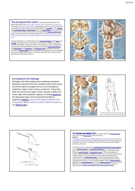
The extrapyramidal system is a neural network located in the<br />
brain that is part of the motor system involved in the coordination of movement.<br />
The system is called "extrapyramidal" to distinguish it from the tracts of the motor<br />
cortex that reach their targets by traveling through the "pyramids" of the medulla.<br />
The pyramidal pathways (corticospinal <strong>and</strong> some corticobulbar tracts) may directly<br />
innervate motor neurons of the spinal cord or brainstem (anterior (ventral) horn<br />
cells or certain cranial nerve nuclei), whereas the extrapyramidal system centers<br />
around the modulation <strong>and</strong> regulation (indirect control) of anterior (ventral) horn<br />
cells.<br />
Extrapyramidal tracts are chiefly found in the reticular formation of the pons <strong>and</strong><br />
medulla, <strong>and</strong> target neurons in the spinal cord involved in reflexes, locomotion,<br />
complex movements, <strong>and</strong> postural control. These tracts are in turn modulated by<br />
various parts of the central nervous system, including the nigrostriatal pathway,<br />
the basal ganglia, the cerebellum, the vestibular nuclei, <strong>and</strong> different sensory<br />
areas of the cerebral cortex. All of these regulatory components can be considered<br />
part of the extrapyramidal system, in that they modulate motor activity without<br />
directly innervating motor neurons.<br />
Corticospinal tract damage<br />
Damage to the descending motor pathways anywhere<br />
along the trajectory from the cerebral cortex to the lower<br />
end of the spinal cord gives rise to a set of symptoms<br />
called the "upper motor neuron syndrome". A few days<br />
after the injury to the upper motor neurons a pattern of<br />
motor signs <strong>and</strong> symptoms appears, including spasticity,<br />
the decreased vigor (<strong>and</strong> increased threshold) of<br />
superficial reflexes, a loss of the ability to perform fine<br />
movements, <strong>and</strong> an extensor plantar response known as<br />
the Babinski sign. [<br />
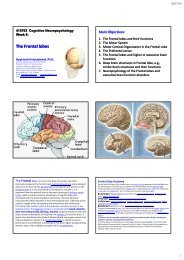
The frontal eye fields (FEF) is a region located in the premotor cortex,<br />
which is part of the frontal cortex of the primate brain.<br />
Function<br />
The cortical area called frontal eye fields (FEF) plays an important role in the control of<br />
visual attention <strong>and</strong> eye movements. Electrical stimulation in the FEF elicits saccadic eye<br />
movements. The FEF have a topographic structure <strong>and</strong> represents saccade targets in<br />
retinotopic coordinates.<br />
The frontal eye field is reported to be activated during the initiation of eye movements,<br />
such as voluntary saccades <strong>and</strong> pursuit eye movements. There is also evidence that it<br />
plays a role in purely sensory processing <strong>and</strong> that it belongs to a “fast brain” system<br />
through a superior colliculus – medial dorsal nucleus –FEF ascending pathway.In<br />
humans, its earliest activations in regard to visual stimuli occur at 45 ms with activations<br />
related to changes in visual stimuli within 45–60 ms (these are comparable with<br />
response times in the primary visual cortex). This fast brain pathway also provides<br />
auditory input at even shorter times starting at 24 ms <strong>and</strong> being affected by auditory<br />
characteristics at 30–60 ms. The FEF constitutes together with the supplementary eye<br />
fields (SEF), the intraparietal sulcus (IPS) <strong>and</strong> the superior colliculus (SC) one of the most<br />
important brain areas involved in the generation <strong>and</strong> control of eye movements,<br />
particularly in the direction contralateral to the frontal eye fields' location.<br />
46