Neurological Examination, clinical cases and neuropsychological ...
Neurological Examination, clinical cases and neuropsychological ...
Neurological Examination, clinical cases and neuropsychological ...

Create successful ePaper yourself
Turn your PDF publications into a flip-book with our unique Google optimized e-Paper software.
23/07/54<br />
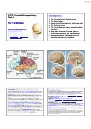
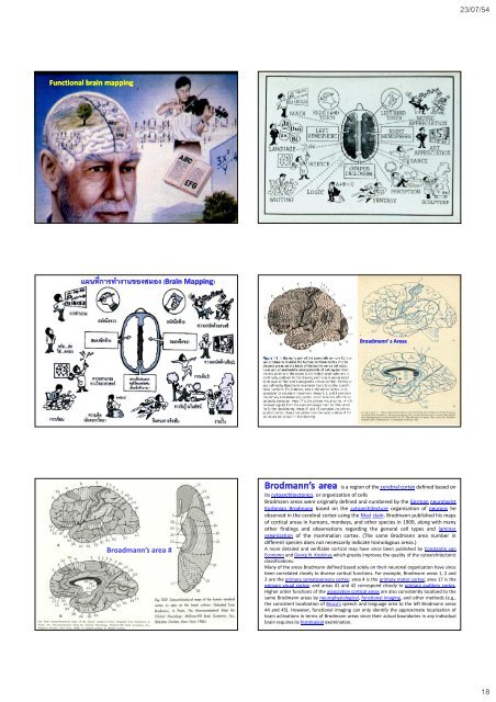
Functional brain mapping<br />
แผนที<br />
่การทํางานของสมอง (Brain Mapping<br />
Brain Mapping)<br />
Broadmann’ s Areas<br />
Broadmann’s area #<br />
Brodmann’s area is a region of the cerebral cortex defined based on<br />
its cytoarchitectonics, or organization of cells<br />
Brodmann areas were originally defined <strong>and</strong> numbered by the German neurologist<br />
Korbinian Brodmann basedonthecytoarchitecture organisation of neurons he<br />
observed in the cerebral cortex using the Nissl stain. Brodmann published his maps<br />
of cortical areas in humans, monkeys, <strong>and</strong> other species in 1909, along with many<br />
other findings <strong>and</strong> observations regarding the general cell types <strong>and</strong> laminar<br />
organization of the mammalian cortex. (The same Brodmann area number in<br />
different species does not necessarily indicate homologous areas.)<br />
A more detailed <strong>and</strong> verifiable cortical map have since been published by Constantin von<br />
Economo <strong>and</strong> Georg N. Koskinas which greatly improves the quality of the cytoarchitectonic<br />
classifications.<br />
Many of the areas Brodmann defined based solely on their neuronal organization have since<br />
been correlated closely to diverse cortical functions. For example, Brodmann areas 1, 2 <strong>and</strong><br />
3aretheprimary somatosensory cortex; area4istheprimary motor cortex; area17isthe<br />
primary visual cortex; <strong>and</strong> areas 41 <strong>and</strong> 42 correspond closely to primary auditory cortex.<br />
Higher order functions of the association cortical areas are also consistently localized to the<br />
same Brodmann areas by neurophysiological, functional imaging, <strong>and</strong> other methods (e.g.,<br />
the consistent localization of Broca's speech <strong>and</strong> language area to the left Brodmann areas<br />
44 <strong>and</strong> 45). However, functional imaging can only identify the approximate localization of<br />
brain activations in terms of Brodmann areas since their actual boundaries in any individual<br />
brain requires its histological examination.<br />
18